
Recombinant Rat Anti-CD8 Antibody (YTS156.7) (CAT#: PABX-035)
Anti-CD8 antibody is a Rat antibody of IgG2b class that binds to an CD8. The mAb is a rat IgG2b against mouse CD8β, which depletes mouse CD8+ T cells in vivo, and blocks interaction between CD8αβ and soluble pMHCI in vitro.
Specific Inquiry

Figure 1 Ca2+ mobilization in OT-I RAG-splenocytes treated with H-2Kb tetramers loaded with the OVA peptide (H-2Kb/OVA) after pre-staining with mAbs 53.6.7 (red line), YTS105.18 (purple line) or YTS156.7.7 (green line).
The tetramers were added at 1 min (arrow) and the sample re-analyzed for a further 4 min. The H-2Kb/OVA tetramer trace is taken from a 5 min time course and overlaid here as a representation of a responding population. Response to the H-2Kb/OVA tetramers in the absence of CD8 antibodies is shown (blue line), as well as response to H-2Kb/SIYR tetramers (black line) as a negative control. All traces indicate the median of the responding population.
Shore, D. A., Issafras, H., Landais, E., Teyton, L., & Wilson, I. A. (2008). The crystal structure of CD8 in complex with YTS156. 7.7 Fab and interaction with other CD8 antibodies define the binding mode of CD8 αβ to MHC class I. Journal of molecular biology, 384(5), 1190-1202.

Figure 2 Equivalent plot to show relative Ca2+ mobilization in OT-I RAG-splenocytes treated with H-2Kb/OVA tetramers after pre-staining now with only the Fabs of mAbs 53.6.7 (red line), YTS105.18 (purple line) or YTS156.7.7 (green line).
The baselines for all traces are normalized to the response to H-2Kb/OVA tetramers in the absence of any Fabs (blue line).
Shore, D. A., Issafras, H., Landais, E., Teyton, L., & Wilson, I. A. (2008). The crystal structure of CD8 in complex with YTS156. 7.7 Fab and interaction with other CD8 antibodies define the binding mode of CD8 αβ to MHC class I. Journal of molecular biology, 384(5), 1190-1202.
SPR

Figure 3 sCD8αβ was determined as having correctly folded native structure by binding to a panel of monoclonal antibodies specific for the CD8α (YTS105.18 and YTS169) and CD8β subunit (YTS156.7)
sCD8αβ was determined as having correctly folded native structure by binding to a panel of monoclonal antibodies specific for the CD8α (YTS105.18 and YTS169) and CD8β subunit (YTS156.7), as determined by surface plasmon resonance (SPR) using a BIAcore 2,000 instrument (BIAcore). mAbs YTS105.18 and YTS169 came from our laboratory (L.T.). Approx. 10,000 response units (RU) of anti-rat IgG Fc (Pierce Biotechnology) was bound to a CM-5 sensor chip by standard amine conjugation. mAbs were passed over the surface of individual flow cells in PBS until an equivalent amount (approx. 250-350 RU) had been captured in each flow cell. Soluble CD8αβ IgSF was subsequently passed over each flow cell in the buffer at concentrations of 0, 3.25, 7.5, 15 and 30 nM. Both CD8 and mAbs were stripped from the surface by injection of 20 mM HCl following each injection.
Shore, D. A., Issafras, H., Landais, E., Teyton, L., & Wilson, I. A. (2008). The crystal structure of CD8 in complex with YTS156. 7.7 Fab and interaction with other CD8 antibodies define the binding mode of CD8 αβ to MHC class I. Journal of molecular biology, 384(5), 1190-1202.
FC

Figure 4 (a) OT-I splenocytes were incubated with a panel of antibodies specific for mouse CD8α (KT12, 53.6.7, YTS105.18) or CD8β (YTS156.7, 53.5.8)
(a) OT-I splenocytes were incubated with a panel of antibodies specific for mouse CD8α (KT12, 53.6.7, YTS105.18) or CD8β (YTS156.7, 53.5.8), before being stained with 50 nM H-2Kb/OVA PE-tetramers (dark line, right panel), or with a PE-conjugated secondary antibody (dark line, left panel). The staining with tetramers of H-2Kb/OVA (right, dark grey) and tetramers of H-2Kb/SIYR (right, light grey) in absence of anti-CD8 antibody, as well as the background of the secondary antibody (left, light grey), are shown as controls. (b) Mean fluorescence of H-2Kb/OVA PE-tetramer staining on OT-I splenocytes in the presence of increasing concentrations of anti-CD8. The experiment was performed as in (a).
Shore, D. A., Issafras, H., Landais, E., Teyton, L., & Wilson, I. A. (2008). The crystal structure of CD8 in complex with YTS156. 7.7 Fab and interaction with other CD8 antibodies define the binding mode of CD8 αβ to MHC class I. Journal of molecular biology, 384(5), 1190-1202.
Specifications
- Immunogen
- The details of the immunogen for this antibody are not available.
- Host Species
- Rat
- Derivation
- Rat
- Type
- IgG
- Specificity
- Tested positive against native Mouse CD8
- Species Reactivity
- Mouse
- Clone
- YTS156. 7
- Applications
- Can be useful in applications such as: Western blot; Enzyme-linked Immunosorbent Assay; Functional Study
Product Property
- Purity
- >95% by SDS-PAGE and HPLC analysis
- Storage
- Store the antibody (in aliquots) at -20°C. Avoid repeated freezing and thawing of samples.
Target
- Alternative Names
- CD8; anti-CD8
Related Resources
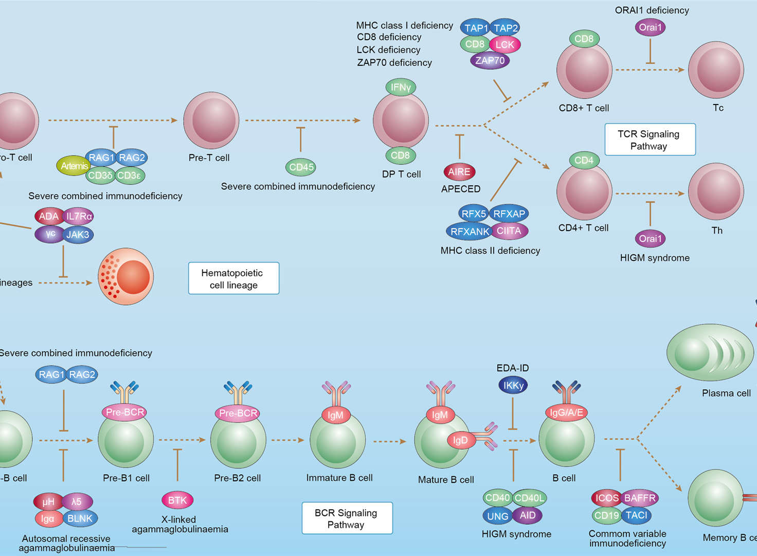 Primary Immunodeficiency
Product Notes
This is a product of Creative Biolabs' Hi-Affi™ recombinant antibody portfolio, which has several benefits including:
• Increased sensitivity
• Confirmed specificity
• High repeatability
• Excellent batch-to-batch consistency
• Sustainable supply
• Animal-free production
See more details about Hi-Affi™ recombinant antibody benefits.
Downloads
Download resources about recombinant antibody development and antibody engineering to boost your research.
See other products for "Clone YTS156. 7"
See other products for "CD8"
Recombinant Antibody
| CAT | Product Name | Application | Type |
|---|---|---|---|
| MOB-0275MZ | Recombinant Mouse Anti-Human CD8 Antibody (clone D9/145B) | IHC | Mouse antibody |
| FAMAB-1680CQ | Human Anti-CD8 Recombinant Antibody (clone G10-1) | Immunotoxin, IP, FC, IF, IHC | Human IgG1 |
| FAMAB-1682CQ | Human Anti-CD8 Recombinant Antibody (clone 14) | FACS, IP, WB, ELISA, IHC | Human IgG1 |
Chimeric Antibody
| CAT | Product Name | Application | Type |
|---|---|---|---|
| PABX-035-S (P) | Recombinant Rat Anti-CD8 Antibody scFv Fragment (YTS156.7) | WB, ELISA, FuncS | scFv |
Mouse Antibody
| CAT | Product Name | Application | Type |
|---|---|---|---|
| PABX-035-F (E) | Recombinant Rat Anti-CD8 Antibody Fab Fragment (YTS156.7) | WB, ELISA, FuncS | Fab |
Customer Reviews and Q&As

There are currently no Customer reviews or questions for PABX-035. Click the button above to contact us or submit your feedback about this product.
View the frequently asked questions answered by Creative Biolabs Support.
For Research Use Only. Not For Clinical Use.
For research use only. Not intended for any clinical use. No products from Creative Biolabs may be resold, modified for resale or used to manufacture commercial products without prior written approval from Creative Biolabs.
Send Inquiry
This site is protected by reCAPTCHA and the Google Privacy Policy and Terms of Service apply.