
Anti-EGFR Recombinant Antibody (Imgatuzumab) (CAT#: TAB-753)
Recombinant humanized (from rat) antibody expressed in CHO binding to human EGFR. Imgatuzumab is a humanized monoclonal antibody designed for the treatment of cancer. It acts as an immunomodulator.
Specific Inquiry
Cyt

Figure 1 In vitro ADCC analysis of imgatuzumab, cetuximab or the combination.
PBMCs as effector cells were added to the untreated H441 (A), H292 (B) or A549 (C) target cells at a ratio of 25:1. ADCC responses were normalized to the ADCC response of the highest imgatuzumab concentration due to variations in responses between PBMCs of different donors. Absolute ADCC responses ranged between 12 and 53%. (*P < 0.05, **P < 0.01, ***P < 0.001 cetuximab vs imgatuzumab). (D) A549 cells were pretreated with imgatuzumab alone (20 µg/mL) or combined with cetuximab (20 µg/mL total) in culture medium for 72 hours. Next, cells with (open triangle and open square) or without (solid triangle and solid square) antibody pretreatment for 72 hours were washed and the ADCC assay was performed using PBMCs as effector cells and A549 cells as target cells at a ratio of 25:1. Data points are mean ± SD (n = 3).
Kol, A., van Scheltinga, A. T., Pool, M., Gerdes, C., de Vries, E., & de Jong, S. (2017). ADCC responses and blocking of EGFR-mediated signaling and cell growth by combining the anti-EGFR antibodies imgatuzumab and cetuximab in NSCLC cells. Oncotarget, 8(28), 45432.
Inhib

Figure 2 In vitro growth-inhibitory effect of anti-EGFR monoclonal antibody treatment.
Cells were seeded in 12-wells plates and allowed to adhere overnight. Next, H292, H322 and A549 cells were treated with the anti-EGFR monoclonal antibodies (20 µg/ mL total) for the indicated days under normal growth conditions without additional EGF (medium with 10% FCS) or in medium containing 10% FCS with 1 or 10 ng/mL EGF. Data points are mean + SD. All experiments were performed in triplicate.
Kol, A., van Scheltinga, A. T., Pool, M., Gerdes, C., de Vries, E., & de Jong, S. (2017). ADCC responses and blocking of EGFR-mediated signaling and cell growth by combining the anti-EGFR antibodies imgatuzumab and cetuximab in NSCLC cells. Oncotarget, 8(28), 45432.
WB

Figure 3 Cellular localization and endocytic trafficking of anti-EGFR monoclonal antibodies in SW-1573.
Kol, A., van Scheltinga, A. T., Pool, M., Gerdes, C., de Vries, E., & de Jong, S. (2017). ADCC responses and blocking of EGFR-mediated signaling and cell growth by combining the anti-EGFR antibodies imgatuzumab and cetuximab in NSCLC cells. Oncotarget, 8(28), 45432.
Inter

Figure 4 Effects of TKI treatment on monoclonal antibody-induced internalization and degradation.
(A) Western blot analysis of the effect of 24 hours anti-EGFR monoclonal antibody treatment (20 µg/mL total) with or without erlotinib (10 µM) on EGFR total protein levels in SW-1573, H292 and A549. Data points are mean + SD. (*P < 0.05, **P < 0.01, ***P < 0.001 compared to untreated control; $ P < 0.05, $ P < 0.01, $$ P < 0.001 compared to erlotinib treated control). (B) SW-1573 cells were pretreated for 24 hours with the EGFR TKI inhibitor erlotinib (10 µM). Cells were subsequently surface labeled with the anti-EGFR monoclonal antibodies (20 µg/mL total) on ice and incubated at 37 °C for 4 hours, and then analyzed for the non-internalized EGFR-antibody complexes (NI) and cell surface expression (SE). The lower the amount of non-internalized EGFR-antibody complexes, the higher the amount of internalized antibodyEGFR complexes. Surface expression of the control and erlotinib treated cells were both set at 100%. 24 hours erlotinib treatment resulted in a slight upregulation (10 – 15 % increase). Data points are mean ± SD (n = 3). (*P < 0.05).
Kol, A., van Scheltinga, A. T., Pool, M., Gerdes, C., de Vries, E., & de Jong, S. (2017). ADCC responses and blocking of EGFR-mediated signaling and cell growth by combining the anti-EGFR antibodies imgatuzumab and cetuximab in NSCLC cells. Oncotarget, 8(28), 45432.
Inter

Figure 5 Influence of monoclonal antibody binding on EGFR internalization and surface expression.
SW-1573 (A) and H292 cells (B) were surface labeled with the anti-EGFR monoclonal antibodies on ice and incubated at 37 °C for the indicated times, and then analyzed for the non-internalized EGFR-antibody complexes (left) and cell surface expression (right). The lower the amount of noninternalized EGFR-antibody complexes, the higher the amount of internalized antibody-EGFR complexes. The surface expression at t=0 was set at 100%. The non-internalized and surface expression were determined as described in Materials and Methods (n=3). Data points are mean ± SD. (**P < 0.01, ***P < 0.001 combination vs imgatuzumab; $ P < 0.01, $$ P < 0.001 combination vs cetuximab; two-way ANOVA followed by Bonferroni post-test).
Kol, A., van Scheltinga, A. T., Pool, M., Gerdes, C., de Vries, E., & de Jong, S. (2017). ADCC responses and blocking of EGFR-mediated signaling and cell growth by combining the anti-EGFR antibodies imgatuzumab and cetuximab in NSCLC cells. Oncotarget, 8(28), 45432.
WB

Figure 6 Western blot analysis of the effect of 24 hours anti-EGFR monoclonal antibody treatment on EGFR total protein levels and downstream signaling under normal growth conditions or 15 min EGF stimulation (10 ng/mL).
The immunoreactive spots were quantified by densitometric analysis and normalized using actin (see also Supplementary Figure 2). Values are expressed as fold increase versus control + SD. All experiments were performed in triplicate.
Kol, A., van Scheltinga, A. T., Pool, M., Gerdes, C., de Vries, E., & de Jong, S. (2017). ADCC responses and blocking of EGFR-mediated signaling and cell growth by combining the anti-EGFR antibodies imgatuzumab and cetuximab in NSCLC cells. Oncotarget, 8(28), 45432.
FC

Figure 7 Effect of anti-EGFR monoclonal antibody treatment on EGFR surface expression levels.
(A) Flow cytometric analysis of imgatuzumab and cetuximab binding alone or in combination in H322, SW-1573, H441, H292 and A549 cells. (B) H322, SW-1573, H292, H441 and A549 cells were treated with the anti-EGFR monoclonal antibodies (20 µg/mL total) for 72 hours. Surface expression levels were determined using flow cytometry. The surface expression in untreated control cells was set at 100% both for the single antibodies and the combination. (*P < 0.05, **P < 0.01 combination vs imgatuzumab; $ P < 0.05, $ P < 0.01, $$ P < 0.001 combination vs cetuximab; unpaired t-test). Data points are mean + SD (n = 3).
Kol, A., van Scheltinga, A. T., Pool, M., Gerdes, C., de Vries, E., & de Jong, S. (2017). ADCC responses and blocking of EGFR-mediated signaling and cell growth by combining the anti-EGFR antibodies imgatuzumab and cetuximab in NSCLC cells. Oncotarget, 8(28), 45432.
IHC

Figure 8 EGFR expression analysis and immunohistochemistry.
(A) Ex vivo tissue analysis. Mean vessel density (MVD), EGFR and hematoxylin and eosin immunohistochemical staining of A431, A549 and H441 tumors. (B) In vitro flow cytometric analysis of EGFR membrane expression in A431, A549 and H441 cells. (C) Ex vivo human EGFR extracellular domain levels in A431, A549 and H441 tumor tissue lysates. (D) EGFR histo-score (H-score) for A431, A549 and H441 tumors. (E) Mean vessel density score for A431, A549 and H441 tumors.
Pool, M., Kol, A., Lub-de Hooge, M. N., Gerdes, C. A., de Jong, S., de Vries, E. G., & van Scheltinga, A. G. T. (2016). Extracellular domain shedding influences specific tumor uptake and organ distribution of the EGFR PET tracer 89Zr-imgatuzumab. Oncotarget, 7(42), 68111.
ELISA

Figure 9 EGFR analysis of plasma, liver and cell lysates.
(A) Human EGFR extracellular domain concentration in plasma of A431, A549 and H441 xenograft bearing and non-tumor bearing control mice. (B) Human EGFR extracellular domain level in liver tissue lysates of A431, A549 and H441 xenograft bearing and non-tumor bearing control mice. (C) Human EGFR extracellular domain concentration in serum-free supernates of confluent cell cultures of A431, A549 and H441 cells. (D) Human EGFR extracellular domain concentration in serum-containing supernates of confluent cell cultures of A431, A549 and H441 cells, only statistical significances against A431 are shown for clarity.
Pool, M., Kol, A., Lub-de Hooge, M. N., Gerdes, C. A., de Jong, S., de Vries, E. G., & van Scheltinga, A. G. T. (2016). Extracellular domain shedding influences specific tumor uptake and organ distribution of the EGFR PET tracer 89Zr-imgatuzumab. Oncotarget, 7(42), 68111.


Specifications
- Immunogen
- The details of the immunogen for this antibody are not available.
- Host Species
- Rat
- Derivation
- Humanized (from rat)
- Type
- IgG1 - kappa
- Species Reactivity
- Human
- Applications
- Neut, ELISA, IF, IP, FuncS, FC, WB, Cyt, Inhib, IHC
- CAS
- 959963-46-3
- Generic Name
- imgatuzumab
- MW
- 145.0 kDa
- Related Disease
- Solid tumors
Product Property
- Purity
- >95.0% as determined by analysis by SDS-PAGE.
- Storage
- Store at -20°C for long-term storage. Store at 2-8°C for up to one month. Avoid freeze/thaw cycles.
Applications
- Application Notes
- The EGFR antibody has been reported in applications of Neut, ELISA, IF, IP, FuncS, FC, WB, Cyt, Inhib, IHC.
Target
- Alternative Names
- imgatuzumab;959963-46-3;RG7160;GA201;RO5083945;ICR62;EGFR;epidermal growth factor receptor;epidermal growth factor receptor (avian erythroblastic leukemia viral (v erb b) oncogene homolog) , ERBB;ERBB1;erythroblastic leukemia viral (v erb b) oncogene homo
- Gene ID
- 1956
- UniProt ID
- P00533
Related Resources
Please refer to Imgatuzumab Overview to learn more about the mechanism of action, clinical projects, and approved drugs of Imgatuzumab.
 Bladder Cancer
 Non-small Cell Lung Cancer
 Pancreatic Cancer
 Hepatocellular Carcinoma
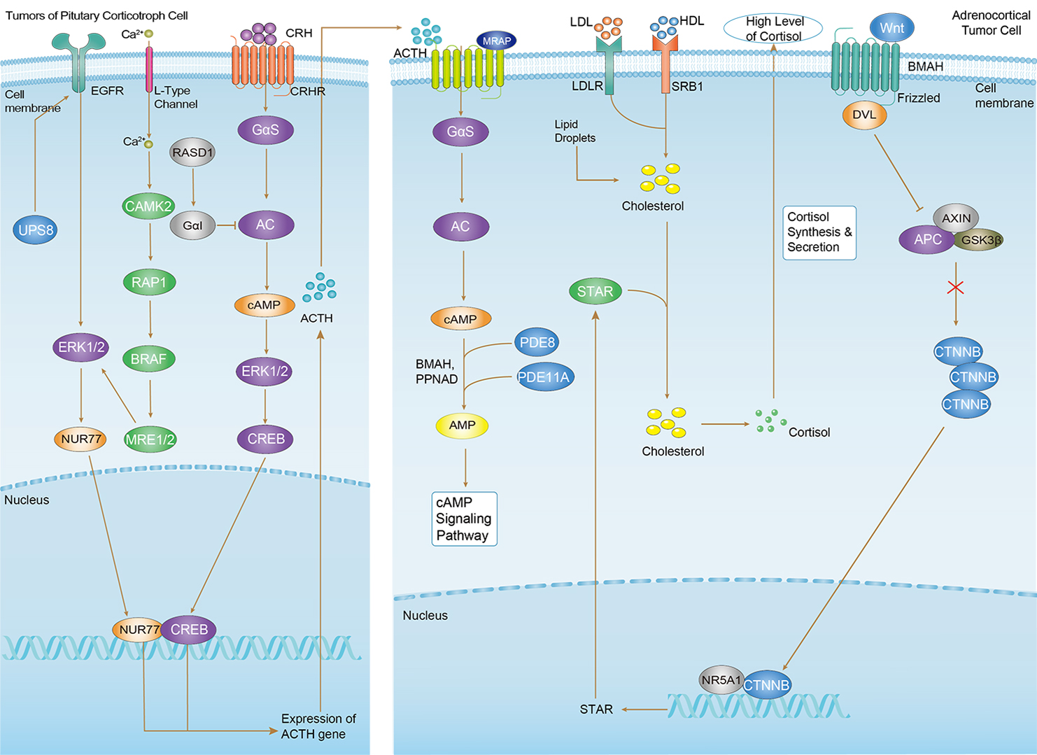 Cushing Syndrome
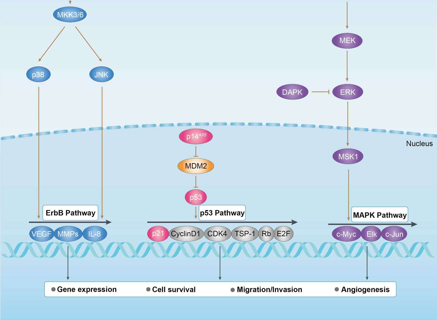
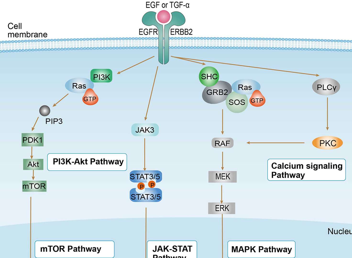
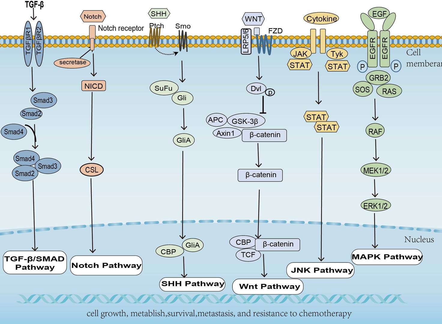
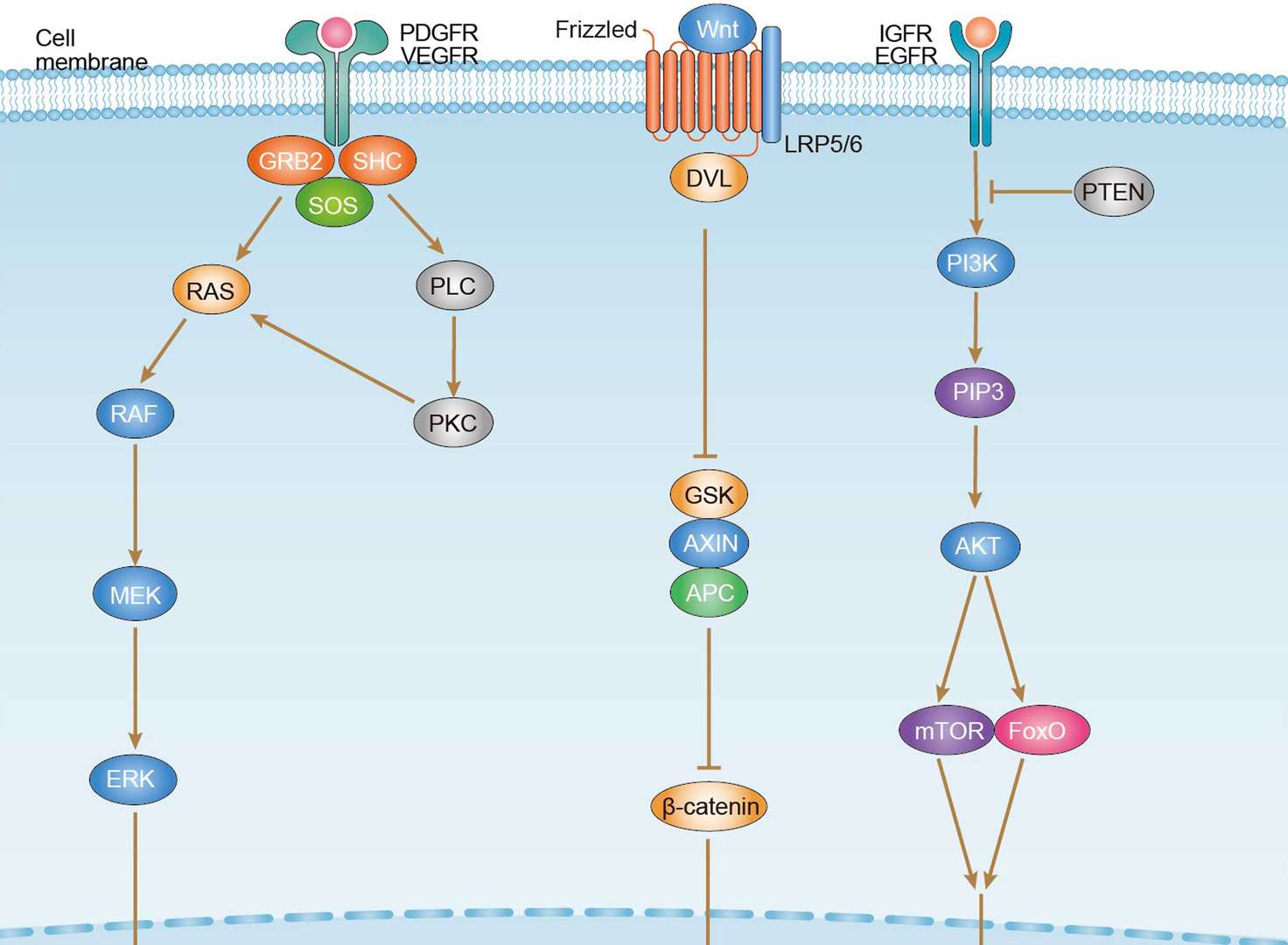
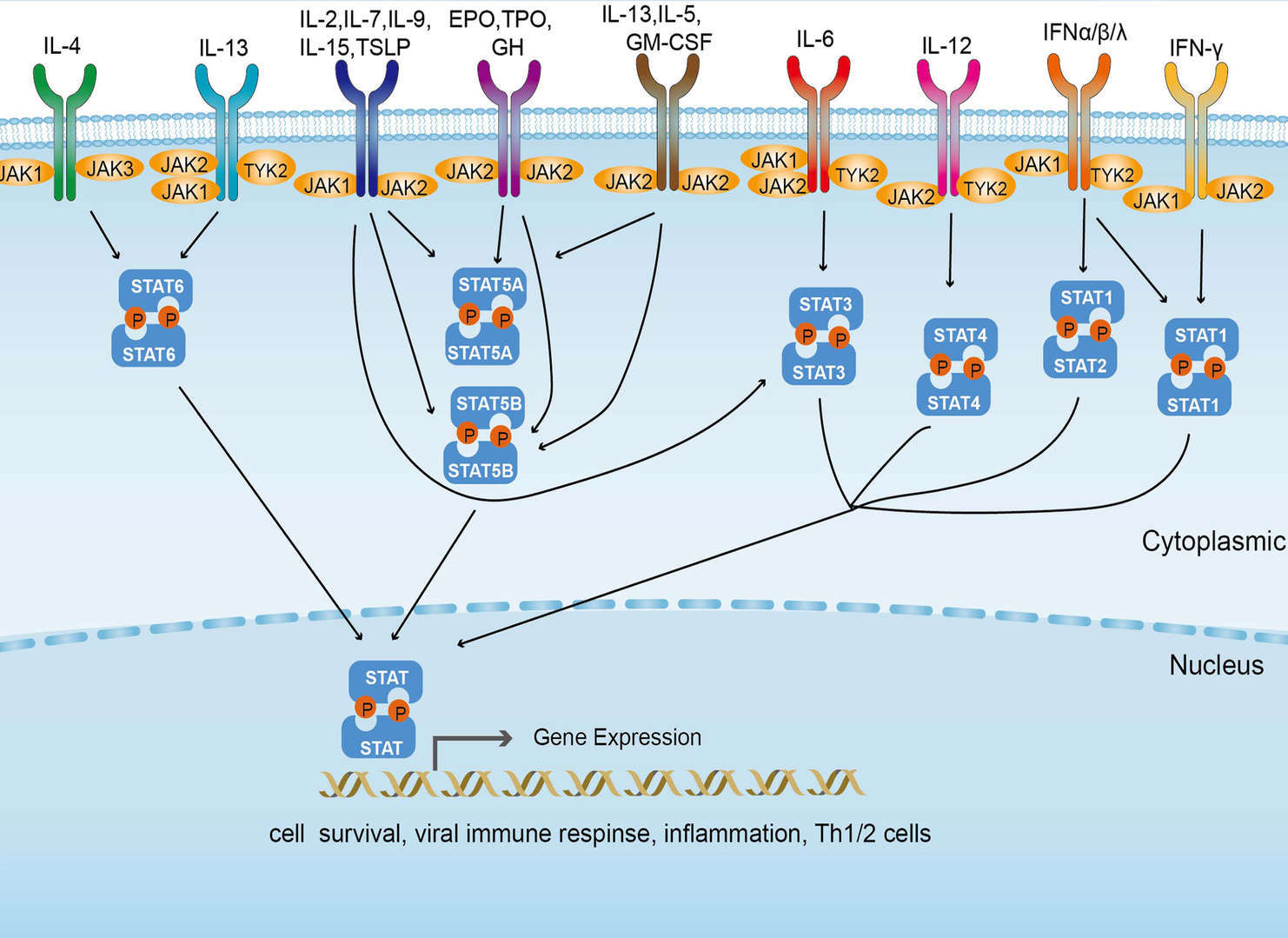 JAK-STAT Signaling Pathway
Product Notes
This is a product of Creative Biolabs' Hi-Affi™ recombinant antibody portfolio, which has several benefits including:
• Increased sensitivity
• Confirmed specificity
• High repeatability
• Excellent batch-to-batch consistency
• Sustainable supply
• Animal-free production
See more details about Hi-Affi™ recombinant antibody benefits.
Downloads
Download resources about recombinant antibody development and antibody engineering to boost your research.
See other products for "Imgatuzumab"
Afuco™ Anti-EGFR ADCC Recombinant Antibody (Imgatuzumab), ADCC EnhancedThis product is an ADCC enhanced antibody produced by our Afuco™ platform. Recombinant humanized (from rat) antibody expressed in CHO binding to human EGFR. Imgatuzumab is a humanized monoclonal antibody designed for the treatment of cancer. It acts as an immunomodulator.
See other products for "EGFR"
Chimeric Antibody
| CAT | Product Name | Application | Type |
|---|---|---|---|
| TAB-H35 | Anti-Human EGFR Recombinant Antibody (Futuximab) | IF, WB, Inhib | IgG1 - kappa |
| TAB-003 | Anti-Human EGFR Recombinant Antibody (Cetuximab) | IF, IP, Neut, FuncS, ELISA, FC, ICC | IgG1 - kappa |
| TAB-0225CL-F(E) | Human Anti-EGFR Recombinant Antibody; Fab Fragment (TAB-0225CL-F(E)) | Block, Inhib, FuncS, Apop, In vivo | Chimeric (Mouse/Human) Fab |
| TAB-308MZ | Human Anti-EGFR Recombinant Antibody (TAB-308MZ) | FuncS | Chimeric antibody (mouse/human) |
| TAB-302MZ-S(P) | Mouse Anti-EGFR Recombinant Antibody; scFv Fragment (TAB-302MZ-S(P)) | ELISA | Mouse scFv |
Recombinant Antibody
| CAT | Product Name | Application | Type |
|---|---|---|---|
| MOB-0242MC | Rabbit Anti-Human EGFR (phospho Y1092) Antibody | IHC, WB | |
| MOB-0243MC | Rabbit Anti-Human EGFR (phospho Y1068) Antibody | IHC, WB | |
| TAB-477CQ | Human Anti-EGFR Recombinant Antibody (clone Laprituximab) | ELISA, IHC, FC, IP, IF, WB | Chimeric (mouse/human) IgG1, κ |
| MOB-052LC | Mouse Anti-EGFR Recombinant Antibody (clone 7H7) | WB, IP | Mouse IgG |
| MOB-053LC | Mouse Anti-EGFR Recombinant Antibody (clone B11) | ELISA, IF, IP, WB | Mouse IgG |
Mouse Antibody
| CAT | Product Name | Application | Type |
|---|---|---|---|
| TAB-0564CL | Mouse Anti-EGFR Recombinant Antibody (TAB-0564CL) | ELISA | Mouse IgG |
| TAB-0565CL | Mouse Anti-EGFR Recombinant Antibody (TAB-0565CL) | ELISA | Mouse IgG |
| TAB-0564CL-S(P) | Mouse Anti-EGFR Recombinant Antibody; scFv Fragment (TAB-0564CL-S(P)) | ELISA | Mouse scFv |
| TAB-0565CL-S(P) | Mouse Anti-EGFR Recombinant Antibody; scFv Fragment (TAB-0565CL-S(P)) | ELISA | Mouse scFv |
| TAB-271MZ-F(E) | Mouse Anti-EGFR Recombinant Antibody; Fab Fragment (TAB-271MZ-F(E)) | FuncS | Mouse Fab |
Humanized Antibody
| CAT | Product Name | Application | Type |
|---|---|---|---|
| TAB-274MZ | Human Anti-EGFR Recombinant Antibody (TAB-274MZ) | FC | Humanized IgG |
| TAB-297MZ | Anti-Human EGFR Recombinant Antibody (H225) | ELISA, WB | Humanized antibody |
| TAB-300MZ | Human Anti-EGFR Recombinant Antibody (TAB-300MZ) | ELISA | Humanized IgG |
| TAB-274MZ-S(P) | Human Anti-EGFR Recombinant Antibody; scFv Fragment (TAB-274MZ-S(P)) | FC | Humanized scFv |
| TAB-297MZ-S(P) | Anti-Human EGFR Recombinant Antibody scFv Fragment (H225) | ELISA, WB | Humanized antibody |
Human Antibody
| CAT | Product Name | Application | Type |
|---|---|---|---|
| TAB-313MZ-S(P) | Anti-Human EGFR Recombinant Antibody scFv Fragment (EG-19-11) | ELISA, FC | Human antibody |
| TAB-314MZ-S(P) | Anti-Human EGFR Recombinant Antibody scFv Fragment (EG-26-11) | ELISA, FC | Human antibody |
| TAB-315MZ-S(P) | Anti-Human EGFR Recombinant Antibody scFv Fragment (528) | WB, FC | Human antibody |
| TAB-321MZ-S(P) | Anti-Human EGFR Recombinant Antibody scFv Fragment (DX 1-4) | SPR | Human antibody |
| TAB-322MZ-S(P) | Anti-Human EGFR Recombinant Antibody scFv Fragment (DX 1-6) | SPR | Human antibody |
Fc Glycosylation
| CAT | Product Name | Application | Type |
|---|---|---|---|
| Gly-055LC | Recombinant Anti-Human EGFR Antibody (Fc glycosylation/High-mannose glycosylated) | ELISA | Chimeric antibody (mouse/human) |
| Gly-144LC | Recombinant Anti-Human EGFR Antibody (Fc glycosylation) | ELISA | Humanized antibody |
High-mannose Glycoform
| CAT | Product Name | Application | Type |
|---|---|---|---|
| Gly-055LC-1 | Recombinant Anti-Human EGFR Antibody (Fc glycosylation/High-mannose glycosylated) | ELISA | Chimeric antibody (mouse/human) |
Deglycosylated Antibody (Non-glycosylated IgGs)
| CAT | Product Name | Application | Type |
|---|---|---|---|
| Gly-167LC | Recombinant Anti-Human EGFR Antibody (Non-glycosylated) | ELISA | Human antibody |
Chicken IgY Antibody
| CAT | Product Name | Application | Type |
|---|---|---|---|
| BRD-0183MZ | Chicken Anti-EGFR Polyclonal IgY | WB | Chicken antibody |
| BRD-0668MZ | Chicken Anti-EGFR Polyclonal IgY | WB | Chicken antibody |
MHC Tetramer for Cancer
| CAT | Product Name | Application | Type |
|---|---|---|---|
| MHC-LC773 | A*0201/Human EGFR (YLNTVQPTCV) MHC Tetramer | FCM | |
| MHC-LC4545 | PE-DQB1*03:02/Human EGFR (SRALEEKKGNYVVTHG) MHC Tetramer | FCM | |
| MHC-YF409 | A*02:01/Human EGF-R-479 (KLFGTSGQKT) MHC Pentamer | FCM |
Neutralizing Antibody
| CAT | Product Name | Application | Type |
|---|---|---|---|
| NEUT-722CQ | Rabbit Anti-EGFR Recombinant Antibody (clone CBL1011) | Neut | Rabbit IgG |
| NEUT-723CQ | Mouse Anti-EGFR Recombinant Antibody (clone CBL931) | WB, IP, IHC, ICC, Neut | Mouse IgG1 |
| NEUT-724CQ | Rabbit Anti-EGFR Recombinant Antibody (clone D1D4J) | IF, FC, WB, IP, Neut | Rabbit IgG |
Rabbit Monoclonal Antibody
| CAT | Product Name | Application | Type |
|---|---|---|---|
| MOR-1101 | Hi-Affi™ Rabbit Anti-EGFR Recombinant Antibody (clone DS1101AB) | IHC-P | Rabbit IgG |
| MOR-4520 | Hi-Affi™ Rabbit Anti-EGFR Recombinant Antibody (clone TH28DS) | IF, ICC, FC | Rabbit IgG |
| MOR-4570 | Hi-Affi™ Rabbit Anti-EGFR Recombinant Antibody (clone TH82DS) | ELISA | Rabbit IgG |
| MOR-4675 | Hi-Affi™ Rabbit Anti-EGFR Recombinant Antibody (clone TH189DS) | WB, IF, ICC, FC | Rabbit IgG |
| MOR-4676 | Hi-Affi™ Rabbit Anti-EGFR Recombinant Antibody (clone TH190DS) | WB, IF, ICC, FC | Rabbit IgG |
Fab Fragment Antibody
| CAT | Product Name | Application | Type |
|---|---|---|---|
| HPAB-0010-YC-F(E) | Mouse Anti-EGFR Recombinant Antibody (clone L211C); Fab Fragment | ELISA, FC | Mouse Fab |
| HPAB-0011-YC-F(E) | Human Anti-EGFR Recombinant Antibody (clone Pep 1); Fab Fragment | FC | Human Fab |
| HPAB-0012-YC-F(E) | Human Anti-EGFR Recombinant Antibody (clone Pep 2); Fab Fragment | FC | Human Fab |
| HPAB-0013-YC-F(E) | Human Anti-EGFR Recombinant Antibody (clone Pep 3); Fab Fragment | FC | Human Fab |
| HPAB-0014-YC-F(E) | Human Anti-EGFR Recombinant Antibody (clone Pep 4); Fab Fragment | FC | Human Fab |
scFv Fragment Antibody
| CAT | Product Name | Application | Type |
|---|---|---|---|
| HPAB-0727-CN-S(P) | Mouse Anti-EGFR Recombinant Antibody (clone EGFR); scFv Fragment | ELISA, FC, IHC, Neut | Mouse scFv |
| HPAB-2822LY-S(P) | Human Anti-EGFR Recombinant Antibody (clone CBL141); scFv Fragment | FC | Human scFv |
| HPAB-0894-WJ-S(P) | Human Anti-EGFR Recombinant Antibody (clone YZ-EGFR V1); scFv Fragment | ELISA, WB | Human scFv |
| HPAB-0895-WJ-S(P) | Human Anti-EGFR Recombinant Antibody (clone YZ-EGFR V2); scFv Fragment | ELISA, WB | Human scFv |
| HPAB-S0023-YC-S(P) | Mouse Anti-EGFR Recombinant Antibody (clone 14E1); scFv Fragment | IB, FuncS | Mouse scFv |
ADCC Enhanced Antibody
| CAT | Product Name | Application | Type |
|---|---|---|---|
| AFC-TAB-165 | Afuco™ Anti-EGFR ADCC Recombinant Antibody (Matuzumab), ADCC Enhanced | Neut, ELISA, IF, IP, FuncS, FC | ADCC enhanced antibody |
| AFC-TAB-464CQ | Afuco™ Anti-EGFR ADCC Recombinant Antibody (Tomuzotuximab), ADCC Enhanced | ELISA, IHC, FC, IP, IF, FuncS | ADCC enhanced antibody |
| AFC-TAB-003 | Afuco™ Anti-EGFR ADCC Recombinant Antibody (Cetuximab), ADCC Enhanced | IF, IP, Neut, FuncS, ELISA, FC | ADCC enhanced antibody |
| AFC-TAB-040 | Afuco™ Anti-EGFR ADCC Recombinant Antibody (Zalutumumab), ADCC Enhanced | ELISA, FC, IP, FuncS, IF, Neut | ADCC enhanced antibody |
| AFC-TAB-119 | Afuco™ Anti-EGFR ADCC Recombinant Antibody (Necitumumab), ADCC Enhanced | FC, IP, ELISA, Neut, FuncS, IF | ADCC enhanced antibody |
Single-domain Antibody
| CAT | Product Name | Application | Type |
|---|---|---|---|
| HPAB-AP881-YC | Recombinant Llama Anti-EGFR Single Domain Antibody (VHH122) | ELISA, FC, IP, FuncS | Llama VHH |
| HPAB-AP882-YC | Recombinant Llama Anti-EGFR Single Domain Antibody (VHH205) | ELISA | Llama VHH |
| HPAB-AP883-YC | Recombinant Llama Anti-EGFR Single Domain Antibody (VHH03) | ELISA | Llama VHH |
| HPAB-AP884-YC | Recombinant Llama Anti-EGFR Single Domain Antibody (VHH139) | ELISA | Llama VHH |
| HPAB-AP885-YC | Recombinant Llama Anti-EGFR Single Domain Antibody (VHH107) | ELISA | Llama VHH |
Customer Reviews and Q&As

There are currently no Customer reviews or questions for TAB-753. Click the button above to contact us or submit your feedback about this product.
View the frequently asked questions answered by Creative Biolabs Support.
For Research Use Only. Not For Clinical Use.
For research use only. Not intended for any clinical use. No products from Creative Biolabs may be resold, modified for resale or used to manufacture commercial products without prior written approval from Creative Biolabs.
Send Inquiry
This site is protected by reCAPTCHA and the Google Privacy Policy and Terms of Service apply.