
Recombinant Anti-Human EGFR VHH Single Domain Antibody (7D12) (CAT#: PNBL-016)
Anti-Human EGFR VHH Single Domain Antibody is a recombinant protein produced in E. coli.
Specific Inquiry

Figure 1 In vitro characterisation of selected monovalent anti‐EGFR sdAbs.
The binding of biotinylated EGF (800 pM) to an EGFR‐ECD‐Fc fusion was tested in the presence of increasing amounts of the 9G8, 7D12, 7C12 or 38G7 sdAbs, or without sdAb. Receptor‐bound EGF was then detected via peroxidase‐coupled streptavidin and staining with OPD/H2O2.
Roovers, R. C., Vosjan, M. J., Laeremans, T., el Khoulati, R., de Bruin, R. C., Ferguson, K. M., ... & van Bergen en Henegouwen, P. M. (2011). A biparatopic anti‐EGFR sdAb efficiently inhibits solid tumour growth. International journal of cancer, 129(8), 2013-2024.

Figure 2 In vitro characterisation of selected monovalent anti‐EGFR sdAbs.
The binding of 7D12 sdAbs to EGFR, expressed on phage, was detected in a 100-fold molar excess of the indicated antibody (cetuximab) or antibody fragments.
Roovers, R. C., Vosjan, M. J., Laeremans, T., el Khoulati, R., de Bruin, R. C., Ferguson, K. M., ... & van Bergen en Henegouwen, P. M. (2011). A biparatopic anti‐EGFR sdAb efficiently inhibits solid tumour growth. International journal of cancer, 129(8), 2013-2024.
Inhib

Figure 3 Inhibition of A431 tumour cell proliferation by monovalent and bivalent sdAbs.
The proliferation of A431 cells in the presence of increasing amounts of sdAb monovalent and bivalent 7D12; was measured using a sulphorhodaminebased stain of total cellular protein after TCA precipitation. Proliferation is plotted as percentage of maximal growth (cells left without treatment). The whole antibody cetuximab was used as ''standard'' in every test. Data points where the value of the control is statistically different from that of the sdAb-treated group are indicated by an asterisk.
Roovers, R. C., Vosjan, M. J., Laeremans, T., el Khoulati, R., de Bruin, R. C., Ferguson, K. M., ... & van Bergen en Henegouwen, P. M. (2011). A biparatopic anti‐EGFR sdAb efficiently inhibits solid tumour growth. International journal of cancer, 129(8), 2013-2024.
Inhib

Figure 4 Inhibition of A431 tumour cell proliferation by monovalent and bivalent sdAbs.
The proliferation of A431 cells in the presence of increasing amounts of sdAb mixtures of monovalent and bivalent 7D12 and 9G8; was measured using a sulphorhodaminebased stain of total cellular protein after TCA precipitation. Proliferation is plotted as percentage of maximal growth (cells left without treatment). The whole antibody cetuximab was used as ''standard'' in every test. Data points where the value of the control is statistically different from that of the sdAb-treated group are indicated by an asterisk; only different between 7D12þ9G8 and control.
Roovers, R. C., Vosjan, M. J., Laeremans, T., el Khoulati, R., de Bruin, R. C., Ferguson, K. M., ... & van Bergen en Henegouwen, P. M. (2011). A biparatopic anti‐EGFR sdAb efficiently inhibits solid tumour growth. International journal of cancer, 129(8), 2013-2024.
FuncS

Figure 6 In vitro optimalisation and characterisation of trivalent biparatopic sdAb CONAN-1.
The proliferation of A431 cells in the presence of increasing amounts of biparatopic sdAb 7D12-9G8 (with linkers varying in length between the two sdAb units) was measured using a sulphorhodamine-based stain of total cellular protein after TCA precipitation. Proliferation is plotted as percentage of maximal growth (cells left without treatment). Error bars have been omitted to increase the clarity of the figure. The different linker lengths between the two sdAbs are indicated.
Roovers, R. C., Vosjan, M. J., Laeremans, T., el Khoulati, R., de Bruin, R. C., Ferguson, K. M., ... & van Bergen en Henegouwen, P. M. (2011). A biparatopic anti‐EGFR sdAb efficiently inhibits solid tumour growth. International journal of cancer, 129(8), 2013-2024.
FuncS

Figure 7 In vitro optimalisation and characterisation of trivalent biparatopic sdAb CONAN-1.
Purified EGFR-ECD-Fc fusion protein was immobilised via a coated anti-human Fc anti-serum, and a concentration range of either bivalent (7D12-9G8) or trivalent (CONAN-1) sdAb was bound. The binding of biotinylated mouse serum albumin (MSA) was subsequently detected with peroxidase-conjugated streptavidin and staining with OPD/H2O2.
Roovers, R. C., Vosjan, M. J., Laeremans, T., el Khoulati, R., de Bruin, R. C., Ferguson, K. M., ... & van Bergen en Henegouwen, P. M. (2011). A biparatopic anti‐EGFR sdAb efficiently inhibits solid tumour growth. International journal of cancer, 129(8), 2013-2024.
FuncS

Figure 8 In vitro optimalisation and characterisation of trivalent biparatopic sdAb CONAN-1.
The proliferation of A431 cells in the presence of increasing amounts of sdAb was measured using the sulphorhodamine B assay. The effect of the presence of human serum albumin (HSA: 1%) in the medium was assessed.
Roovers, R. C., Vosjan, M. J., Laeremans, T., el Khoulati, R., de Bruin, R. C., Ferguson, K. M., ... & van Bergen en Henegouwen, P. M. (2011). A biparatopic anti‐EGFR sdAb efficiently inhibits solid tumour growth. International journal of cancer, 129(8), 2013-2024.
FuncS

Figure 9 In vitro specificity of ⁹⁹ᵐTc-7C12 and ⁹⁹ᵐTc-7D12 on A431 cell line after 2-h incubation on ice.
Binding of labeled compound was blocked by 1,000-fold excess of unlabeled 99mTc-7C12, ⁹⁹ᵐTc-7D12, or cetuximab.
Gainkam, L. O. T., Huang, L., Caveliers, V., Keyaerts, M., Hernot, S., Vaneycken, I., ... & Lahoutte, T. (2008). Comparison of the biodistribution and tumor targeting of two 99mTc-labeled anti-EGFR sdAbs in mice, using pinhole SPECT/micro-CT. Journal of Nuclear Medicine, 49(5), 788-795.
FuncS

Figure 10 Tumor uptake (%IA/g) of ⁹⁹ᵐTc-7D12 expressed as function of tumor weight in nu/nu mouse A431 xenografts.
Animals were dissected at 1.5 h after injection of tracer. Tumor uptake, in %IA/g, decreased with increasing tumor size.
Gainkam, L. O. T., Huang, L., Caveliers, V., Keyaerts, M., Hernot, S., Vaneycken, I., ... & Lahoutte, T. (2008). Comparison of the biodistribution and tumor targeting of two 99mTc-labeled anti-EGFR sdAbs in mice, using pinhole SPECT/micro-CT. Journal of Nuclear Medicine, 49(5), 788-795.
FuncS

Figure 11 Blood clearance.
Non–tumor-bearing nu/nu mice (n53) received intravenous injection of ⁹⁹ᵐTc-7C12 or 99mTc7D12. Blood samples were collected at indicated time points and analyzed with g-counter. Half-life was determined using biexponential nonlinear regression fit (GraphPad Prism).
Gainkam, L. O. T., Huang, L., Caveliers, V., Keyaerts, M., Hernot, S., Vaneycken, I., ... & Lahoutte, T. (2008). Comparison of the biodistribution and tumor targeting of two 99mTc-labeled anti-EGFR sdAbs in mice, using pinhole SPECT/micro-CT. Journal of Nuclear Medicine, 49(5), 788-795.

Figure 12 7D12 and 425scFv retain binding capacity to EGFR after coupling to liposomes and EGFR downregulation is a specific feature of sdAb-liposomes.
The binding of 7D12 to EGFR was tested in the presence of: EGa1 sdAb (EGa1), EGa1-liposomes (EGa1-L 0.4 and EGa1-L 0.8), 7D12 sdAb (7D12), 7D12- liposomes (7D12-L 0.4), 425 single chain variable fragment (425scFv), 425scFvliposomes (425scFv-L 0.4), Erbitux, and liposomes (L). Bound phage were detected with a HRP-coupled anti-M13 monoclonal antibody and staining with OPD/H2O.
Oliveira, S., Schiffelers, R. M., van der Veeken, J., van der Meel, R., Vongpromek, R., en Henegouwen, P. M. V. B., ... & Roovers, R. C. (2010). Downregulation of EGFR by a novel multivalent sdAb-liposome platform. Journal of Controlled Release, 145(2), 165-175.

Figure 13 7D12 and 425scFv retain binding capacity to EGFR after coupling to liposomes and EGFR downregulation is a specific feature of sdAb-liposomes.
The binding of EGa1-phage to EGFR was tested in the presence of: EGa1 sdAb (EGa1), EGa1-liposomes (EGa1-L 0.4 and EGa1-L 0.8), 7D12 sdAb (7D12), 7D12- liposomes (7D12-L 0.4), 425 single chain variable fragment (425scFv), 425scFvliposomes (425scFv-L 0.4), Erbitux, and liposomes (L). Bound phage were detected with a HRP-coupled anti-M13 monoclonal antibody and staining with OPD/H2O.
Oliveira, S., Schiffelers, R. M., van der Veeken, J., van der Meel, R., Vongpromek, R., en Henegouwen, P. M. V. B., ... & Roovers, R. C. (2010). Downregulation of EGFR by a novel multivalent sdAb-liposome platform. Journal of Controlled Release, 145(2), 165-175.

Figure 14 7D12 and 425scFv retain binding capacity to EGFR after coupling to liposomes and EGFR downregulation is a specific feature of sdAb-liposomes.
Cells were incubated for 4 h at 37 °C with different liposome formulations: EGa1-L, 7D12-L and 425scFv-L. After the incubation with liposomes at 0.5, 0.25 and 0.125 mM TL concentrations, cells were further incubated for 2 days. Thereafter, the level of cell surface EGFR was determined.
Oliveira, S., Schiffelers, R. M., van der Veeken, J., van der Meel, R., Vongpromek, R., en Henegouwen, P. M. V. B., ... & Roovers, R. C. (2010). Downregulation of EGFR by a novel multivalent sdAb-liposome platform. Journal of Controlled Release, 145(2), 165-175.


Specifications
- Immunogen
- Human epidermal growth factor receptor
- Host Species
- Llama
- Derivation
- Llama
- Type
- Llama VHH
- Species Reactivity
- Human
- Clone
- 7D12
- Applications
- WB, ELISA
Product Property
- Purity
- >95% by SDS-PAGE and HPLC analysis
- Storage
- Store the antibody (in aliquots) at -20°C. Avoid repeated freezing and thawing of samples.
Target
- Alternative Names
- EGFR; epidermal growth factor receptor; ERBB; HER1; mENA; ERBB1; PIG61; NISBD2; proto-oncogene c-ErbB-1; cell growth inhibiting protein 40; erb-b2 receptor tyrosine kinase 1; cell proliferation-inducing protein 61; receptor tyrosine-protein kinase erbB-1; avian erythroblastic leukemia viral (v-erb-b) oncogene homolog
- Gene ID
- 1956
- UniProt ID
- P00533
Related Resources
 Bladder Cancer
 Non-small Cell Lung Cancer
 Pancreatic Cancer
 Hepatocellular Carcinoma
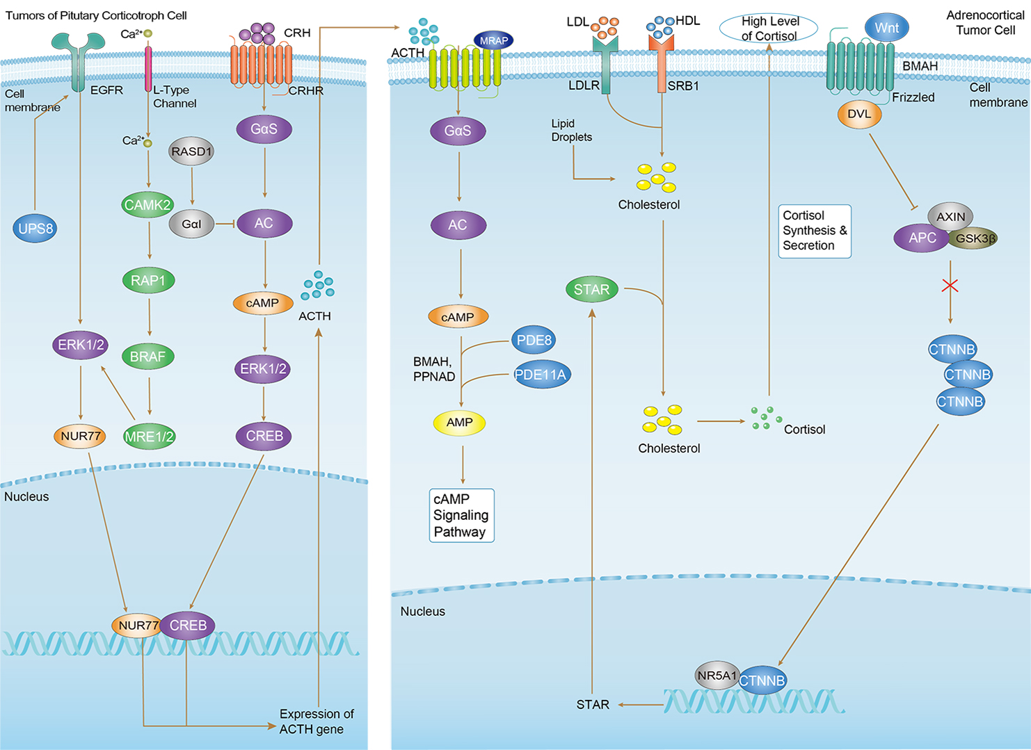 Cushing Syndrome
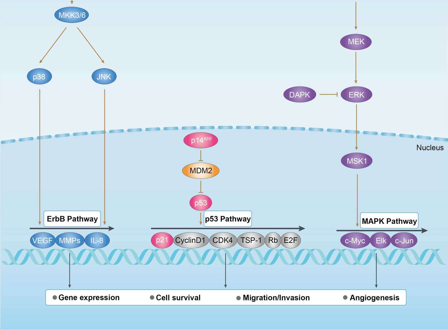
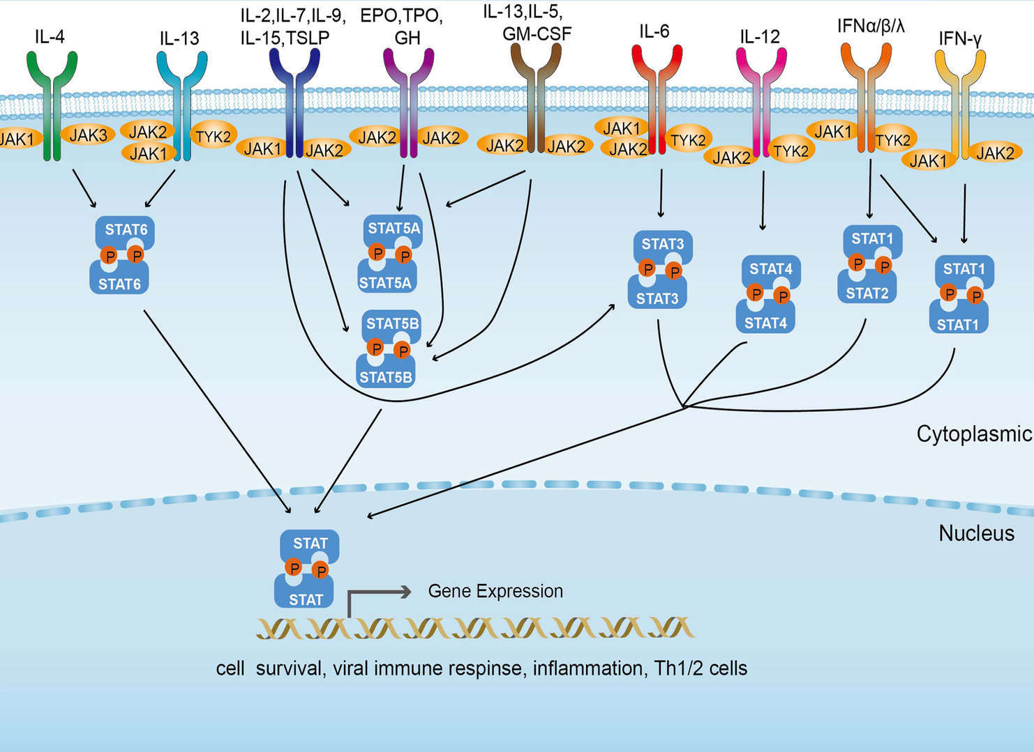 JAK-STAT Signaling Pathway
Product Notes
This is a product of Creative Biolabs' Hi-Affi™ recombinant antibody portfolio, which has several benefits including:
• Increased sensitivity
• Confirmed specificity
• High repeatability
• Excellent batch-to-batch consistency
• Sustainable supply
• Animal-free production
See more details about Hi-Affi™ recombinant antibody benefits.
Downloads
Download resources about recombinant antibody development and antibody engineering to boost your research.
See other products for "EGFR"
Recombinant Antibody
| CAT | Product Name | Application | Type |
|---|---|---|---|
| MOB-1078z | Mouse Anti-EGFR Recombinant Antibody (clone 42C11) | WB, ELISA, FC, IF, IHC, FuncS | Mouse IgG1, κ |
| MOB-0242MC | Rabbit Anti-Human EGFR (phospho Y1092) Antibody | IHC, WB | |
| MOB-0243MC | Rabbit Anti-Human EGFR (phospho Y1068) Antibody | IHC, WB | |
| PABW-088 | Mouse Anti-EGFR Recombinant Antibody (clone 7A7) | ELISA, WB | Mouse IgG |
| PABL-462 | Human Anti-EGFR Recombinant Antibody (clone C225) | FC | Human IgG |
Chimeric Antibody
| CAT | Product Name | Application | Type |
|---|---|---|---|
| TAB-H35 | Anti-Human EGFR Recombinant Antibody (Futuximab) | IF, WB, Inhib | IgG1 - kappa |
| TAB-272MZ | Human Anti-EGFR Recombinant Antibody (TAB-272MZ) | FuncS | Chimeric (mouse/human) IgG |
| TAB-302MZ | Human Anti-EGFR Recombinant Antibody (TAB-302MZ) | ELISA | Chimeric (mouse/human) IgG1 |
| TAB-308MZ | Human Anti-EGFR Recombinant Antibody (TAB-308MZ) | FuncS | Chimeric antibody (mouse/human) |
| TAB-302MZ-S(P) | Mouse Anti-EGFR Recombinant Antibody; scFv Fragment (TAB-302MZ-S(P)) | ELISA | Mouse scFv |
Humanized Antibody
| CAT | Product Name | Application | Type |
|---|---|---|---|
| TAB-165 | Anti-Human EGFR Recombinant Antibody (Matuzumab) | Neut, ELISA, IF, IP, FuncS, FC, ICC | IgG1 |
| TAB-710 | Anti-EGFR Recombinant Antibody (Nimotuzumab) | ELISA, IP, FC, FuncS, Neut, IF, IHC | IgG1 - kappa |
| TAB-274MZ | Human Anti-EGFR Recombinant Antibody (TAB-274MZ) | FC | Humanized IgG |
| TAB-297MZ | Anti-Human EGFR Recombinant Antibody (H225) | ELISA, WB | Humanized antibody |
| TAB-298MZ | Anti-Human EGFR Recombinant Antibody (Hu225) | ELISA, WB | Humanized antibody |
Fab Fragment Antibody
| CAT | Product Name | Application | Type |
|---|---|---|---|
| PFBL-080 | Human Anti-EGFR Recombinant Antibody; Fab Fragment (PFBL-080) | ELISA, WB, FuncS | Human Fab |
| PFBL-459 | Human Anti-EGFR Recombinant Antibody (clone C225); Fab Fragment | FC | Human Fab |
| PFBL-460 | Human Anti-EGFR Recombinant Antibody (clone h-R3); Fab Fragment | ELISA, WB, FuncS | Humanized Fab |
| HPAB-0010-YC-F(E) | Mouse Anti-EGFR Recombinant Antibody (clone L211C); Fab Fragment | ELISA, FC | Mouse Fab |
| HPAB-0011-YC-F(E) | Human Anti-EGFR Recombinant Antibody (clone Pep 1); Fab Fragment | FC | Human Fab |
Single-domain Antibody
| CAT | Product Name | Application | Type |
|---|---|---|---|
| PNBL-017 | Recombinant Anti-Human EGFR VHH Single Domain Antibody (9G8) | FuncS, ELISA, IF | Llama VHH |
| PNBL-018 | Recombinant Anti-Human EGFR VHH Single Domain Antibody (EgA1) | FuncS, SPR | Llama VHH |
| HPAB-AP881-YC | Recombinant Llama Anti-EGFR Single Domain Antibody (VHH122) | ELISA, FC, IP, FuncS | Llama VHH |
| HPAB-AP882-YC | Recombinant Llama Anti-EGFR Single Domain Antibody (VHH205) | ELISA | Llama VHH |
| HPAB-AP883-YC | Recombinant Llama Anti-EGFR Single Domain Antibody (VHH03) | ELISA | Llama VHH |
Human Antibody
| CAT | Product Name | Application | Type |
|---|---|---|---|
| TAB-299MZ-S(P) | Human Anti-EGFR Recombinant Antibody; scFv Fragment (TAB-299MZ-S(P)) | Internalization Assay | Human scFv |
| TAB-313MZ-S(P) | Anti-Human EGFR Recombinant Antibody scFv Fragment (EG-19-11) | ELISA, FC | Human antibody |
| TAB-314MZ-S(P) | Anti-Human EGFR Recombinant Antibody scFv Fragment (EG-26-11) | ELISA, FC | Human antibody |
| TAB-315MZ-S(P) | Anti-Human EGFR Recombinant Antibody scFv Fragment (528) | WB, FC | Human antibody |
| TAB-321MZ-S(P) | Anti-Human EGFR Recombinant Antibody scFv Fragment (DX 1-4) | SPR | Human antibody |
Mouse Antibody
| CAT | Product Name | Application | Type |
|---|---|---|---|
| TAB-310MZ-S(P) | Mouse Anti-EGFR Recombinant Antibody; scFv Fragment (TAB-310MZ-S(P)) | ELISA | Mouse scFv |
| TAB-311MZ-S(P) | Mouse Anti-EGFR Recombinant Antibody; scFv Fragment (TAB-311MZ-S(P)) | ELISA | Mouse scFv |
| TAB-312MZ-S(P) | Mouse Anti-EGFR Recombinant Antibody; scFv Fragment (TAB-312MZ-S(P)) | ELISA | Mouse scFv |
| TAB-316MZ-S(P) | Anti-Human EGFR Recombinant Antibody scFv Fragment (07D06) | WB, MTT assay | |
| TAB-317MZ-S(P) | Anti-Human EGFR Recombinant Antibody scFv Fragment (12D03) | WB, MTT assay |
Fc Glycosylation
| CAT | Product Name | Application | Type |
|---|---|---|---|
| Gly-055LC | Recombinant Anti-Human EGFR Antibody (Fc glycosylation/High-mannose glycosylated) | ELISA | Chimeric antibody (mouse/human) |
| Gly-144LC | Recombinant Anti-Human EGFR Antibody (Fc glycosylation) | ELISA | Humanized antibody |
High-mannose Glycoform
| CAT | Product Name | Application | Type |
|---|---|---|---|
| Gly-055LC-1 | Recombinant Anti-Human EGFR Antibody (Fc glycosylation/High-mannose glycosylated) | ELISA | Chimeric antibody (mouse/human) |
Deglycosylated Antibody (Non-glycosylated IgGs)
| CAT | Product Name | Application | Type |
|---|---|---|---|
| Gly-167LC | Recombinant Anti-Human EGFR Antibody (Non-glycosylated) | ELISA | Human antibody |
Chicken IgY Antibody
| CAT | Product Name | Application | Type |
|---|---|---|---|
| BRD-0183MZ | Chicken Anti-EGFR Polyclonal IgY | WB | Chicken antibody |
| BRD-0668MZ | Chicken Anti-EGFR Polyclonal IgY | WB | Chicken antibody |
MHC Tetramer for Cancer
| CAT | Product Name | Application | Type |
|---|---|---|---|
| MHC-LC773 | A*0201/Human EGFR (YLNTVQPTCV) MHC Tetramer | FCM | |
| MHC-LC4545 | PE-DQB1*03:02/Human EGFR (SRALEEKKGNYVVTHG) MHC Tetramer | FCM | |
| MHC-YF409 | A*02:01/Human EGF-R-479 (KLFGTSGQKT) MHC Pentamer | FCM |
Neutralizing Antibody
| CAT | Product Name | Application | Type |
|---|---|---|---|
| NEUT-722CQ | Rabbit Anti-EGFR Recombinant Antibody (clone CBL1011) | Neut | Rabbit IgG |
| NEUT-723CQ | Mouse Anti-EGFR Recombinant Antibody (clone CBL931) | WB, IP, IHC, ICC, Neut | Mouse IgG1 |
| NEUT-724CQ | Rabbit Anti-EGFR Recombinant Antibody (clone D1D4J) | IF, FC, WB, IP, Neut | Rabbit IgG |
Rabbit Monoclonal Antibody
| CAT | Product Name | Application | Type |
|---|---|---|---|
| MOR-1101 | Hi-Affi™ Rabbit Anti-EGFR Recombinant Antibody (clone DS1101AB) | IHC-P | Rabbit IgG |
| MOR-4520 | Hi-Affi™ Rabbit Anti-EGFR Recombinant Antibody (clone TH28DS) | IF, ICC, FC | Rabbit IgG |
| MOR-4675 | Hi-Affi™ Rabbit Anti-EGFR Recombinant Antibody (clone TH189DS) | WB, IF, ICC, FC | Rabbit IgG |
| MOR-4676 | Hi-Affi™ Rabbit Anti-EGFR Recombinant Antibody (clone TH190DS) | WB, IF, ICC, FC | Rabbit IgG |
| MOR-0033-FY | Rabbit Anti-EGFR Recombinant Antibody (clone AFY0004) | ICC, IHC, WB | Rabbit IgG |
ADCC Enhanced Antibody
| CAT | Product Name | Application | Type |
|---|---|---|---|
| AFC-TAB-165 | Afuco™ Anti-EGFR ADCC Recombinant Antibody (Matuzumab), ADCC Enhanced | Neut, ELISA, IF, IP, FuncS, FC | ADCC enhanced antibody |
| AFC-TAB-464CQ | Afuco™ Anti-EGFR ADCC Recombinant Antibody (Tomuzotuximab), ADCC Enhanced | ELISA, IHC, FC, IP, IF, FuncS | ADCC enhanced antibody |
| AFC-TAB-003 | Afuco™ Anti-EGFR ADCC Recombinant Antibody (Cetuximab), ADCC Enhanced | IF, IP, Neut, FuncS, ELISA, FC | ADCC enhanced antibody |
| AFC-TAB-040 | Afuco™ Anti-EGFR ADCC Recombinant Antibody (Zalutumumab), ADCC Enhanced | ELISA, FC, IP, FuncS, IF, Neut | ADCC enhanced antibody |
| AFC-TAB-119 | Afuco™ Anti-EGFR ADCC Recombinant Antibody (Necitumumab), ADCC Enhanced | FC, IP, ELISA, Neut, FuncS, IF | ADCC enhanced antibody |
scFv Fragment Antibody
| CAT | Product Name | Application | Type |
|---|---|---|---|
| HPAB-2059-FY-S(P) | Mouse Anti-EGFR Recombinant Antibody (clone L2-12B); scFv Fragment | FC, ELISA | Mouse scFv |
| HPAB-2060-FY-S(P) | Mouse Anti-EGFR Recombinant Antibody (clone L3-11D); scFv Fragment | FC, ELISA | Mouse scFv |
| HPAB-1530WJ-S(P) | Mouse Anti-EGFR Recombinant Antibody; scFv Fragment (clone 340) | ELISA, WB | Mouse scFv |
| HPAB-1531WJ-S(P) | Human Anti-EGFR Recombinant Antibody; scFv Fragment (clone hu340) | ELISA, WB | Human scFv |
| HPAB-1563WJ-S(P) | Human Anti-EGFR Recombinant Antibody; scFv Fragment (clone chMint5) | ELISA, WB | Human scFv |
Customer Reviews and Q&As

There are currently no Customer reviews or questions for PNBL-016. Click the button above to contact us or submit your feedback about this product.
View the frequently asked questions answered by Creative Biolabs Support.
For Research Use Only. Not For Clinical Use.
For research use only. Not intended for any clinical use. No products from Creative Biolabs may be resold, modified for resale or used to manufacture commercial products without prior written approval from Creative Biolabs.
Send Inquiry
This site is protected by reCAPTCHA and the Google Privacy Policy and Terms of Service apply.