
Anti-Human ErbB2 Recombinant Antibody (Trastuzumab) (CAT#: TAB-005)
Recombinant monoclonal antibody to Human ErbB2. Trastuzumab (INN; trade names Herclon, trastuzumab) is a monoclonal antibody that interferes with the HER2/neu receptor. Its main use is to treat certain breast cancers.
The HER receptors are proteins that are embedded in the cell membrane and communicate molecular signals from outside the cell (molecules called EGFs) to inside the cell, and turn genes on and off. The HER proteins stimulate cell proliferation. In some cancers, notably certain types of breast cancer, HER2 is over-expressed, and causes cancer cells to reproduce uncontrollably.
Specific Inquiry

Figure 1 t-DARPP and ERBB2 are significantly overexpressed in adenocarcinomas.
left, cell viability of OE19 and OE33 cells in response to trastuzumab treatment was evaluated by Trypan blue staining. OE19 cells were 2-fold more sensitive to trastuzumab than OE33 cells (P < 0.001). D, right, Western blot analysis shows higher protein expression of ERBB2 in OE19 cells than OE33 cells. In contrast, t-DARPP expression was undetectable in OE19 cells but highly expressed in OE33 cells.
Hong, J., Katsha, A., Lu, P., Shyr, Y., Belkhiri, A., & El-Rifai, W. (2012). Regulation of ERBB2 receptor by t-DARPP mediates trastuzumab resistance in human esophageal adenocarcinoma. Cancer research.

Figure 2 t-DARPP expression blocks trastuzumab-induced apoptosis.
A, apoptosis in OE19 cells infected with control (10 MOI) or t-DARPP (10 MOI) recombinant adenoviruses after treatment with vehicle or trastuzumab (20 μg/mL) for 48 hours was determined by Annexin-V/PI staining and FACS analysis. B, Western blot analysis of caspase-3, cleaved caspase-3, and t-DARPP proteins in OE19 cells infected with control or t-DARPP adenoviruses following treatments as described in A. C, apoptosis in parental and trastuzumab-resistant OE19 cells after treatment with vehicle or trastuzumab (20 μg/mL) for 48 hours was evaluated by Annexin-V/PI staining and FACS analysis. D, Western blot analysis of caspase-3, cleaved caspase-3, and t-DARPP proteins in parental and trastuzumab-resistant OE19 cells after treatments as described in C. These data indicate that endogenous and exogenous t-DARPP expression counteracted trastuzumab-induced apoptosis in OE19 cells.
Hong, J., Katsha, A., Lu, P., Shyr, Y., Belkhiri, A., & El-Rifai, W. (2012). Regulation of ERBB2 receptor by t-DARPP mediates trastuzumab resistance in human esophageal adenocarcinoma. Cancer research.

Figure 3 t-DARPP associates with ERBB2 and interferes with trastuzumab/ERBB2 protein interaction.
A, Western blot analysis of coimmunoprecipitated exogenous t-DARPP and endogenous ERBB2 proteins with M2-flag or trastuzumab antibodies in OE19 cells infected with t-DARPP-flag adenovirus (10 MOI). These data show protein association of ERBB2 with t-DARPP. B, Western blot analysis of immunoprecipitated endogenous ERBB2 protein with trastuzumab antibody in OE19 cells infected with control (10 MOI) or t-DARPP (10 MOI) adenoviruses. Pulled-down ERBB2 band intensity was depicted as a ratio relative to input ERBB2 protein. The results show that exogenous t-DARPP expression blocked binding of trastuzumab to ERBB2 receptor relative to control. C, Western blot analysis of immunoprecipitated endogenous ERBB2 protein with trastuzumab antibody in parental or trastuzumab-resistant OE19 cells. The band intensity of immunoprecipitated ERBB2 protein was shown as a ratio relative to input ERBB2. These data indicate that endogenous t-DARPP expression in trastuzumab-resistant cells was associated with a significant decrease in trastuzumab/ERBB2 protein interaction relative to control.
Hong, J., Katsha, A., Lu, P., Shyr, Y., Belkhiri, A., & El-Rifai, W. (2012). Regulation of ERBB2 receptor by t-DARPP mediates trastuzumab resistance in human esophageal adenocarcinoma. Cancer research.

Figure 4 t-DARPP overexpression promotes tumor growth and blocks response to trastuzumab treatment in vivo.
The H&E staining at the end of trastuzumab treatment shows effectively diminished control tumors leaving necrotic and fibrotic lesions (left), whereas t-DARPP tumors were unaffected (right).
Hong, J., Katsha, A., Lu, P., Shyr, Y., Belkhiri, A., & El-Rifai, W. (2012). Regulation of ERBB2 receptor by t-DARPP mediates trastuzumab resistance in human esophageal adenocarcinoma. Cancer research.

Figure 5 Enhanced internalization of ErbB2 in caveolin-1 expressing SKBR-3 cells.
(A) SKBR-3/Cav-1 cells were incubated with 1 μM of EC-eGFP at 37°C for 5 min and internalization was assessed using anti-ErbB2 monoclonal antibody. Cell surface binding of EC-eGFP (green) with ErbB2 (red) was shown in the merged picture (yellow). (B) enhanced endocytosis of Ec-eGFP (green) and Intracellular co-localization with ErbB2 (red) was observed after 15 min of incubation at 37°C. (C) Parental SKBR-3 cells incubated with EC-eGFP for 15 min at 37°C were processed for endocytosis analysis as above using anti ErbB2 antibody (red). (D) SKBR-3/Cav-1 cells incubated with Trastuzumab (green) for 5 min at 37°C were processed and stained with anti-caveolin-1 antibody (blue); cell surface binding of Trastuzumab with caveolin-1 was shown (opaque) in the merged image. (E) Internalization and localization of Trastuzumab (green) with caveolin-1 (blue) in SKBR-3/Cav-1 cells after 15 min of stimulation at 37°C. (F) Parental SKBR-3 cells incubated similarly with Trastuzumab for 15 min were processed for endocytosis analysis as detailed in the material and methods section. Scale bar: 20 μm.
Sekhar, S. C., Kasai, T., Satoh, A., Shigehiro, T., Mizutani, A., Murakami, H., ... & Seno, M. (2013). Identification of caveolin-1 as a potential causative factor in the generation of trastuzumab resistance in breast cancer cells. Journal of Cancer, 4(5), 391.

Figure 6 Ligand induced ErbB2 internalization after EC-eGFP and Trastuzumab treatment.
SKBR-3 cells were surface biotinylated to monitor internalization. (A) Biotinylated caveolin-1 expressing SKBR-3 cells were stimulated with 1 μM EC-eGFP and Trastuzumab for 15, 30 and 60 min at 37°C, 4°C or left untreated for 1 h at 37°C and 4°C. Densitometry was performed using Image J software. (B) Wild type SKBR-3 cells were incubated with Ec-eGFP and Trastuzumab as described above for 15-60 min. Cell lysates prepared from treated samples were then pull-downed with avidin agarose and subjected to western blotting using an anti-ErbB2 antibody. Endocytosed transferrin receptor was also monitored simultaneously as internal control. The results are expressed as the mean SD of three individual experiments. Since there was no significant internalization observed in parental SKBR-3 cells, densitometry was performed only for caveolin-1 expressing SKBR-3 cells.
Sekhar, S. C., Kasai, T., Satoh, A., Shigehiro, T., Mizutani, A., Murakami, H., ... & Seno, M. (2013). Identification of caveolin-1 as a potential causative factor in the generation of trastuzumab resistance in breast cancer cells. Journal of Cancer, 4(5), 391.

Figure 7 Fc receptor mediated ADCC in SKBR-3 cells.
ADCC activity mediated by Trastuzumab and EC-Fc against cell surface ErbB2 in SKBR-3, SKBR-3/Cav-1 and SKOV-3 cells was measured using human PBMC's as effector cells at an effector: target cell ratio of (A) 25:1 (B) 50:1 and (C) 100:1, with standard LDH assay as described in materials and methods. Data are expressed as the mean of (±) SD (n=3). Student's t- test (two tailed) was used to compare the ADCC response in SKBR-3/Cav-1 and SKOV-3 with parental SKBR-3. Differences were statistically significant at P < 0.05.
Sekhar, S. C., Kasai, T., Satoh, A., Shigehiro, T., Mizutani, A., Murakami, H., ... & Seno, M. (2013). Identification of caveolin-1 as a potential causative factor in the generation of trastuzumab resistance in breast cancer cells. Journal of Cancer, 4(5), 391.


Specifications
- Immunogen
- NIH 3T3/HER2-3400 cells are used to immunize BALB/c mice.
- Host Species
- Mouse
- Derivation
- Humanized (from mouse)
- Type
- IgG1 - kappa
- Specificity
- Tested positive against native human antigen
- Species Reactivity
- Human
- Applications
- FC, IP, ELISA, Neut, FuncS, IF, IHC
- Trade name
- Herclon, trastuzumab
- CAS
- 180288-69-1
- Generic Name
- Trastuzumab
- Biological Half-Life
- 2-12 days
- ATC Code
- L01XC03
- DrugBank
- DB00072
- UNII
- P188ANX8CK
- ChEMBL
- CHEMBL1201585
- MW
- 145,531.5 g/mol
- Related Disease
- Breast cancers overexpressing ERBB2, metastatic
Product Property
- Purity
- >95.0% as determined by Analysis by RP-HPLC & analysis by SDS-PAGE.
- Storage
- 4°C. For long term storage, aliquot and store at -20°C. Repeated thawing and freezing must be avoided.
Applications
- Application Notes
- The ERBB2 antibody has been reported in applications of WB, Inhib, IP, H&E staining, IF, Internalization, ADCC.
WB: Protein samples lysed in Laemmlli sample buffer and were then boiled for 5 min. Equal quantities of protein lysates were then analyzed on SDS-PAGE and transferred to PVDF membrane. The membranes were blocked with 10% skim milk, incubated with primary antibody in 0.4% skim milk/TBS for 2h, washed with TBST followed by appropriate secondary antibody in 0.4% skim milk/TBS incubation for 1 h. The proteins were visualized using Western lighting plus-ECL reagent in Light-Capture II cooled CCD camera system.
IF: For confocal microscopic observation, SKBR-3 cells and caveolin-1 transfected SKBR-3 cells were grown on 18 mm cover slips. Cells were then fixed with 4% PFA for 10 min, permeabilized with 0.1% Triton X-100/PBS and blocked with 1% BSA-PBS for 30 min at RT. For caveolin-1 localization, cells were incubated with 1:500 diluted anti-caveolin-1 antibody in 0.25% BSA/PBS for 1 h at RT, followed by staining with Alexa 555 labeled anti-rabbit IgG for 30 min. After wash, the cover slips were mounted with DAPI (Vector Laboratories and observed by confocal microscope IX81 with 60x magnification lens.
ADCC: Caveolin-1 expressing and wild type SKBR-3 and SKOV-3 cells that had been cultured in 96 well plates were incubated with 1 µM of EC-Fc and Trastuzumab for 4 h at 37˚C.
Target
- Alternative Names
- Trastuzumab;Herclon, trastuzumab;180288-69-1;Herclon;DB00072ERBB2;v-erb-b2 erythroblastic leukemia viral oncogene homolog 2, neuro/glioblastoma derived oncogene homolog (avian);NGL, v erb b2 avian erythroblastic leukemia viral oncogene homolog 2 (neuro/glio
- Gene ID
- 2064
- UniProt ID
- P04626
Related Resources
 Bladder Cancer
 Non-small Cell Lung Cancer
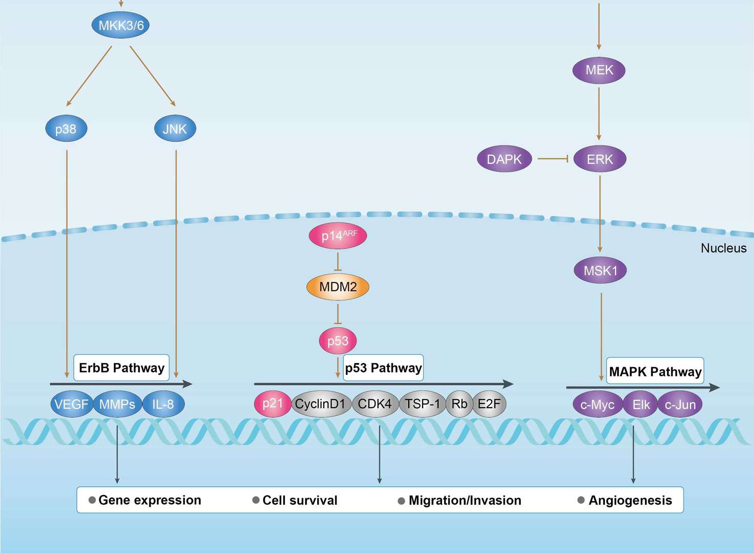
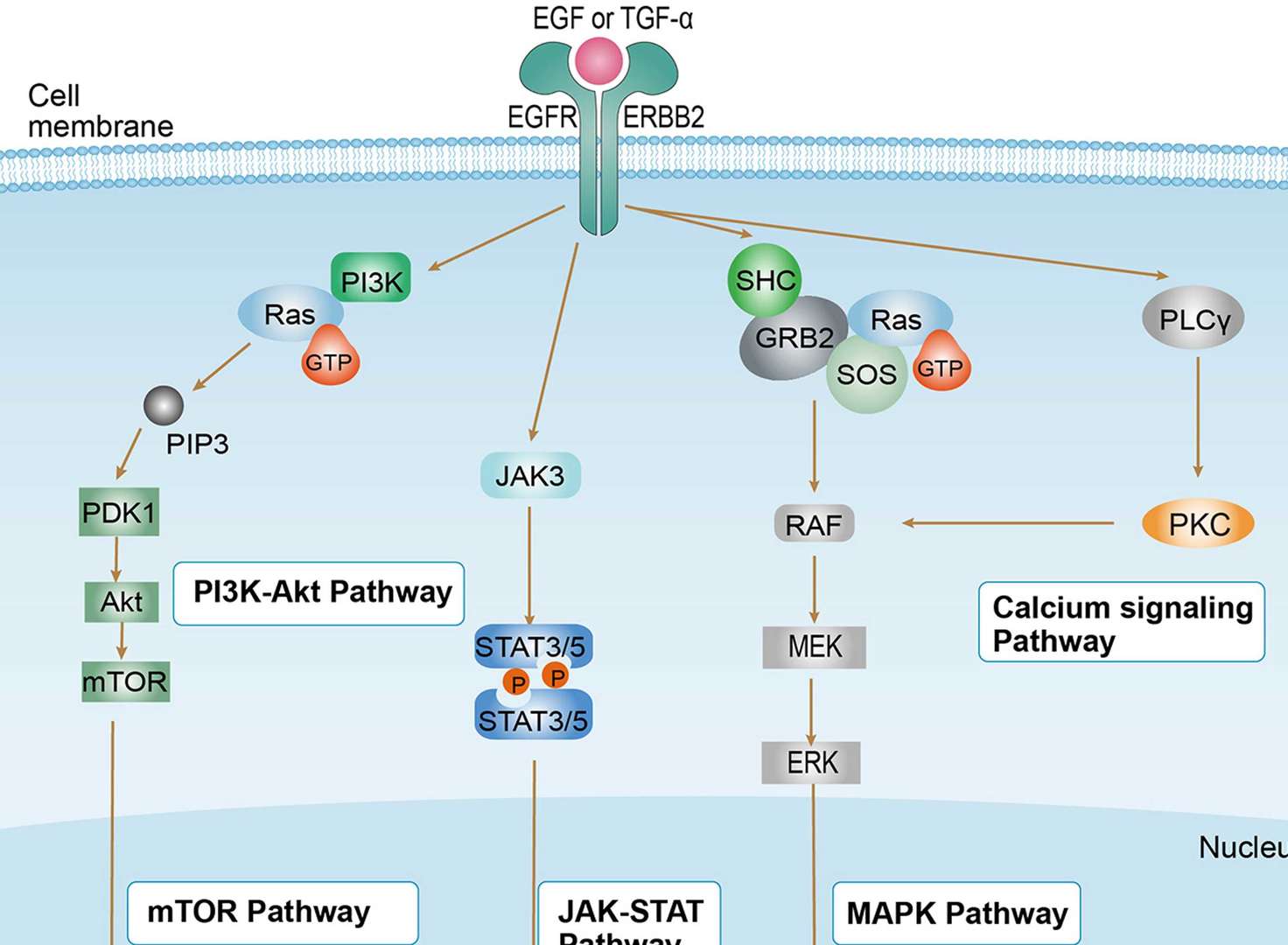
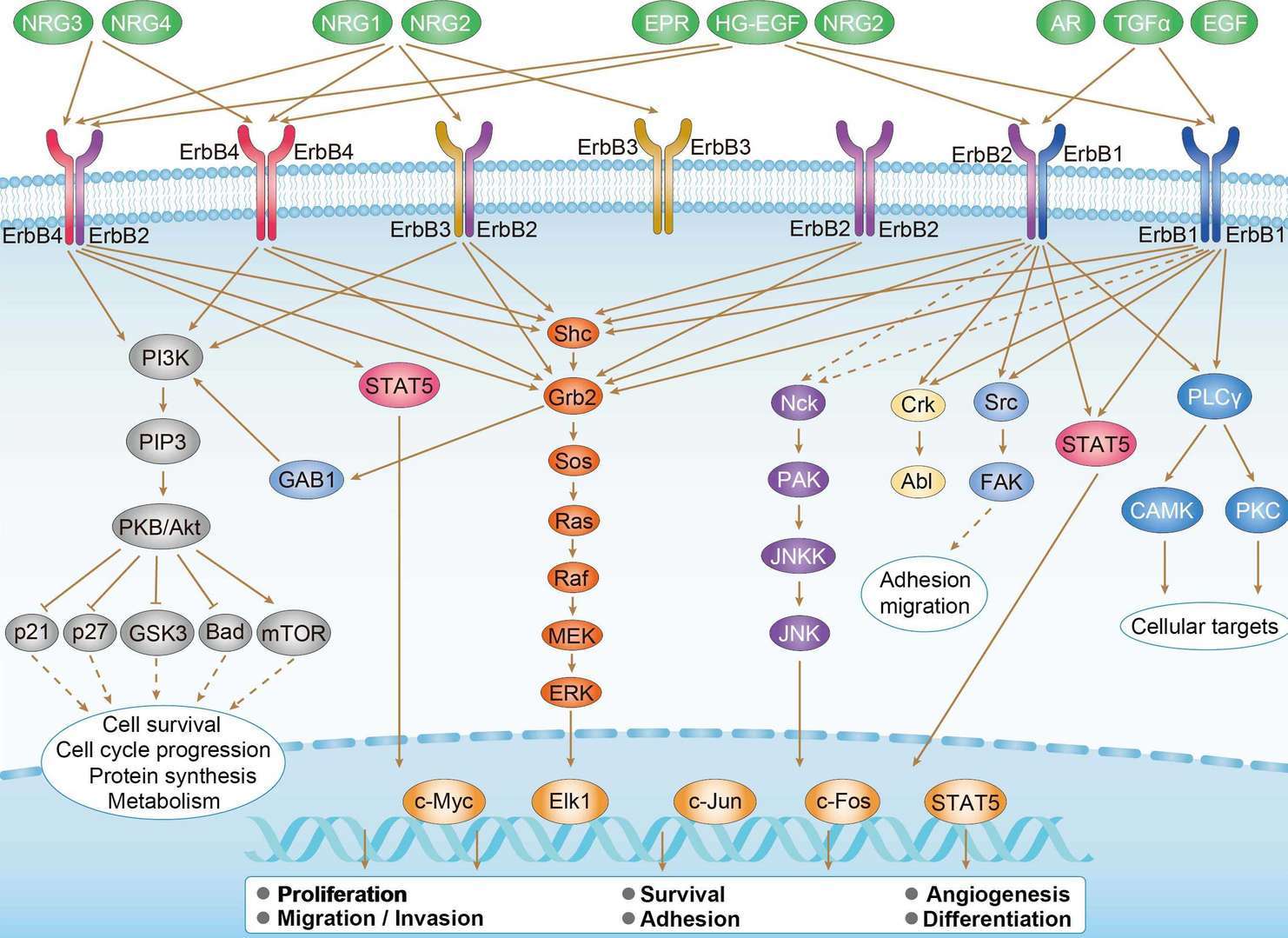 ErbB Signaling Pathway
Product Notes
This is a product of Creative Biolabs' Hi-Affi™ recombinant antibody portfolio, which has several benefits including:
• Increased sensitivity
• Confirmed specificity
• High repeatability
• Excellent batch-to-batch consistency
• Sustainable supply
• Animal-free production
See more details about Hi-Affi™ recombinant antibody benefits.
Downloads
Download resources about recombinant antibody development and antibody engineering to boost your research.
See other products for "Trastuzumab"
Afuco™ Anti-ERBB2 ADCC Recombinant Antibody (Trastuzumab), ADCC EnhancedThis product is an ADCC enhanced antibody produced by our Afuco™ platform. Recombinant monoclonal antibody to Human ErbB2. Trastuzumab (INN; trade names Herclon, trastuzumab) is a monoclonal antibody that interferes with the HER2/neu receptor. Its main use is to treat certain breast cancers.
The HER receptors are proteins that are embedded in the cell membrane and communicate molecular signals from outside the cell (molecules called EGFs) to inside the cell, and turn genes on and off. The HER proteins stimulate cell proliferation. In some cancers, notably certain types of breast cancer, HER2 is over-expressed, and causes cancer cells to reproduce uncontrollably.
See other products for "ERBB2"
Single-domain Antibody
| CAT | Product Name | Application | Type |
|---|---|---|---|
| NABG-059 | Recombinant Anti-Human ERBB2 VHH Single Domain Antibody | IHC, FC, CA, FuncS | Llama VHH |
| PNBL-066 | Recombinant Anti-HER2 VHH Single Domain Antibody (Gr3) | SPR | Llama VHH |
| TAB-032CT | Anti-Human HER2/neu Therapeutic Single Domain Antibody | WB | Single domain antibody |
| TAB-046CT | Anti-Human HER2/neu Therapeutic Single Domain Antibody (Gr3) | ELISA, Inhibition, FC | Single domain antibody |
| TAB-047CT | Anti-Human HER2/neu Therapeutic Single Domain Antibody (Gr6) | WB | Single domain antibody |
Intrabody
| CAT | Product Name | Application | Type |
|---|---|---|---|
| IAB-B008(A) | Recombinant Anti-human ERBB2 Intrabody [(D-Arg)9] | IF, FC, FuncS | scFv-(D-Arg)9 |
| IAB-B008(G) | Recombinant Anti-human ERBB2 Intrabody [+36 GFP] | WB, ICC, FuncS | scFv-(+36GFP) |
| IAB-B008(T) | Recombinant Anti-human ERBB2 Intrabody [Tat] | ICC, Neut, FuncS | scFv-Tat |
Chimeric Antibody
| CAT | Product Name | Application | Type |
|---|---|---|---|
| TAB-761 | Anti-ERBB2 Recombinant Antibody (Margetuximab) | IF, IP, Neut, FuncS, ELISA, FC, WB | IgG1 - kappa |
| TAB-049CT | Anti-Human HER2/neu Recombinant Antibody (ChA21) | IHC, Inhib, WB, Activ, FC | Chimeric antibody (mouse/human) |
| TAB-049CT-S(P) | Anti-Human HER2/neu Recombinant Antibody scFv Fragment (ChA21) | WB | Chimeric antibody (mouse/human) |
| TAB-049CT-F(E) | Anti-Human HER2/neu Recombinant Antibody Fab Fragment (ChA21) | WB | Chimeric antibody (mouse/human) |
Immunotoxin
| CAT | Product Name | Application | Type |
|---|---|---|---|
| AGTO-G022E | Anti-ERBB2 immunotoxin 4D5 (scFv)-PE | Cytotoxicity assay, Function study | |
| AGTO-G022D | Anti-ERBB2 immunotoxin 4D5 (scFv)-DT | Cytotoxicity assay, Function study | |
| AGTO-G022G | Anti-ERBB2 immunotoxin 4D5 (scFv)-Gel | Cytotoxicity assay, Function study | |
| AGTO-G022R | Anti-ERBB2 immunotoxin 4D5 (scFv)-RTA | Cytotoxicity assay, Function study | |
| AGTO-G022S | Anti-ERBB2 immunotoxin 4D5 (scFv)-Sap | Cytotoxicity assay, Function study |
Fab Fragment Antibody
| CAT | Product Name | Application | Type |
|---|---|---|---|
| PFBL-085 | Human Anti-ERBB2 Recombinant Antibody (PFBL-085) | ELISA, WB, FuncS | Human Fab |
| PFBL-463 | Human Anti-ERBB2 Recombinant Antibody (clone mAb37); Fab Fragment | WB | Human Fab |
| PSBW-161 | Mouse Anti-ERBB2 Recombinant Antibody (clone chA21); scFv Fragment | FC, WB | Mouse scFv |
| HPAB-0608LY-F(E) | Human Anti-ERBB2 Recombinant Antibody; Fab Fragment (HPAB-0608LY-F(E)) | ELISA, Inhib | Humanized Fab |
| HPAB-0609LY-F(E) | Human Anti-ERBB2 Recombinant Antibody; Fab Fragment (HPAB-0609LY-F(E)) | ELISA, Inhib | Humanized Fab |
Human Antibody
| CAT | Product Name | Application | Type |
|---|---|---|---|
| TAB-0193CL-F(E) | Human Anti-ERBB2 Recombinant Antibody; Fab Fragment (TAB-0193CL-F(E)) | ELISA, Cyt, Internalization | Human Fab |
| TAB-0194CL-F(E) | Human Anti-ERBB2 Recombinant Antibody; Fab Fragment (TAB-0194CL-F(E)) | ELISA, Cyt, Internalization | Human Fab |
| TAB-042CT-F(E) | Anti-Human HER2/neu Recombinant Antibody Fab Fragment (CRX01) | WB | Human antibody |
| PABX-081-F (E) | Recombinant Human Anti-HER2 Antibody Fab Fragment (Gr3 ) | WB, ELISA, FuncS | Fab |
| PABX-082-F (E) | Recombinant Human Anti-HER2 Antibody Fab Fragment (Gr6) | WB, ELISA, FuncS | Fab |
Humanized Antibody
| CAT | Product Name | Application | Type |
|---|---|---|---|
| TAB-033CT | Human Anti-ERBB2 Recombinant Antibody (TAB-033CT) | ELISA, WB | Human IgG1, κ |
| TAB-033CT-S(P) | Human Anti-ERBB2 Recombinant Antibody; scFv Fragment (TAB-033CT-S(P)) | ELISA, WB | Human scFv |
| TAB-038CT-S(P) | Human Anti-ERBB2 Recombinant Antibody; scFv Fragment (TAB-038CT-S(P)) | WB | Humanized scFv |
| TAB-039CT-S(P) | Anti-Human HER2/neu Recombinant Antibody scFv Fragment (h1E11) | WB | Humanized antibody |
| TAB-041CT-S(P) | Human Anti-ERBB2 Recombinant Antibody; scFv Fragment (TAB-041CT-S(P)) | ELISA, WB | Humanized scFv |
Mouse Antibody
| CAT | Product Name | Application | Type |
|---|---|---|---|
| TAB-034CT-S(P) | Anti-Human HER2/neu Recombinant Antibody scFv Fragment (pertuzumab ) | WB | |
| TAB-270CQ | Mouse Anti-ERBB2 Recombinant Antibody (TAB-270CQ) | WB, Inhib | Mouse IgG |
| TAB-270CQ-S(P) | Mouse Anti-ERBB2 Recombinant Antibody; scFv Fragment (TAB-270CQ-S(P)) | WB, Inhib | Mouse scFv |
| TAB-270CQ-F(E) | Mouse Anti-ERBB2 Recombinant Antibody; Fab Fragment (TAB-270CQ-F(E)) | WB, Inhib | Mouse Fab |
| PABX-082-S (P) | Recombinant Human Anti-HER2 Antibody scFv Fragment (Gr6) | WB, ELISA, FuncS | scFv |
Fab Glycosylation
| CAT | Product Name | Application | Type |
|---|---|---|---|
| Gly-024LC | Recombinant Anti-Human ERBB2 Antibody (Fab glycosylation) | ELISA | Mouse antibody |
| Gly-039LC | Recombinant Anti-Human ERBB2 Antibody (Fab glycosylation) | ELISA | Human antibody |
Fc Glycosylation
| CAT | Product Name | Application | Type |
|---|---|---|---|
| Gly-117LC | Recombinant Anti-Human ERBB2 Antibody (Fc glycosylation) | ELISA | Humanized antibody |
| Gly-118LC | Recombinant Anti-Human ERBB2 Antibody (Fc glycosylation) | ELISA | Mouse antibody |
| Gly-145LC | Recombinant Anti-Human ERBB2 Antibody (Fc glycosylation) | ELISA | Human antibody |
| Gly-146LC | Recombinant Anti-Human ERBB2 Antibody (Fc glycosylation) | ELISA | Human antibody |
| Gly-147LC | Recombinant Anti-Human ERBB2 Antibody (Fc glycosylation) | ELISA | Human antibody |
Deglycosylated Antibody (Non-glycosylated IgGs)
| CAT | Product Name | Application | Type |
|---|---|---|---|
| Gly-177LC | Recombinant Anti-Human ERBB2 Antibody (Non-glycosylated) | ELISA | Humanized antibody |
Chicken IgY Antibody
| CAT | Product Name | Application | Type |
|---|---|---|---|
| BRD-0195MZ | Chicken Anti-ERBB2 Polyclonal IgY | WB | Chicken antibody |
| BRD-0695MZ | Chicken Anti-ERBB2 Polyclonal IgY | WB | Chicken antibody |
| BRD-0779MZ | Chicken Anti-ErbB2 Polyclonal IgY | ICC, IF, IHC, IP | Chicken antibody |
Blocking Antibody
| CAT | Product Name | Application | Type |
|---|---|---|---|
| NEUT-737CQ | Mouse Anti-ERBB2 Recombinant Antibody (clone 7.16.4) | Block, IP, IF, FC | Mouse IgG2a |
Rabbit Monoclonal Antibody
| CAT | Product Name | Application | Type |
|---|---|---|---|
| MOR-1178 | Hi-Affi™ Rabbit Anti-ERBB2 Recombinant Antibody (clone DS1178AB) | IHC-P, IHC-Fr | Rabbit IgG |
| MOR-4586 | Hi-Affi™ Rabbit Anti-ERBB2 Recombinant Antibody (clone TH99DS) | WB | Rabbit IgG |
| MOR-4587 | Hi-Affi™ Rabbit Anti-ERBB2 Recombinant Antibody (clone TH100DS) | WB | Rabbit IgG |
MHC Tetramer for Cancer
| CAT | Product Name | Application | Type |
|---|---|---|---|
| MHC-LC2349 | APC-A*02:01/Human ERBB2 (HLYQGCQVV) MHC Tetramer | FCM | |
| MHC-LC2351 | APC-A*02:01/Human ERBB2 (PLTSIISAV) MHC Tetramer | FCM | |
| MHC-YF221 | A*02:01/Human Her-2/neu (KIFGSLAFL) MHC Monomer | MHC Multimer | |
| MHC-YF222 | A*02:01/Human Her-2/neu (RLLQETELV) MHC Monomer | MHC Multimer | |
| MHC-YF584 | H-2Kd/Human Neu/Her-2/Erbb2 proto-oncoprotein (TYVPANASL) MHC Pentamer | FCM |
Recombinant Antibody
| CAT | Product Name | Application | Type |
|---|---|---|---|
| HPAB-M0510-YC | Mouse Anti-ERBB2 Recombinant Antibody (HPAB-M0510-YC) | FC, FuncS | Mouse IgG2a |
| HPAB-M0073-YC | Human Anti-ERBB2 Recombinant Antibody (HPAB-M0073-YC) | ELISA, FC | Human IgG |
| HPAB-M0074-YC | Human Anti-ERBB2 Recombinant Antibody (HPAB-M0074-YC) | ELISA, FC | Human IgG |
| HPAB-M0075-YC | Human Anti-ERBB2 Recombinant Antibody (HPAB-M0075-YC) | ELISA, FC | Human IgG |
| HPAB-M0076-YC | Human Anti-ERBB2 Recombinant Antibody (HPAB-M0076-YC) | ELISA, FC | Human IgG |
ADCC Enhanced Antibody
| CAT | Product Name | Application | Type |
|---|---|---|---|
| AFC-TAB-468CQ | Afuco™ Anti-ERBB2 ADCC Recombinant Antibody (Timigutuzumab), ADCC Enhanced | ELISA, IHC, FC, IP, IF, FuncS | ADCC enhanced antibody |
| AFC-TAB-053 | Afuco™ Anti-ERBB2 ADCC Recombinant Antibody (Pertuzumab), ADCC Enhanced | FuncS, IF, Neut, ELISA, FC, IP | ADCC enhanced antibody |
| AFC-TAB-761 | Afuco™ Anti-ERBB2 ADCC Recombinant Antibody (Margetuximab), ADCC Enhanced | IF, IP, Neut, FuncS, ELISA, FC | ADCC enhanced antibody |
| AFC-TAB-005 | Afuco™ Anti-ERBB2 ADCC Recombinant Antibody (Trastuzumab), ADCC Enhanced | FC, IP, ELISA, Neut, FuncS, IF | ADCC enhanced antibody |
scFv Fragment Antibody
| CAT | Product Name | Application | Type |
|---|---|---|---|
| HPAB-2957LY-S(P) | Human Anti-ERBB2 Recombinant Antibody (clone P2h2); scfv Fragment | ELISA, FC | Humanized scfv |
| HPAB-AP784-YC-S(P) | Mouse Anti-ERBB2 Recombinant Antibody (clone F/2A); scFv Fragment | ELISA | Mouse scFv |
| FAMAB-0172JF-S(P) | Human Anti-ERBB2 Recombinant Antibody (clone C6H2); scFv Fragment | ELISA, FuncS | Human scFv |
| FAMAB-0173JF-S(P) | Human Anti-ERBB2 Recombinant Antibody (clone C6H211); scFv Fragment | ELISA, FuncS | Human scFv |
| PSBS-0070 | Human Anti-ERBB2 Recombinant Antibody; scFv Fragment (PSBS-0070) | ELISA, FuncS | Human scFv |
Customer Reviews and Q&As

There are currently no Customer reviews or questions for TAB-005. Click the button above to contact us or submit your feedback about this product.
View the frequently asked questions answered by Creative Biolabs Support.
For Research Use Only. Not For Clinical Use.
For research use only. Not intended for any clinical use. No products from Creative Biolabs may be resold, modified for resale or used to manufacture commercial products without prior written approval from Creative Biolabs.
Send Inquiry
This site is protected by reCAPTCHA and the Google Privacy Policy and Terms of Service apply.