
Afuco™ Anti-Human MET ADCC Recombinant Antibody (ABT-700), ADCC Enhanced (CAT#: AFC-501CL)
Anti-MET ADCC Enhanced Antibody (ABT-700) is an ADCC enhanced antibody produced by our Afuco™ platform. ABT-700 is an anti-c-Met antibody with significant preclinical single-agent activity against MA human xenograft tumors. ABT-700 was well tolerated and appeared to have substantial single-agent activity. In this small cohort of gastric patients MA appears to be more common in treatment-refractory tumors than in primary untreated tumors, suggesting that screening efforts should focus on this treatment-refractory patient population.
Specific Inquiry

Figure 1 ABT-700 specifically binds cellular c-Met and antagonizes c-Met signaling in both HGF-dependent and -independent settings.
A FACS analysis of ABT-700 binding to MCF7 transfectants. Stable human c-Met or vector control transfectants of human MCF7 breast cancer cells were incubated with increasing amounts of ABT-700 and bound ABT-700 was detected by FACS with secondary anti-human IgG conjugated with Alexa 488. b ELISA quantification of phospho-c-Met in A549 cells. A549 cells grown in a 96-well plate were pre-incubated for one hour with antibodies in a dose-range as shown, followed by stimulation with 1 nM HGF for 10 min. Total cell lysates were made and phospho-c-Met was detected by ELISA. c ELISA quantification of phospho-c-Met in SNU5 cells. SNU5 cells grown in a 96-well plate were incubated with antibodies in a dose-range as shown for 6 h. Total cell lysates were made and subjected to ELISA for phospho-c-Met. The value of cells in media alone was used as 100 % of control. d ELISA quantification of total c-Met in SNU5 cells. SNU5 cells grown in a 96-well plate were incubated with antibodies in a dose-range as shown for 6 h. Total cell lysates were made and c-Met level was determined by ELISA. The value of cells in media alone was used as 100 % of control. e Western blot analysis of U87MG cell lysates. U87MG cells grown in a 12-well plate were treated with antibodies as shown at 10 μg/mL for 10 min, 1 h or 6 h. Total cell lysates were analyzed for c-Met and other phosphorylated targets as shown. Western blot analysis of Hs746T cell lysates. Hs746T cells grown in a 12-well pate were treated with antibodies as shown at 10 μg/mL for 6 h. Total cell lysates were analyzed for c-Met and other phosphorylated targets as shown. f Western blot analysis of SNU620 cell lysates. SNU620 cells grown in a 12-well pate were treated with antibodies as shown at 10 μg/mL for 24 h. Total cell lysates were analyzed for c-Met and other phosphorylated targets as shown. g Inhibition of proliferation of SNU620 cells. SNU620 cells were plated in a 96-well plate and treated with antibodies in a dose range as shown for 3 days. Quantification of live cells at the end of incubation was done with Cell-titer Glo reagents.
Wang, J., Goetsch, L., Tucker, L., Zhang, Q., Gonzalez, A., Vaidya, K. S., ... & Pestova, E. (2016). Anti-c-Met monoclonal antibody ABT-700 breaks oncogene addiction in tumors with MET amplification. BMC cancer, 16(1), 105.

Figure 2 ABT-700 inhibits proliferation and induces apoptosis in MET amplified tumor cells.
A MET status and dependence in a panel of 35 human cancer lines. Average of MET/CEP7 was calculated in 20 random cells and the red symbol indicates cell lines harboring MET amplification (MET/CEP 7 ratio ≥2). MET dependence was indicated by the maximal inhibition of proliferation of each cell line grown under regular medium containing FBS by the selective c-Met kinase inhibitor PF-4217903 at 1 μM for 3 days. Additional details are summarized in Table 1. b Image of FISH analysis of MET in SNU5 cells. Red represents signal of MET while green represents CEP 7. FISH images of additional cell lines are shown in Additional file 3: Figure S1. c Immune-blots of total and phosphorylated c-Met protein from total cell lysate of A549 and SNU5 cells as described in the legend of Fig. 1. d and e Inhibition of SNU5 proliferation. SNU5 cells were plated in 96-well plate and treated with antibodies or antibody fragments in a dose range as shown for 3 days. Quantification of live cells at the end of incubation was done with Cell-titer Glo reagents. Data are from one representative experiment. f Immune-blots of signaling and apoptosis pathway molecules in SNU5 cells treated with 10 μg/mL ABT-700 for 24h as described in the legend of Fig. 1. g Dual PI and Annexin V FACS analysis of SNU5 cells treated with 10 μg/mL ABT-700 for 24h. The percent of Annexin V positive apoptotic cells is shown. Data are from one experiment that was reproduced in independent experiments
Wang, J., Goetsch, L., Tucker, L., Zhang, Q., Gonzalez, A., Vaidya, K. S., ... & Pestova, E. (2016). Anti-c-Met monoclonal antibody ABT-700 breaks oncogene addiction in tumors with MET amplification. BMC cancer, 16(1), 105.

Figure 3 ABT-700 shows antitumor activity in preclinical models of tumor xenografts of human cancer cells harboring MET amplification.
A Tumor growth curves of SNU5 gastric cancer treated with ABT-700. SCID mice with established tumors were treated with ABT-700 in a dose response administered by intra-peritoneal injections every 21 days. Each group had 6 mice; all ABT-700 treatment groups show significant difference (P value <0.0001 indicated by ****) when compared to the control group (day 4–28); high dose (10–40 mg/kg) groups are significant different from 5 mg/kg group (P value <0.0001 indicated by ++++) for day 4–63. b IHC analysis of SNU5 tumors treated with ABT-700. As in (a), tumors from mice treated with a single dose of ABT-700 at 10 mg/kg or vehicle control for 7 or 21 days were harvested and subjected to IHC analysis for markers as indicated. Representative images of one tumor are shown. c Tumor growth curves of EBC1 tumor xenograft model. SCID mice with established tumors were treated with ABT-700 in dose response administered by intra-peritoneal injections every 21 days. Each group had 5 mice; all ABT-700 treatment groups show significant difference (P value <0.0001 indicated by ****) when compared to the control group (day 4–31); high dose (20–40 mg/kg) groups are significant different from 10 mg/kg group (P value <0.0001 indicated by ++++) for day 4–42. d Tumor growth curves of SNU620 gastric cancer treated with ABT-700. SCID mice with established tumors were treated with ABT-700 in dose response administered by intra-peritoneal injections every 21 days. Each group had 6 mice; all ABT-700 treatment groups show significant difference (P value <0.0001 indicated by ****) when compared to the control group (day 4–28); there was no significant difference among treatment groups (day 4–28). e Survival curves of mice with metastatic EBC1 tumors treated with ABT-700. Primary tumors established after subcutaneous inoculation of EBC1 cells were surgically removed and ABT-700 at 10 mg/kg, Q21D, was administered with several schedules (start and end day) as shown. Growth of metastases in the lung caused death of animals and mortality was monitored over 530 days. Each group had 7 mice; comparison of survival curves by Log-rank (Mantel-Cox) test showed significance with P value <0.01 indicated by ** or <0.05 indicated by * as compared to the control group
Wang, J., Goetsch, L., Tucker, L., Zhang, Q., Gonzalez, A., Vaidya, K. S., ... & Pestova, E. (2016). Anti-c-Met monoclonal antibody ABT-700 breaks oncogene addiction in tumors with MET amplification. BMC cancer, 16(1), 105.

Figure 4 Combination of ABT-700 and chemotherapies exhibits enhanced antitumor activity in preclinical tumor models.
A Tumor growth curves of Hs746T gastric cancer treated with ABT-700 in combination with docetaxel. ABT-700 at 10 mg/kg was administered twice a week for the duration of the experiment either alone or in combination. Docetaxel at a dose of 7.5 mg/kg was administered once at the start of dosing either alone or in combination. A human IgG control antibody was used as a negative control agent for ABT-700. Each group had 10 mice; there was no significant difference among the vehicle and isotype control groups; all treatment groups showed significant difference (P value <0.0001 indicated by ****) when compared to the control groups (day 4–11); there was a significant difference (P value <0.0001 indicated by ++++) between ABT-700 or docetaxel single agent and the combination group (day 4–28). b Tumor growth curves of EBC1 xenografts treated with ABT-700 in combination with gemcitabine. Athymic mice with established tumors were treated with ABT-700 at 20 mg/kg by i.p. injections every 21 days during the course of study either alone or in combination. Single dose of gemcitabine at 138.5 mg/kg was given on day 0 either alone or in combination. Each group had 5 mice; all treatment groups showed significant difference (P value <0.0001 indicated by ****) when compared to the control groups (day 6–24); there was a significant difference (P value <0.0001 indicated by ++++) between ABT-700 or gemcitabine single agent and the combination group (day 6–51). c Tumor growth curves of NCI-H441 NSCLC treated with m224G11 in combination with Navelbine. Athymic nude mice with established tumors were treated i.p. either with a loading dose of 2 mg of antibody/mouse and then twice a week with 1 mg of antibody/mouse until Day 33 either alone or in combination. Navelbine was dosed (on D0, D7, and D14) at 8 mg/kg by i.p. injections either alone or in combination. A third group administered with the combination treatment was also included. Each group had 6 mice; all treatment groups showed significant difference (P value <0.0001 indicated by ****) when compared to the control groups (day 3–36); there was a significant difference (P value <0.0001 indicated by ++++) between ABT-700 or navelbine single agent and the combination group (day 3–58). d Tumor growth curves of U87MG glioma treated with m224G11 in combination with TMZ (temozolomide). Athymic nude mice with established tumors were treated i.p. with a loading dose of 2 mg of antibody/mouse and then twice a week with 1 mg of antibody/mouse until Day 33 either alone or in combination. TMZ was given (on D0, D7, and D14) at 5 mg/kg by i.p. injections either alone or in combination. A third group administered with the combine treatment was included. Each group had 6 mice; all treatment groups showed significant difference (P value <0.0001 indicated by ****) when compared to the control groups (day 4–14); there was a significant difference (P value <0.0001 indicated by ++++) between ABT-700 or TMZ single agent and the combination group (day 4–35)
Wang, J., Goetsch, L., Tucker, L., Zhang, Q., Gonzalez, A., Vaidya, K. S., ... & Pestova, E. (2016). Anti-c-Met monoclonal antibody ABT-700 breaks oncogene addiction in tumors with MET amplification. BMC cancer, 16(1), 105.

Figure 5 MET amplification as detected by FISH analysis in human gastric cancer specimens.
A Venn diagram showing amplification frequency in a tissue microarray of gastric cancer samples of Asian patients. MET amplification was defined as average of MET/CEP 7 ratio ≥2 in chromosomally abnormal cells selected for enumeration. HER2 amplification was defined similarly but with the ratio of HER2/CEP 17 ≥ 2. b Composite image of a MET amplified gastric cancer sample. Insert shows clusters of amplified MET genes in the nuclei of tumor cells
Wang, J., Goetsch, L., Tucker, L., Zhang, Q., Gonzalez, A., Vaidya, K. S., ... & Pestova, E. (2016). Anti-c-Met monoclonal antibody ABT-700 breaks oncogene addiction in tumors with MET amplification. BMC cancer, 16(1), 105.


Specifications
- Host Species
- Humanized
- Derivation
- Humanized
- Type
- ADCC enhanced antibody
- Species Reactivity
- Human
- Related Disease
- Cancer; Solid Tumors
Product Property
- Purity
- >97%, by SDS-PAGE under reducing conditions
- Storage
- Store at -20°C for long-term storage. Store at 4°C for up to one month. Avoid freeze/thaw cycles.
Target
- Alternative Names
- MET; MET proto-oncogene, receptor tyrosine kinase; HGFR; AUTS9; RCCP2; c-Met; hepatocyte growth factor receptor; SF receptor; HGF receptor; HGF/SF receptor; proto-oncogene c-Met; scatter factor receptor; tyrosine-protein kinase Met; met proto-oncogene tyrosine kinase
- Gene ID
- 4233
- UniProt ID
- A0A024R759
Related Resources
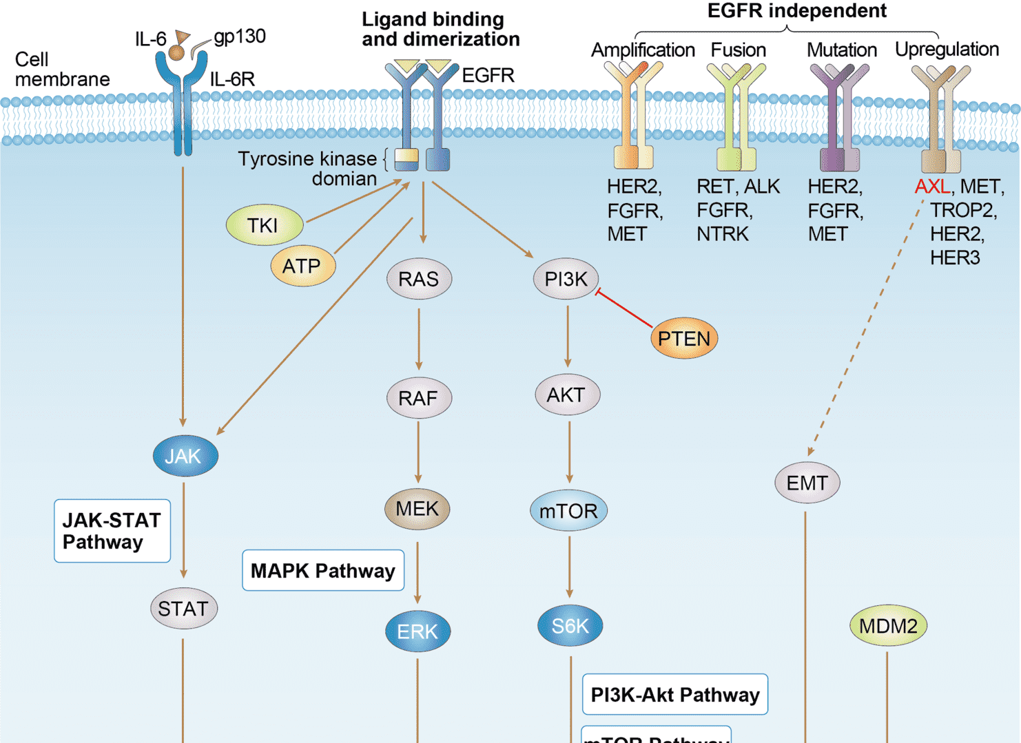 EGFR Tyrosine Kinase Inhibitor Resistance
Product Notes
This is a product of Creative Biolabs' Hi-Affi™ recombinant antibody portfolio, which has several benefits including:
• Increased sensitivity
• Confirmed specificity
• High repeatability
• Excellent batch-to-batch consistency
• Sustainable supply
• Animal-free production
See more details about Hi-Affi™ recombinant antibody benefits.
Downloads
Download resources about recombinant antibody development and antibody engineering to boost your research.
See other products for "MET"
Immunotoxin
| CAT | Product Name | Application | Type |
|---|---|---|---|
| AGTO-G049E | Anti-MET immunotoxin (Fab)-PE | Cytotoxicity assay, Function study |
Humanized Antibody
| CAT | Product Name | Application | Type |
|---|---|---|---|
| TAB-521CL | Anti-Human MET Recombinant Antibody (ABT-700) | WB, FuncS | Antibody |
| TAB-026MZ-F(E) | Anti-Human MET Recombinant Antibody Fab Fragment (73R009) | WB | Humanized antibody |
| TAB-0881CLV | Human Anti-MET Recombinant Antibody (TAB-0881CLV) | ELISA, FuncS | Humanized IgG1 |
| TAB-0883CLV | Human Anti-MET Recombinant Antibody (TAB-0883CLV) | ELISA, FuncS | Humanized IgG1 |
| TAB-0884CLV | Human Anti-MET Recombinant Antibody (TAB-0884CLV) | ELISA, FuncS | Humanized IgG1 |
ADCC Enhanced Antibody
| CAT | Product Name | Application | Type |
|---|---|---|---|
| PABZ-155 | Afuco™ Anti-MET ADCC Recombinant Antibody (Onartuzumab), ADCC Enhanced | ELISA, Neut | ADCC enhanced antibody |
| AFC-TAB-H24 | Afuco™ Anti-MET ADCC Recombinant Antibody (Emibetuzumab), ADCC Enhanced | FuncS, IF, Neut, ELISA, FC, IP | ADCC enhanced antibody |
Chimeric Antibody
| CAT | Product Name | Application | Type |
|---|---|---|---|
| TAB-002MZ | Anti-Human MET Recombinant Antibody (MvDN30) | IHC, PET imaging | Chimeric antibody (mouse/human) |
| TAB-002MZ-S(P) | Anti-Human MET Recombinant Antibody scFv Fragment (MvDN30) | IHC, PET imaging | Chimeric antibody (mouse/human) |
| TAB-0880CLV | Human Anti-MET Recombinant Antibody (TAB-0880CLV) | ELISA, FuncS | Chimeric (mouse/human) IgG1 |
| TAB-0880CLV-F(E) | Human Anti-MET Recombinant Antibody; Fab Fragment (TAB-0880CLV-F(E)) | ELISA, FuncS | Chimeric (mouse/human) Fab |
| TAB-0880CLV-S(P) | Mouse Anti-MET Recombinant Antibody; scFv Fragment (TAB-0880CLV-S(P)) | ELISA, FuncS | Mouse scFv |
Mouse Antibody
| CAT | Product Name | Application | Type |
|---|---|---|---|
| TAB-027MZ | Mouse Anti-MET Recombinant Antibody (TAB-027MZ) | WB, IP, IF | Mouse lgG1 |
| TAB-028MZ | Mouse Anti-MET Recombinant Antibody (TAB-028MZ) | IP | Mouse lgG1 |
| TAB-029MZ | Mouse Anti-MET Recombinant Antibody (TAB-029MZ) | IP, WB | Mouse lgG1 |
| TAB-030MZ | Mouse Anti-MET Recombinant Antibody (TAB-030MZ) | FuncS, IP, IF, FC | Mouse lgG1 |
| TAB-031MZ | Mouse Anti-MET Recombinant Antibody (TAB-031MZ) | IF, FC, WB, IP | Mouse lgG1 |
Human Antibody
| CAT | Product Name | Application | Type |
|---|---|---|---|
| TAB-034MZ-S(P) | Anti-Human MET Recombinant Antibody scFv Fragment (PGIA-1-A1) | WB, ELISA | Human antibody |
| TAB-035MZ-S(P) | Anti-Human MET Recombinant Antibody scFv Fragment (PGIA-3-A9) | WB, ELISA | Human antibody |
| TAB-036MZ-S(P) | Anti-Human MET Recombinant Antibody scFv Fragment (PGIA-3-A11) | WB, ELISA | Human antibody |
| TAB-037MZ-S(P) | Anti-Human MET Recombinant Antibody scFv Fragment (PGIA-3-B2) | WB, ELISA | Human antibody |
| TAB-038MZ-S(P) | Anti-Human MET Recombinant Antibody scFv Fragment (PGIA-4-A5) | WB, ELISA | Human antibody |
Recombinant Antibody
| CAT | Product Name | Application | Type |
|---|---|---|---|
| MOB-1226CT | Recombinant Mouse anti-Human MET Monoclonal antibody (9G22) | IHC-Fr, IHC-P | |
| HPAB-0030-WJ | Human Anti-MET Recombinant Antibody (HPAB-0030-WJ) | ELISA, WB | Human IgG |
| HPAB-1298LY | Human Anti-MET Recombinant Antibody (HPAB-1298LY) | ELISA, IP | Human IgG2 |
| HPAB-1299LY | Human Anti-MET Recombinant Antibody (HPAB-1299LY) | ELISA, IP | Human IgG2 |
| HPAB-1300LY | Human Anti-MET Recombinant Antibody (HPAB-1300LY) | ELISA, IP | Human IgG2 |
Neutralizing Antibody
| CAT | Product Name | Application | Type |
|---|---|---|---|
| NEUT-1726CQ | Recombinant Rabbit Anti-MET Antibody (CBL1008) | Neut | IgG |
Rabbit Monoclonal Antibody
| CAT | Product Name | Application | Type |
|---|---|---|---|
| MOR-2216 | Hi-Affi™ Recombinant Rabbit Anti-MET Monoclonal Antibody (DS2216AB) | ICC/IF, IHC-P, WB | IgG |
| MOR-4519 | Hi-Affi™ Recombinant Rabbit Anti-MET Monoclonal Antibody (TH27DS) | WB, IF, ICC | IgG |
| MOR-4645 | Hi-Affi™ Recombinant Rabbit Anti-MET Monoclonal Antibody (TH158DS) | WB, IF, ICC, FC, ELISA | IgG |
| MOR-4694 | Hi-Affi™ Recombinant Rabbit Anti-MET Monoclonal Antibody (TH208DS) | WB, ELISA | IgG |
Fab Fragment Antibody
| CAT | Product Name | Application | Type |
|---|---|---|---|
| HPAB-0760-CN-F(E) | Mouse Anti-MET Recombinant Antibody; Fab Fragment (HPAB-0760-CN-F(E)) | WB, ELISA, IHC, IP | Mouse Fab |
| HPAB-0761-CN-F(E) | Human Anti-MET Recombinant Antibody; Fab Fragment (HPAB-0761-CN-F(E)) | WB, ELISA, IHC, IP | Human Fab |
| HPAB-0762-CN-F(E) | Human Anti-MET Recombinant Antibody; Fab Fragment (HPAB-0762-CN-F(E)) | WB, ELISA, IHC, IP | Human Fab |
| HPAB-0030-WJ-F(E) | Human Anti-MET Recombinant Antibody; Fab Fragment (HPAB-0030-WJ-F(E)) | ELISA, WB | Human Fab |
| HPAB-1308LY-F(E) | Human Anti-MET Recombinant Antibody; Fab Fragment (HPAB-1308LY-F(E)) | ELISA, IP | Human Fab |
scFv Fragment Antibody
| CAT | Product Name | Application | Type |
|---|---|---|---|
| HPAB-N0176-YC-S(P) | Mouse Anti-MET Recombinant Antibody; scFv Fragment (HPAB-N0176-YC-S(P)) | ELISA, FC | Mouse scFv |
| HPAB-N0177-YC-S(P) | Human Anti-MET Recombinant Antibody; scFv Fragment (HPAB-N0177-YC-S(P)) | ELISA, FC | Humanized scFv |
| HPAB-0714-FY-F(E) | Mouse Anti-MET Recombinant Antibody (clone AB013); scFv Fragment | ELISA | Mouse scFv |
| HPAB-0486-FY-S(P) | Mouse Anti-MET Recombinant Antibody; scFv Fragment (HPAB-0486-FY-S(P)) | Inhib | Mouse scFv |
| HPAB-0487-FY-S(P) | Human Anti-MET Recombinant Antibody; scFv Fragment (HPAB-0487-FY-S(P)) | Inhib | Human scFv |
MHC Tetramer for Cancer
| CAT | Product Name | Application | Type |
|---|---|---|---|
| MHC-YF474 | A*02:01/Human Hepatocyte growth factor receptor (YVDPVITSI) MHC Pentamer | FCM |
Customer Reviews and Q&As

There are currently no Customer reviews or questions for AFC-501CL. Click the button above to contact us or submit your feedback about this product.
View the frequently asked questions answered by Creative Biolabs Support.
For Research Use Only. Not For Clinical Use.
For research use only. Not intended for any clinical use. No products from Creative Biolabs may be resold, modified for resale or used to manufacture commercial products without prior written approval from Creative Biolabs.
Send Inquiry
This site is protected by reCAPTCHA and the Google Privacy Policy and Terms of Service apply.