
Mouse Anti-MYC Recombinant Antibody (clone 9E10) (CAT#: PABL-281)
Recombinant mouse antibody (9E10) is capable of binding to MYC. The murine monoclonal antibody 9E10, which was raised against a peptide from the C-terminal region of the human c-myc protooncogene product, is widely used in cell biology and protein engineering as a detection or purification tool for proteins that are fused with the corresponding epitope as an affinity tag.
Specific Inquiry

Figure 1 MYC expression in normal human colon.
Representative haematoxylin and eosin staining, in‐situ hybridization (MYCmRNA, pink) and immunohistochemical staining with Y69 (N‐terminal MYC), 9E10 (C‐terminal MYC) and Ki67 antibodies (brown) in normal human colon. The lower panels focus on the surface epithelium, which appears negative for Y69 and Ki67 expression, but strongly positive for 9E10 expression. Scale bars: 100 μm (upper panels) and 50 μm (lower panels).
Baker, A. M., Van Noorden, S., Rodriguez‐Justo, M., Cohen, P., Wright, N. A., & Lampert, I. A. (2016). Distribution of the c‐MYC gene product in colorectal neoplasia. Histopathology,69(2), 222-229.

Figure 2 MYC expression in conventional adenomas.
A, Representative haematoxylin and eosin (H&E) staining, in‐situhybridization (MYC mRNA, pink) and immunohistochemical staining with Y69 (N‐terminal MYC), 9E10 (C‐terminalMYC) and Ki67 antibodies (brown) in adenoma with low‐grade dysplasia. A 'reverse gradient' of MYC mRNA and Y69 expression, with highest expression at the luminal surface, is apparent. Scale bars: 200 μm. B, Representative H&E staining, in‐situ hybridization (MYC mRNA, pink) and immunohistochemical staining with Y69 (N‐terminal MYC), 9E10 (C‐terminal MYC) and Ki67 antibodies (brown) in adenoma, focusing on a gland with a highly proliferative phenotype. Again, a reverse gradient is apparent, although, in the proliferative gland, the MYC mRNA, Y69 and Ki67 expression can be seen extending to the base of the crypt, in contrast to the surrounding glands and normal crypts. Scale bars: 500 μm (upper panels) and 200 μm (lower panels). C, Representative H&E staining, in‐situ hybridization (MYC mRNA, pink) and immunohistochemical staining with Y69 (N‐terminal MYC), 9E10 (C‐terminal MYC) and Ki67 antibodies (brown) in high‐grade dysplasia. Extensive positivity for MYC mRNA, Y69 and Ki67 is apparent, whereas 9E10 staining is remarkably weak. Scale bars: 200 μm.
Baker, A. M., Van Noorden, S., Rodriguez‐Justo, M., Cohen, P., Wright, N. A., & Lampert, I. A. (2016). Distribution of the c‐MYC gene product in colorectal neoplasia. Histopathology,69(2), 222-229.

Figure 3 MYC expression in traditional serrated adenomas.
Representative haematoxylin and eosin staining, in‐situhybridization (MYC mRNA, pink) and immunohistochemical staining with Y69 (N‐terminal MYC), 9E10 (C‐terminalMYC) and Ki67 antibodies (brown) in a traditional serrated adenoma. Strong positivity for MYC mRNA, Y69 and Ki67 is apparent in ectopic crypt foci. Scale bars: 200 μm (upper panels) and 100 μm (lower panels).
Baker, A. M., Van Noorden, S., Rodriguez‐Justo, M., Cohen, P., Wright, N. A., & Lampert, I. A. (2016). Distribution of the c‐MYC gene product in colorectal neoplasia. Histopathology,69(2), 222-229.

Figure 4 MYC expression in hyperplastic polyps.
Representative haematoxylin and eosin staining, in‐situ hybridization (MYCmRNA, pink) and immunohistochemical staining with Y69 (N‐terminal MYC), 9E10 (C‐terminal MYC) and Ki67 antibodies (brown) in a hyperplastic polyp. The lower panels focus on the surface epithelium, which appears negative for Y69 and Ki67 expression, but strongly positive for 9E10 expression. Scale bars: 500 μm (upper panels) and 100 μm (lower panels).
Baker, A. M., Van Noorden, S., Rodriguez‐Justo, M., Cohen, P., Wright, N. A., & Lampert, I. A. (2016). Distribution of the c‐MYC gene product in colorectal neoplasia. Histopathology,69(2), 222-229.

Figure 5 MYC expression in invasive carcinoma.
A, Representative haematoxylin and eosin (H&E) staining, in‐situhybridization (MYC mRNA, pink) and immunohistochemical staining with Y69 (N‐terminal MYC), 9E10 (C‐terminalMYC) and Ki67 antibodies (brown) in a region of invasive carcinoma. The lower panels highlight the reciprocal relationship between the expression of MYC mRNA, Y69, and Ki67, and the staining of the 9E10 antibody. B, Representative H&E staining, in‐situ hybridization (MYC mRNA, pink) and immunohistochemical staining with Y69 (N‐terminal MYC), 9E10 (C‐terminal MYC) and Ki67 antibodies (brown) in a region of invasive carcinoma. The lower panels focus on an invasive gland showing flattened non‐proliferative cells at the invasive edge, which are notably Y69‐negative and 9E10‐positive. Scale bars: 200 μm (upper panels) and 50 μm (lower panels).
Baker, A. M., Van Noorden, S., Rodriguez‐Justo, M., Cohen, P., Wright, N. A., & Lampert, I. A. (2016). Distribution of the c‐MYC gene product in colorectal neoplasia. Histopathology,69(2), 222-229.

Figure 6 Immunofluorescence staining of methanol-fixed COS cells transfected with c-Myc fusion protein showing cytoplasmic staining.

Figure 7 Western blot analysis of c-Myc expression in HeLa (A), Jurkat (B) and K-562 (C) whole cell lysates.


Specifications
- Immunogen
- Human v-myc avian myelocytomatosis viral oncogene homolog
- Host Species
- Mouse
- Derivation
- Mouse
- Type
- Mouse IgG
- Specificity
- Human MYC
- Species Reactivity
- Human
- Clone
- 9E10
- Applications
- WB, IHC, FuncS
Product Property
- Purity
- >95% as determined by SDS-PAGE and HPLC analysis
- Concentration
- Please refer to the vial label for the specific concentration.
- Buffer
- PBS
- Preservative
- No preservatives
- Storage
- Centrifuge briefly prior to opening vial. Store at +4°C short term (1-2 weeks). Aliquot and store at -20°C long term. Avoid repeated freeze/thaw cycles.
Target
- Alternative Names
- MYC; v-myc avian myelocytomatosis viral oncogene homolog; MRTL; MYCC; c-Myc; bHLHe39; myc proto-oncogene protein; proto-oncogene c-Myc; transcription factor p64; class E basic helix-loop-helix protein 39; avian myelocytomatosis viral oncogene homolog; v-myc myelocytomatosis viral oncogene homolog; myc-related translation/localization regulatory factor
- Gene ID
- 4609
- UniProt ID
- P01106
Related Resources
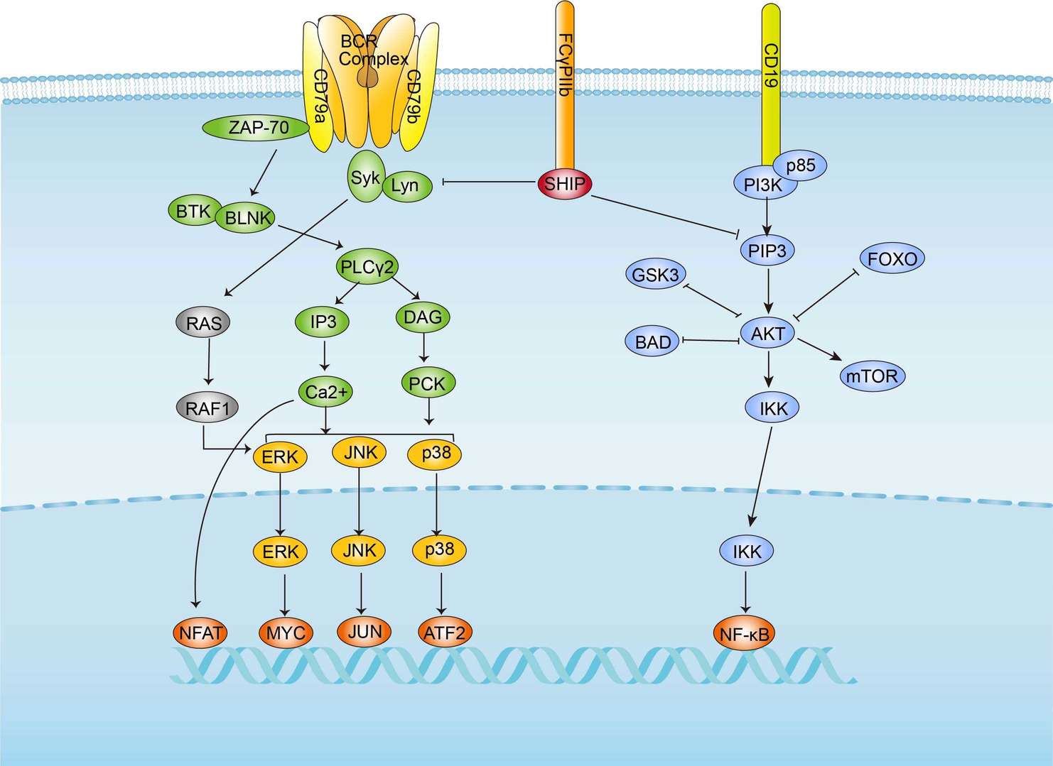 BCR Signaling Pathway
 Bladder Cancer
 Breast Cancer
 Colorectal Cancer
 Gastric Cancer
Product Notes
This is a product of Creative Biolabs' Hi-Affi™ recombinant antibody portfolio, which has several benefits including:
• Increased sensitivity
• Confirmed specificity
• High repeatability
• Excellent batch-to-batch consistency
• Sustainable supply
• Animal-free production
See more details about Hi-Affi™ recombinant antibody benefits.
Downloads
Download resources about recombinant antibody development and antibody engineering to boost your research.
See other products for "Clone 9E10"
See other products for "MYC"
scFv Fragment Antibody
| CAT | Product Name | Application | Type |
|---|---|---|---|
| PSBL-281 | Mouse Anti-MYC Recombinant Antibody (clone 9E10); scFv Fragment | WB, IHC, FuncS | Mouse scFv |
| FAMAB-0112-CN-S(P) | Mouse Anti-MYC Recombinant Antibody (clone CT14); scFv Fragment | ELISA, WB, FC | Mouse scFv |
Fab Fragment Antibody
| CAT | Product Name | Application | Type |
|---|---|---|---|
| PFBL-281 | Mouse Anti-MYC Recombinant Antibody (clone 9E10); Fab Fragment | WB, IHC, FuncS | Mouse Fab |
| FAMAB-0112-CN-F(E) | Mouse Anti-MYC Recombinant Antibody (clone CT14); Fab Fragment | ELISA, WB, FC | Mouse Fab |
Mouse Antibody
| CAT | Product Name | Application | Type |
|---|---|---|---|
| MOB-0400MC | Anti-c-Myc antibody (HRP) | ELISA, IHC-P, IHC-Fr, WB |
Chicken IgY Antibody
| CAT | Product Name | Application | Type |
|---|---|---|---|
| BRD-0380MZ | Chicken Anti-c-Myc (ab1) Polyclonal IgY | Indirect ELISA, WB | Chicken antibody |
| BRD-0381MZ | Chicken Anti-c-Myc (ab2) Polyclonal IgY | WB | Chicken antibody |
Rabbit Monoclonal Antibody
| CAT | Product Name | Application | Type |
|---|---|---|---|
| MOR-2336 | Hi-Affi™ Recombinant Rabbit Anti-MYC Monoclonal Antibody (DS2336AB) | WB, IP, ICC | IgG |
| MOR-4554 | Hi-Affi™ Recombinant Rabbit Anti-MYC Monoclonal Antibody (TH64DS) | WB, IF, ICC, FC | IgG |
| MOR-0050-FY | Rabbit Anti-MYC Recombinant Antibody (clone AFY0021) | ICC, IHC, WB | Rabbit IgG |
Recombinant Antibody
| CAT | Product Name | Application | Type |
|---|---|---|---|
| ZG-0433F | Mouse Anti-MYC Recombinant Antibody (ZG-0433F) | WB, ELISA | Mouse IgG |
| ZG-0258U | Mouse Anti-MYC Recombinant Antibody (clone 5H5G5) | ELISA, WB | Mouse IgG1 |
| VS3-XY1126 | Mouse Anti-MYC Recombinant Antibody (clone 6G6) | WB, IP, IF, IHC, FC | Mouse IgG1 |
| VS3-XY1127 | Mouse Anti-MYC Recombinant Antibody (clone 7E10) | ELISA, WB | Mouse IgG1 |
| VS-0223-WK12 | Rabbit Anti-MYC Recombinant Antibody (VS-0223-WK12) | WB, IP, ICC, IHC, IHC-P | Rabbit IgG |
Customer Reviews and Q&As

There are currently no Customer reviews or questions for PABL-281. Click the button above to contact us or submit your feedback about this product.
View the frequently asked questions answered by Creative Biolabs Support.
For Research Use Only. Not For Clinical Use.
For research use only. Not intended for any clinical use. No products from Creative Biolabs may be resold, modified for resale or used to manufacture commercial products without prior written approval from Creative Biolabs.
Send Inquiry
This site is protected by reCAPTCHA and the Google Privacy Policy and Terms of Service apply.