
Human Anti-ERBB2 Recombinant Antibody (clone bH1) (CAT#: PABZ-041)
Recombinant Human Antibody (bH1) is capable of binding to ERBB2, expressed in HEK 293 cells. Expressed as the combination of a heavy chain (HC) containing VH from anti-ERBB2 mAb and CH1-3 region of human IgG1 and a light chain (LC) encoding VL from anti-ERBB2 mAb and CL of human kappa light chain. Exists as a disulfide linked dimer of the HC and LC hetero-dimer under non-reducing condition.
Specific Inquiry

Figure 1 Analysis of BH-antibody affinities for recombinant ERBB2 by ELISA.
50 ng/well ERBB2 ECD-coated Nunc-polysorb plates were incubated with increasing concentrations of purified anti-ERBB2 antibodies. Strength of antibody binding was colorimetrically detected (OD 405 nm) by the alkaline phosphatase activity following the incubation with alkaline phosphatase-conjugated anti-mouse IgG antibodies. T Bars: SD.
Ceran, C. (2012). Novel monoclonal antibodies targeting conformational ERBB2 epitopes (Doctoral dissertation, Bilkent University).

Figure 2 Analysis of BH-antibody affinities for cellular ERBB2 by Flow Cytometry (B).
~3x10⁵ T47D (left column) or ~5x10⁵ BT-474 (right column) cells were incubated with increasing amount of BH-antibodies (0.5 – 2 μg antibody/sample) in 100 μl. Anti-ERBB2 incubation of paraformaldehyde fixed single cell suspensions was followed by Alexa-488-conjugated secondary antibodies and fluorescence intensities were measured using flow cytometry. Each graph shows fluorescence intensities of cells stained with different primary antibodies used at same concentration. Maximum fluorescence emissions at 488 nm are shown on x-axis (FL1-H) and cell counts are shown on y-axis. IgG1-isotype control antibody (IgG) at equal concentrations were used as negative controls. (-) indicates the control samples which were incubated only with Alexa-488-conjugated secondary antibodies without any prior primary antibody incubation.
Ceran, C. (2012). Novel monoclonal antibodies targeting conformational ERBB2 epitopes (Doctoral dissertation, Bilkent University).

Figure 3 Analysis of cellular ERBB2 by indirect immunofluorescence using BH-antibodies.
A) For FISH analysis, human breast cancer cells with different ERBB2 levels were harvested at confluence, fixed and subjected to dual-color FISH using Texas Red-labeled ERBB2 (red) and FITC-labeled chromosome 17 α-satellite CEP17 (green) DNA probes. B-G) For immunofluorescence assays, cells were seeded on coverslips on 6-well plates and incubated 36 hours in cell culture medium at 37°C. Paraformaldehyde fixed and permeabilised cells were incubated with 20 μg/ml BH-antibodies for two hours. Photographs are representative of merged images of BH-antibody binding detected using Alexa-488-conjugated anti-mouse IgG and nuclei counterstained with DAPI. (-) indicates the control samples where the cells were incubated only with Alexa-488-conjugated secondary antibodies without any prior primary antibody incubation.
Ceran, C. (2012). Novel monoclonal antibodies targeting conformational ERBB2 epitopes (Doctoral dissertation, Bilkent University).

Figure 4 Immunoprecipitation of endogenous ERBB2 by BH-antibodies.
Endogenously expressed ERBB2 present in 2 mg T47D RIPA lysate was immunoprecipitated by 20 μg BH-antibodies or Trastuzumab (Tzm). Antigen-antibody complexes that were captured onto Protein G-conjugated beads were eluted by boiling in SDS-PAGE loading dye (left panel). ERBB2 proteins that were uncaptured by the antibody-bead complex remained in the flow through (right panel). Proteins resolved under denaturing conditions were subjected to western blot assay using CB11 antibody. Trastuzumab was used as a positive control for ERBB2 immunoprecipitation. (-) is the negative control where T47D lysate was incubated with Protein-G beads in the absence of any antibody. ERBB2 protein band is detected at 185 kDa.
Ceran, C. (2012). Novel monoclonal antibodies targeting conformational ERBB2 epitopes (Doctoral dissertation, Bilkent University).

Figure 5 Analysis of senescence induction upon BH1 treatment in the presence or absence of TNF-α.
SK-BR-3 cells plated on coverslips in 12-well dishes were treated with 5 μg/ml BH1, Trastuzumab or IgG1 isotype control antibody with or without 1000 U/ml TNF-α. After 6 day treatment, cells were formaldehyde fixed and SAβ-gal stained for 12 hours. Nuclei was counterstained with nuclear fast red and and cells were observed under light microscopy. SAβ-gal positive cells contain dark blue spots suggesting a senescent phenotype. 100 μM CPT (camptothecin) and 50 ng/ml Adriamycin were used as positive controls of senescence induction.
Ceran, C. (2012). Novel monoclonal antibodies targeting conformational ERBB2 epitopes (Doctoral dissertation, Bilkent University).


Specifications
- Immunogen
- Human erb-b2 receptor tyrosine kinase 2
- Host Species
- Human
- Type
- Human IgG
- Specificity
- Human ERBB2
- Species Reactivity
- Human
- Clone
- bH1
- Applications
- WB, IF, FuncS
Product Property
- Purity
- >95% as determined by SDS-PAGE and HPLC analysis
- Concentration
- Please refer to the vial label for the specific concentration.
- Storage
- Centrifuge briefly prior to opening vial. Store at +4°C short term (1-2 weeks). Aliquot and store at -20°C long term. Avoid repeated freeze/thaw cycles.
Applications
- Application Notes
- The antibody bH1 has been reported in applications of Enzyme-linked Immunosorbent Assay, Flow Cytometry, Immunofluorescence, Immunoprecipitation, Western Blot, Functional Assay. It's recommended that the optimal antibody concentration, dilution, incubition time etc. are best to be carefully titrated in specific assays.
Immunofluorescence: The reported concentration of bH1 for immunofluorescence was 10 μg/ml.
Target
- Alternative Names
- ERBB2; erb-b2 receptor tyrosine kinase 2; NEU; NGL; HER2; TKR1; CD340; HER-2; MLN 19; HER-2/neu; receptor tyrosine-protein kinase erbB-2; herstatin; p185erbB2; proto-oncogene Neu; c-erb B2/neu protein; proto-oncogene c-ErbB-2; metastatic lymph node gene 19 protein; human epidermal growth factor receptor 2; neuro/glioblastoma derived oncogene homolog; tyrosine kinase-type cell surface receptor HER2; neuroblastoma/glioblastoma derived oncogene homolog; v-erb-b2 avian erythroblastic leukemia viral oncoprotein 2; v-erb-b2 avian erythroblastic leukemia viral oncogene homolog 2; v-erb-b2 erythroblastic leukemia viral oncogene homolog 2, neuro/glioblastoma derived oncogene homolog
- Gene ID
- 2064
- UniProt ID
- P04626
Related Resources
 Bladder Cancer
 Non-small Cell Lung Cancer
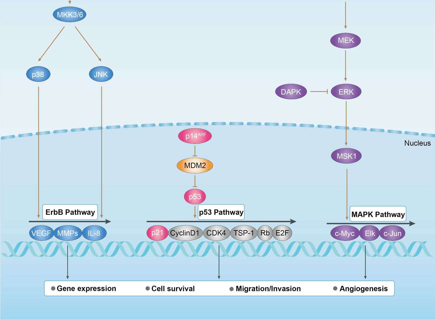
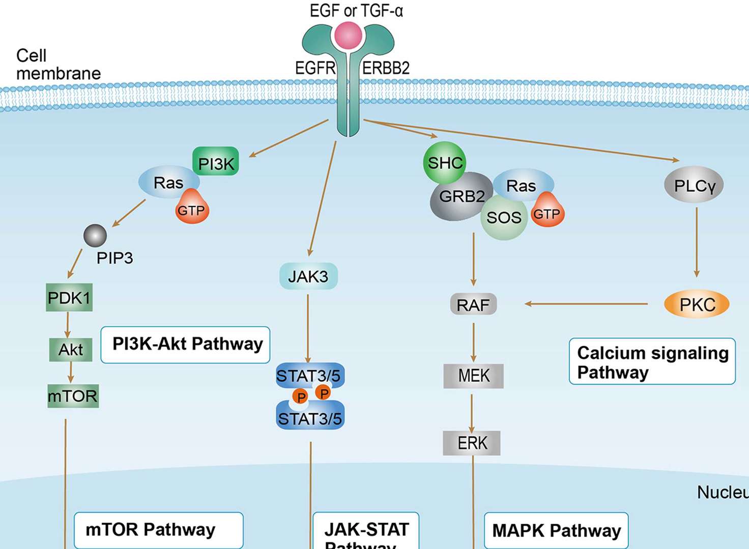
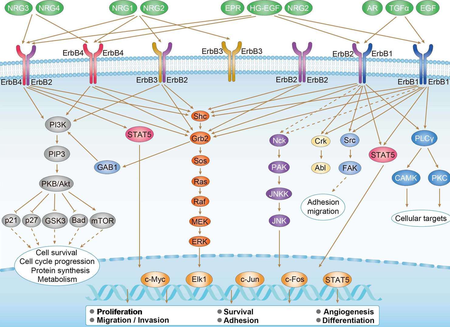 ErbB Signaling Pathway
Product Notes
This is a product of Creative Biolabs' Hi-Affi™ recombinant antibody portfolio, which has several benefits including:
• Increased sensitivity
• Confirmed specificity
• High repeatability
• Excellent batch-to-batch consistency
• Sustainable supply
• Animal-free production
See more details about Hi-Affi™ recombinant antibody benefits.
Downloads
Download resources about recombinant antibody development and antibody engineering to boost your research.
See other products for "Clone bH1"
See other products for "ERBB2"
Single-domain Antibody
| CAT | Product Name | Application | Type |
|---|---|---|---|
| NABG-059 | Recombinant Anti-Human ERBB2 VHH Single Domain Antibody | IHC, FC, CA, FuncS | Llama VHH |
| PNBL-066 | Recombinant Anti-HER2 VHH Single Domain Antibody (Gr3) | SPR | Llama VHH |
| PNBL-067 | Recombinant Anti-HER2 VHH Single Domain Antibody (Gr6) | SPR | Llama VHH |
| HPAB-1026WJ | Recombinant Camelid Anti-ERBB2 Single Domain Antibody | ELISA | Camelid VHH |
| HPAB-0725-YJ-VHH | Camelid Anti-ERBB2 Recombinant Single Domain Antibody (SR-87) | ELISA | Camelid VHH |
Intrabody
| CAT | Product Name | Application | Type |
|---|---|---|---|
| IAB-B008(A) | Recombinant Anti-human ERBB2 Intrabody [(D-Arg)9] | IF, FC, FuncS | scFv-(D-Arg)9 |
| IAB-B008(G) | Recombinant Anti-human ERBB2 Intrabody [+36 GFP] | WB, ICC, FuncS | scFv-(+36GFP) |
| IAB-B008(T) | Recombinant Anti-human ERBB2 Intrabody [Tat] | ICC, Neut, FuncS | scFv-Tat |
Chimeric Antibody
| CAT | Product Name | Application | Type |
|---|---|---|---|
| TAB-761 | Anti-ERBB2 Recombinant Antibody (Margetuximab) | IF, IP, Neut, FuncS, ELISA, FC, WB | IgG1 - kappa |
| TAB-049CT | Anti-Human HER2/neu Recombinant Antibody (ChA21) | IHC, Inhib, WB, Activ, FC | Chimeric antibody (mouse/human) |
| TAB-049CT-S(P) | Anti-Human HER2/neu Recombinant Antibody scFv Fragment (ChA21) | WB | Chimeric antibody (mouse/human) |
| TAB-049CT-F(E) | Anti-Human HER2/neu Recombinant Antibody Fab Fragment (ChA21) | WB | Chimeric antibody (mouse/human) |
Immunotoxin
| CAT | Product Name | Application | Type |
|---|---|---|---|
| AGTO-G022E | Anti-ERBB2 immunotoxin 4D5 (scFv)-PE | Cytotoxicity assay, Function study | |
| AGTO-G022D | Anti-ERBB2 immunotoxin 4D5 (scFv)-DT | Cytotoxicity assay, Function study | |
| AGTO-G022G | Anti-ERBB2 immunotoxin 4D5 (scFv)-Gel | Cytotoxicity assay, Function study | |
| AGTO-G022R | Anti-ERBB2 immunotoxin 4D5 (scFv)-RTA | Cytotoxicity assay, Function study | |
| AGTO-G022S | Anti-ERBB2 immunotoxin 4D5 (scFv)-Sap | Cytotoxicity assay, Function study |
Fab Fragment Antibody
| CAT | Product Name | Application | Type |
|---|---|---|---|
| PSBW-161 | Mouse Anti-ERBB2 Recombinant Antibody (clone chA21); scFv Fragment | FC, WB | Mouse scFv |
| HPAB-0608LY-F(E) | Human Anti-ERBB2 Recombinant Antibody; Fab Fragment (HPAB-0608LY-F(E)) | ELISA, Inhib | Humanized Fab |
| HPAB-0609LY-F(E) | Human Anti-ERBB2 Recombinant Antibody; Fab Fragment (HPAB-0609LY-F(E)) | ELISA, Inhib | Humanized Fab |
| HPAB-0610LY-F(E) | Human Anti-ERBB2 Recombinant Antibody; Fab Fragment (HPAB-0610LY-F(E)) | ELISA, Inhib | Humanized Fab |
| HPAB-0193-WJ-F(E) | Mouse Anti-ERBB2 Recombinant Antibody (clone 6H8); Fab Fragment | ELISA | Mouse Fab |
Human Antibody
| CAT | Product Name | Application | Type |
|---|---|---|---|
| TAB-0191CL-F(E) | Human Anti-ERBB2 Recombinant Antibody; Fab Fragment (TAB-0191CL-F(E)) | ELISA, Cyt, Internalization | Human Fab |
| TAB-042CT-S(P) | Anti-Human HER2/neu Recombinant Antibody scFv Fragment (CRX01) | WB | Human antibody |
| TAB-043CT-S(P) | Human Anti-ERBB2 Recombinant Antibody; scFv Fragment (TAB-043CT-S(P)) | ELISA, Inhibion | Human scFv |
| TAB-044CT-S(P) | Anti-Human HER2/neu Recombinant Antibody scFv Fragment (MIL5) | WB, FC, IF, Funcs | Human antibody |
| TAB-045CT-S(P) | Anti-Human HER2/neu Recombinant Antibody scFv Fragment (Erbicin) | ELISA, Inhibition | Human antibody |
Humanized Antibody
| CAT | Product Name | Application | Type |
|---|---|---|---|
| TAB-050CT | Anti-Human HER2/neu Recombinant Antibody (HuA21) | Inhib, Cty, IHC, WB, IF, FC, ELISA | Humanized antibody |
| TAB-053CT | Human Anti-ERBB2 Recombinant Antibody (TAB-053CT) | ELISA, Inhib, FC | Humanized IgG |
| TAB-033CT-S(P) | Human Anti-ERBB2 Recombinant Antibody; scFv Fragment (TAB-033CT-S(P)) | ELISA, WB | Human scFv |
| TAB-038CT-S(P) | Human Anti-ERBB2 Recombinant Antibody; scFv Fragment (TAB-038CT-S(P)) | WB | Humanized scFv |
| TAB-039CT-S(P) | Anti-Human HER2/neu Recombinant Antibody scFv Fragment (h1E11) | WB | Humanized antibody |
Mouse Antibody
| CAT | Product Name | Application | Type |
|---|---|---|---|
| TAB-034CT-S(P) | Anti-Human HER2/neu Recombinant Antibody scFv Fragment (pertuzumab ) | WB | |
| TAB-036CT-S(P) | Anti-Human HER2/neu Recombinant Antibody scFv Fragment (6B2) | ELISA, WB | |
| TAB-037CT-S(P) | Anti-Human HER2/neu Recombinant Antibody scFv Fragment (7C2/7F3) | ELISA, WB | |
| TAB-270CQ | Mouse Anti-ERBB2 Recombinant Antibody (TAB-270CQ) | WB, Inhib | Mouse IgG |
| PABX-082-S (P) | Recombinant Human Anti-HER2 Antibody scFv Fragment (Gr6) | WB, ELISA, FuncS | scFv |
Fab Glycosylation
| CAT | Product Name | Application | Type |
|---|---|---|---|
| Gly-024LC | Recombinant Anti-Human ERBB2 Antibody (Fab glycosylation) | ELISA | Mouse antibody |
| Gly-039LC | Recombinant Anti-Human ERBB2 Antibody (Fab glycosylation) | ELISA | Human antibody |
Fc Glycosylation
| CAT | Product Name | Application | Type |
|---|---|---|---|
| Gly-117LC | Recombinant Anti-Human ERBB2 Antibody (Fc glycosylation) | ELISA | Humanized antibody |
| Gly-118LC | Recombinant Anti-Human ERBB2 Antibody (Fc glycosylation) | ELISA | Mouse antibody |
| Gly-145LC | Recombinant Anti-Human ERBB2 Antibody (Fc glycosylation) | ELISA | Human antibody |
| Gly-146LC | Recombinant Anti-Human ERBB2 Antibody (Fc glycosylation) | ELISA | Human antibody |
| Gly-147LC | Recombinant Anti-Human ERBB2 Antibody (Fc glycosylation) | ELISA | Human antibody |
Deglycosylated Antibody (Non-glycosylated IgGs)
| CAT | Product Name | Application | Type |
|---|---|---|---|
| Gly-177LC | Recombinant Anti-Human ERBB2 Antibody (Non-glycosylated) | ELISA | Humanized antibody |
Chicken IgY Antibody
| CAT | Product Name | Application | Type |
|---|---|---|---|
| BRD-0195MZ | Chicken Anti-ERBB2 Polyclonal IgY | WB | Chicken antibody |
| BRD-0695MZ | Chicken Anti-ERBB2 Polyclonal IgY | WB | Chicken antibody |
| BRD-0779MZ | Chicken Anti-ErbB2 Polyclonal IgY | ICC, IF, IHC, IP | Chicken antibody |
Blocking Antibody
| CAT | Product Name | Application | Type |
|---|---|---|---|
| NEUT-737CQ | Mouse Anti-ERBB2 Recombinant Antibody (clone 7.16.4) | Block, IP, IF, FC | Mouse IgG2a |
Rabbit Monoclonal Antibody
| CAT | Product Name | Application | Type |
|---|---|---|---|
| MOR-1178 | Hi-Affi™ Rabbit Anti-ERBB2 Recombinant Antibody (clone DS1178AB) | IHC-P, IHC-Fr | Rabbit IgG |
| MOR-4586 | Hi-Affi™ Rabbit Anti-ERBB2 Recombinant Antibody (clone TH99DS) | WB | Rabbit IgG |
| MOR-4587 | Hi-Affi™ Rabbit Anti-ERBB2 Recombinant Antibody (clone TH100DS) | WB | Rabbit IgG |
MHC Tetramer for Cancer
| CAT | Product Name | Application | Type |
|---|---|---|---|
| MHC-LC2349 | APC-A*02:01/Human ERBB2 (HLYQGCQVV) MHC Tetramer | FCM | |
| MHC-LC2351 | APC-A*02:01/Human ERBB2 (PLTSIISAV) MHC Tetramer | FCM | |
| MHC-LC2742 | PE-A*02:01/Human ERBB2 (HLYQGCQV) MHC Tetramer | FCM | |
| MHC-YF221 | A*02:01/Human Her-2/neu (KIFGSLAFL) MHC Monomer | MHC Multimer | |
| MHC-YF222 | A*02:01/Human Her-2/neu (RLLQETELV) MHC Monomer | MHC Multimer |
Recombinant Antibody
| CAT | Product Name | Application | Type |
|---|---|---|---|
| FAMAB-0098YC | Mouse Anti-ERBB2 Recombinant Antibody (clone E402) | Cyt | Mouse IgG1, κ |
| FAMAB-0099YC | Human Anti-ERBB2 Recombinant Antibody (clone CH402) | Cyt | Chimeric (mouse/human) IgG1 |
| FAMAB-0132CQ | Mouse Anti-ERBB2 Recombinant Antibody (clone SER4) | ELISA | Mouse IgG1 |
| HPAB-0010-FY | Human Anti-ERBB2 Recombinant Antibody (HPAB-0010-FY) | ELISA, FC, ADCC | Human IgG1, κ |
| HPAB-0011-FY | Mouse Anti-ERBB2 Recombinant Antibody (HPAB-0011-FY) | Inhib, ADCC, FuncS | Mouse IgG1, κ |
ADCC Enhanced Antibody
| CAT | Product Name | Application | Type |
|---|---|---|---|
| AFC-TAB-468CQ | Afuco™ Anti-ERBB2 ADCC Recombinant Antibody (Timigutuzumab), ADCC Enhanced | ELISA, IHC, FC, IP, IF, FuncS | ADCC enhanced antibody |
| AFC-TAB-053 | Afuco™ Anti-ERBB2 ADCC Recombinant Antibody (Pertuzumab), ADCC Enhanced | FuncS, IF, Neut, ELISA, FC, IP | ADCC enhanced antibody |
| AFC-TAB-761 | Afuco™ Anti-ERBB2 ADCC Recombinant Antibody (Margetuximab), ADCC Enhanced | IF, IP, Neut, FuncS, ELISA, FC | ADCC enhanced antibody |
| AFC-TAB-005 | Afuco™ Anti-ERBB2 ADCC Recombinant Antibody (Trastuzumab), ADCC Enhanced | FC, IP, ELISA, Neut, FuncS, IF | ADCC enhanced antibody |
scFv Fragment Antibody
| CAT | Product Name | Application | Type |
|---|---|---|---|
| HPAB-S0027-YC-S(P) | Human Anti-ERBB2 Recombinant Antibody (clone F5); scFv Fragment | ELISA, Inhib, Internalization | Human scFv |
| HPAB-S0028-YC-S(P) | Human Anti-ERBB2 Recombinant Antibody (clone C1); scFv Fragment | ELISA, Inhib, Internalization | Human scFv |
| HPAB-1855-FY-F(E) | Human Anti-ERBB2 Recombinant Antibody; Fab Fragment (HPAB-1855-FY-F(E)) | ELISA | Humanized Fab |
| FAMAB-0359JF-S(P) | Human Anti-ERBB2 Recombinant Antibody (clone FRc13); scFv Fragment | DB, ELISA, FuncS | Human scFv |
| PSBS-0070 | Human Anti-ERBB2 Recombinant Antibody; scFv Fragment (PSBS-0070) | ELISA, FuncS | Human scFv |
Customer Reviews and Q&As

There are currently no Customer reviews or questions for PABZ-041. Click the button above to contact us or submit your feedback about this product.
View the frequently asked questions answered by Creative Biolabs Support.
For Research Use Only. Not For Clinical Use.
For research use only. Not intended for any clinical use. No products from Creative Biolabs may be resold, modified for resale or used to manufacture commercial products without prior written approval from Creative Biolabs.
Send Inquiry
This site is protected by reCAPTCHA and the Google Privacy Policy and Terms of Service apply.