Rabbit Anti-PAK1 Recombinant Antibody (VS3-CJ183)
CAT#: VS3-CJ183
This product is a rabbit antibody that recognizes Phospho-PAK1 (S144)+PAK2 (S141)+PAK3 (S139).


Specifications
- Immunogen
- KLH-conjugated synthetic peptide
- Host Species
- Rabbit
- Type
- Rabbit IgG
- Specificity
- Human, Mouse and Rat PAK1
- Species Reactivity
- Human, Mouse, Rat
- Applications
- WB, ICC, IF, IHC, IP, FC
- Conjugate
- Unconjugated
Product Property
- Purification
- Protein A affinity purified
- Purity
- >95% as determined by SDS-PAGE
- Buffer
- 40% Glycerol, 1% BSA, TBS, pH7.4.
- Preservative
- 0.05% sodium azide
- Storage
- Store at 4°C for short term. Aliquot and store at -20°C for long term. Avoid repeated freeze/thaw cycles.
- Shipping
- Ice packs
Applications
- Application Notes
- This antibody has been tested for use in Western Blot, Immunocytochemistry, Immunofluorescence, Immunohistochemistry, Immunoprecipitation, Flow Cytometry.
Target
- Alternative Names
- IDDMSSD; p65-PAK; PAKalpha; alpha-PAK
- Gene ID
- 5058
- UniProt ID
- Q13153
- Sequence Similarities
- Belongs to the protein kinase superfamily. STE Ser/Thr protein kinase family. STE20 subfamily.
- Cellular Localization
- Cell junction, Cell membrane, Cell projection, Chromosome, Cytoplasm, Cytoskeleton, Membrane, Nucleus
- Post Translation Modifications
- Autophosphorylated in trans, meaning that in a dimer, one kinase molecule phosphorylates the other one. Activated by autophosphorylation at Thr-423 in response to a conformation change, triggered by interaction with GTP-bound CDC42 or RAC1. Activated by phosphorylation at Thr-423 by BRSK2 and by PDPK1. Phosphorylated by JAK2 in response to PRL; this increases PAK1 kinase activity. Phosphorylated at Ser-21 by PKB/AKT; this reduces interaction with NCK1 and association with focal adhesion sites. Upon DNA damage, phosphorylated at Thr-212 and translocates to the nucleoplasm (PubMed:23260667).
Phosphorylated at tyrosine residues, which can be enhanced by NTN1 (By similarity).
- Protein Refseq
- NP_001122092.1; NP_002567.3
- Function
- Protein kinase involved in intracellular signaling pathways downstream of integrins and receptor-type kinases that plays an important role in cytoskeleton dynamics, in cell adhesion, migration, proliferation, apoptosis, mitosis, and in vesicle-mediated transport processes (PubMed:11896197, PubMed:30290153).
Can directly phosphorylate BAD and protects cells against apoptosis. Activated by interaction with CDC42 and RAC1. Functions as GTPase effector that links the Rho-related GTPases CDC42 and RAC1 to the JNK MAP kinase pathway. Phosphorylates and activates MAP2K1, and thereby mediates activation of downstream MAP kinases. Involved in the reorganization of the actin cytoskeleton, actin stress fibers and of focal adhesion complexes. Phosphorylates the tubulin chaperone TBCB and thereby plays a role in the regulation of microtubule biogenesis and organization of the tubulin cytoskeleton. Plays a role in the regulation of insulin secretion in response to elevated glucose levels. Part of a ternary complex that contains PAK1, DVL1 and MUSK that is important for MUSK-dependent regulation of AChR clustering during the formation of the neuromuscular junction (NMJ). Activity is inhibited in cells undergoing apoptosis, potentially due to binding of CDC2L1 and CDC2L2. Phosphorylates MYL9/MLC2. Phosphorylates RAF1 at 'Ser-338' and 'Ser-339' resulting in: activation of RAF1, stimulation of RAF1 translocation to mitochondria, phosphorylation of BAD by RAF1, and RAF1 binding to BCL2. Phosphorylates SNAI1 at 'Ser-246' promoting its transcriptional repressor activity by increasing its accumulation in the nucleus. In podocytes, promotes NR3C2 nuclear localization. Required for atypical chemokine receptor ACKR2-induced phosphorylation of LIMK1 and cofilin (CFL1) and for the up-regulation of ACKR2 from endosomal compartment to cell membrane, increasing its efficiency in chemokine uptake and degradation. In synapses, seems to mediate the regulation of F-actin cluster formation performed by SHANK3, maybe through CFL1 phosphorylation and inactivation. Plays a role in RUFY3-mediated facilitating gastric cancer cells migration and invasion (PubMed:25766321).
In response to DNA damage, phosphorylates MORC2 which activates its ATPase activity and facilitates chromatin remodeling (PubMed:23260667).
In neurons, plays a crucial role in regulating GABA(A) receptor synaptic stability and hence GABAergic inhibitory synaptic transmission through its role in F-actin stabilization (By similarity).
In hippocampal neurons, necessary for the formation of dendritic spines and excitatory synapses; this function is dependent on kinase activity and may be exerted by the regulation of actomyosin contractility through the phosphorylation of myosin II regulatory light chain (MLC) (By similarity).
Along with GIT1, positively regulates microtubule nucleation during interphase (PubMed:27012601).
Customer Review
There are currently no Customer reviews or questions for VS3-CJ183. Click the button above to contact us or submit your feedback about this product.
Submit Your Publication
Published with our product? Submit your paper and receive a 10% discount on your next order! Share your research to earn exclusive rewards.
Related Diseases
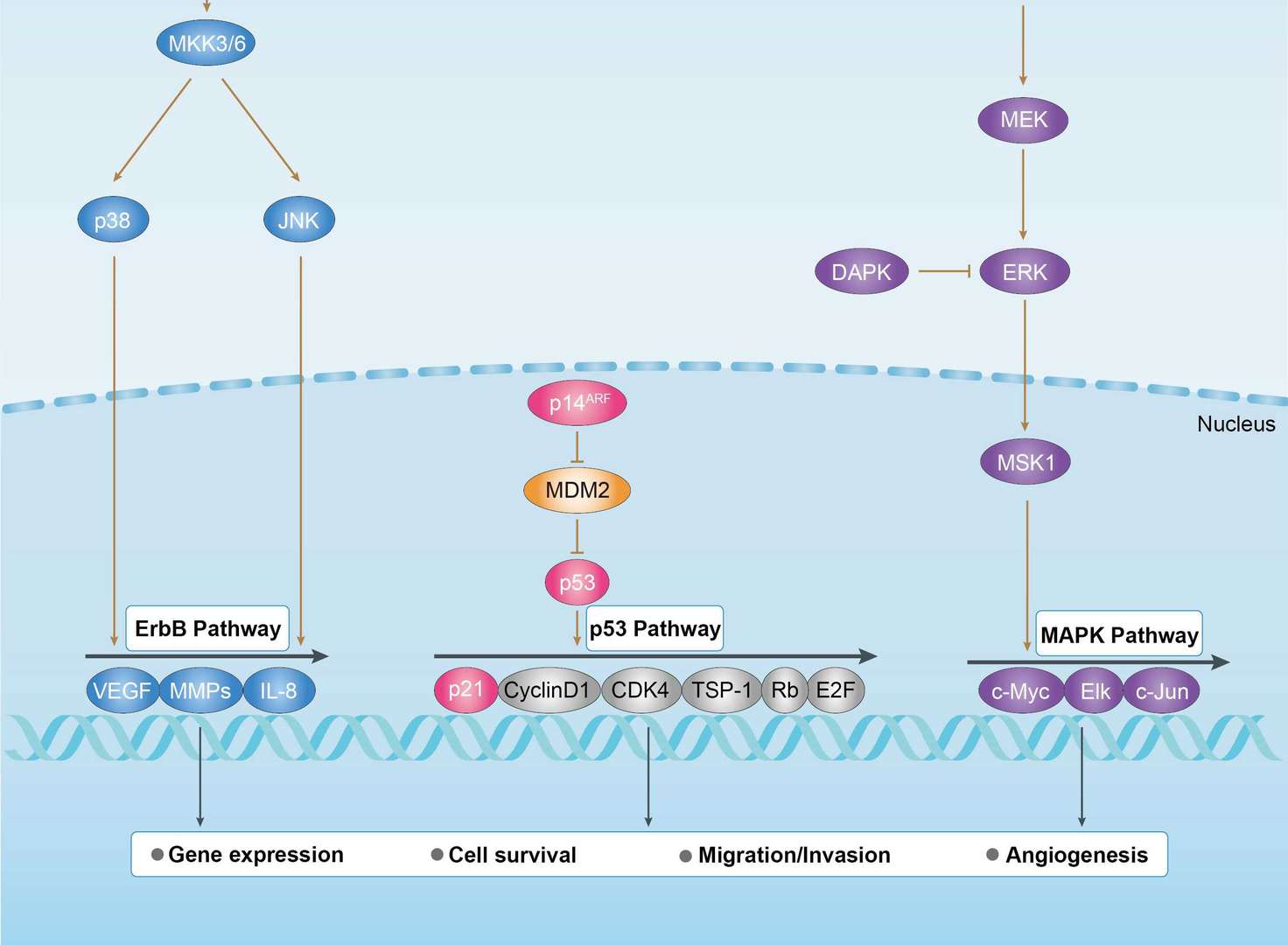
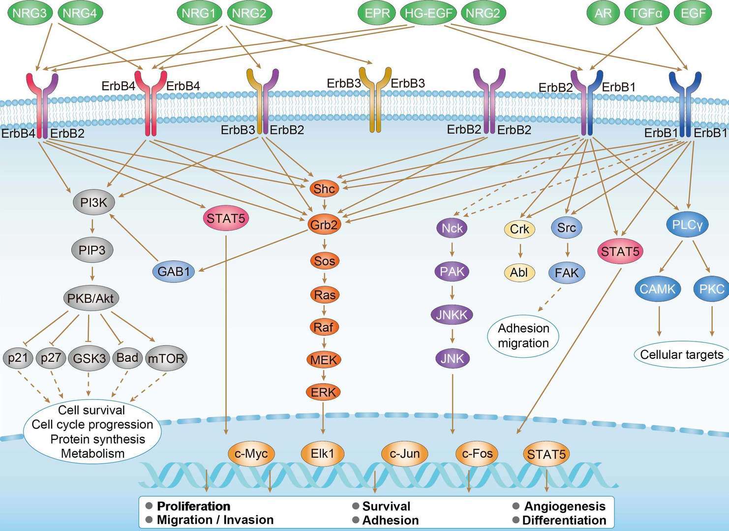
Related Signaling Pathways
Downloadable Resources
Download resources about recombinant antibody development and antibody engineering to boost your research.
Product Notes
This is a product of Creative Biolabs' Hi-Affi™ recombinant antibody portfolio, which has several benefits including:
• Increased sensitivity
• Confirmed specificity
• High repeatability
• Excellent batch-to-batch consistency
• Sustainable supply
• Animal-free production
See more details about Hi-Affi™ recombinant antibody benefits.
Datasheet
MSDS
COA
Certificate of Analysis LookupTo download a Certificate of Analysis, please enter a lot number in the search box below. Note: Certificate of Analysis not available for kit components.
Protocol & Troubleshooting
We have outlined the assay protocols, covering reagents, solutions, procedures, and troubleshooting tips for common issues in order to better assist clients in conducting experiments with our products. View the full list of Protocol & Troubleshooting.
Isotype Control
- CAT
- Product Name
Secondary Antibody
- CAT
- Product Name
See other products for "PAK1"
Select a product category from the dropdown menu below to view related products.
| CAT | Product Name | Application | Type |
|---|---|---|---|
| MOB-2175z | Mouse Anti-PAK1 Recombinant Antibody (clone 3A7) | WB, ELISA, IHC | Mouse IgG1, κ |
| MOB-1535CT | Recombinant Mouse anti-Human PAK1 Monoclonal antibody (EML2308) | IHC-P, WB | |
| MRO-2355-CN | Recombinant Rabbit Anti-PAK1 (phosphorylated Ser144+Ser141+Ser139) Monoclonal Antibody (CBACN-644) | WB, IF, IHC, IP, FC | Rabbit IgG |
| VS3-FY1090 | Recombinant Rabbit Anti-PAK1 Antibody (clone R02-5G3) | WB, IHC-Fr, IHC-P, ICC, IF, IP | Rabbit IgG |
| VS3-FY1091 | Recombinant Rabbit Anti-PAK1 Antibody (clone R08-5I5) | WB, ICC, IF, IP | Rabbit IgG |
| CAT | Product Name | Application | Type |
|---|---|---|---|
| MOR-2582 | Hi-Affi™ Recombinant Rabbit Anti-PAK1 Monoclonal Antibody (DS2582AB) | IP, WB | IgG |
| CAT | Product Name | Application | Type |
|---|---|---|---|
| VS-0724-YC307 | AbPlus™ Anti-Pak1 Magnetic Beads (VS-0724-YC307) | IP, Protein Purification |
| CAT | Product Name | Application | Type |
|---|---|---|---|
| VS-0325-XY1556 | Anti-PAK1 Immunohistochemistry Kit | IHC | |
| VS-0525-XY5164 | Anti-Mouse PAK1 Immunohistochemistry Kit | IHC | |
| VS-0525-XY5163 | Anti-Human PAK1 Immunohistochemistry Kit | IHC | |
| VS-0525-XY5165 | Anti-Rat PAK1 Immunohistochemistry Kit | IHC |
Popular Products

Application: IF, IP, Neut, FuncS, ELISA, FC, ICC

Application: WB, ELISA, Neut, FuncS

Application: WB, ELISA, FC, IHC, IF, IP
-3.jpg)
Application: ELISA, WB

Application: ELISA, WB

Application: WB, IHC-Fr, IHC-P, ICC, IF
For research use only. Not intended for any clinical use. No products from Creative Biolabs may be resold, modified for resale or used to manufacture commercial products without prior written approval from Creative Biolabs.
This site is protected by reCAPTCHA and the Google Privacy Policy and Terms of Service apply.


 Bladder Cancer
 ErbB Signaling Pathway


