


ATG Family
Representative AMPK Targets Full List of AMPK Targets Tested Data-Supported Products for Targeting AMPK
The ATG (Autophagy-related) family of proteins plays a crucial role in autophagy, a cellular degradation and recycling process that is essential for maintaining cellular homeostasis, responding to nutrient stress, and managing cellular damage. Autophagy involves the degradation of unnecessary or dysfunctional cellular components through the lysosomal machinery, helping to clear aggregated proteins, eliminate damaged organelles, and provide cellular building blocks and energy under nutrient starvation conditions. The ATG proteins are integral to the initiation, nucleation, expansion, and completion of the autophagosome, which is the key structure in autophagy. This family includes more than 30 proteins that work in concert to regulate the autophagic process. These proteins can be broadly categorized based on their function in different steps of autophagy: Initiation, Nucleation, Expansion and Completion, and Recycling. The regulation of autophagy and the ATG proteins is interconnected with several signaling pathways, including those mediated by AMPK, which activates autophagy by inhibiting mTOR, and p53, which can induce autophagy under stress conditions. Additionally, ATG proteins are involved in selective autophagy processes like mitophagy, pexophagy, and xenophagy, which target specific cellular components for degradation.
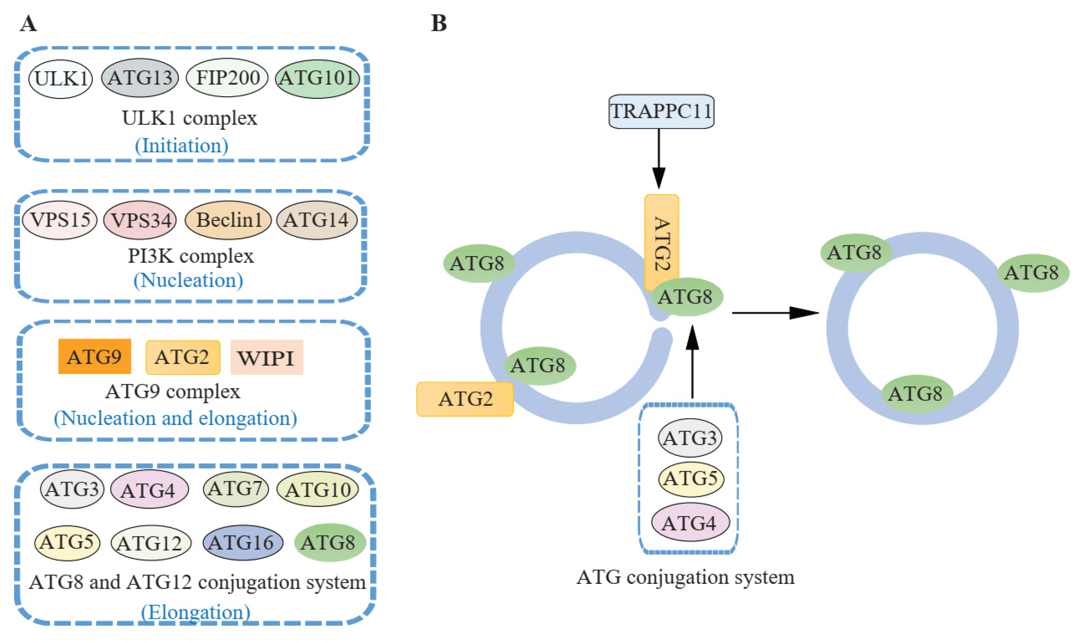 Figure 1 ATG proteins involved in autophagosome closure. (Jiang, 2021)
Representative AMPK Targets
ATG5
ATG5, or Autophagy-related 5, is a crucial protein involved in the autophagy pathway. The ATG5 protein is integral to the formation of the autophagosome, a key structure in autophagy, where it conjugates with ATG12 and associates with ATG16L1 to form the ATG12–ATG5-ATG16L1 complex. This complex is vital for the expansion of the autophagosome membrane. The activity of ATG5 is particularly important during cellular stress conditions, such as nutrient starvation, where it helps to ensure cellular survival by recycling intracellular components to generate new building blocks and energy. Moreover, ATG5 is involved in cellular processes that regulate immune responses, where it contributes to the elimination of intracellular pathogens and the presentation of antigens, playing a role in maintaining immune homeostasis. Dysregulation or defects in the autophagy process, involving ATG5, have been linked to various diseases, including neurodegenerative diseases, cancers, and infectious diseases. In neurodegeneration, impaired autophagy can lead to the accumulation of toxic protein aggregates, while in cancer, the role of autophagy is more complex, acting as a tumor suppressor in some contexts and promoting tumor survival in others. This dual role makes ATG5 a potential therapeutic target, with strategies aimed at modulating its activity to enhance or inhibit autophagy based on the specific disease context.
Recommended Mouse Anti-ATG5 mAb (CAT#: ZG-0038J)
 Figure 2 Mouse Anti-ATG5 Recombinant Antibody (clone 3C7) in IHC. Immunohistochemical analysis of paraffin-embedded Rat-testis tissue. 1.ATG5 Mouse Monoclonal Antibody was diluted at 1:200 (4°C, overnight). 2, Sodium citrate pH 6.0 was used for Antibody retrieval(>98°C, 20min). 3, Secondary Antibody was diluted at 1:200 (room temperature, 30min). Negative control was used by secondary Antibody only.
Recommended Mouse Anti-ATG5 mAb (CAT#: ZG-0039J)
 Figure 3 Mouse Anti-ATG5 Recombinant Antibody (clone 8B4) in IHC. Immunohistochemical analysis of paraffin-embedded Human Breast Carcinoma Tissue using ATG5 Mouse mAb diluted at 1:200.
ATG7
ATG7, or Autophagy-related 7, acts as an E1-like activating enzyme in the autophagy machinery, facilitating the conjugation of other ATG proteins necessary for autophagosome formation, specifically ATG8/LC3 to phosphatidylethanolamine and the ATG12-ATG5 conjugate formation, which are critical steps in the autophagic process. This protein is ubiquitously expressed and is essential not only for the routine turnover of cellular components but also for survival during nutrient deprivation by recycling intracellular resources. It plays a protective role in various stress conditions, including hypoxia, infection, and oxidative stress. The activity of ATG7 is crucial in processes beyond mere recycling; it's involved in immune responses through the regulation of inflammation and immunity mechanisms, and it also participates in the removal of invasive pathogens and the presentation of antigens. Dysfunction in ATG7 and the autophagy pathway is linked to numerous diseases, including neurodegenerative disorders like Parkinson’s and Alzheimer’s disease, where a buildup of protein aggregates can be detrimental. In cancer, similar to other autophagy-related genes, ATG7 can have dual roles: suppressing tumor initiation while potentially supporting tumor growth and resistance to therapy in established cancers. This complex role makes ATG7 a target of interest for therapeutic strategies aiming to manipulate autophagy for disease treatment.
Recommended Mouse Anti-ATG7 mAb (CAT#: ZG-0040J)
 Figure 4 Mouse Anti-ATG7 Recombinant Antibody (clone 3D6) in IHC. Immunohistochemical analysis of paraffin-embedded Rat-lung tissue. 1.ATG7 Mouse Monoclonal Antibody was diluted at 1:200 (4°C, overnight). 2, Sodium citrate pH 6.0 was used for Antibody retrieval(>98°C, 20min). 3, Secondary Antibody was diluted at 1:200 (room temperature, 30min). Negative control was used by secondary Antibody only.
BECN1
BECN1, or Beclin 1, is a key regulator of autophagy, a critical cellular process for maintaining homeostasis through the degradation and recycling of cellular components. Beclin 1 is integral to the initiation phase of autophagy, primarily involved in the nucleation of autophagosomal membranes. It functions as part of the class III phosphatidylinositol 3-kinase (PI3K) complex, which is essential for the early steps of autophagosome formation. This protein is not only pivotal in autophagy but also plays significant roles in other important cellular processes, including apoptosis and cell proliferation. The activity of BECN1 is tightly regulated by interactions with various cofactors that either promote or inhibit its activity. For instance, its interaction with BCL-2, an anti-apoptotic protein, can inhibit autophagy by preventing the activation of the BECN1-dependent PI3K complex. Conversely, dissociation of BECN1 from BCL-2 is a critical step in the induction of autophagy, particularly under stress conditions such as nutrient deprivation or oxidative stress. BECN1 has been implicated in a range of diseases, including cancer, neurodegenerative diseases, and infections. In cancer, for example, reduced expression or mutations in the BECN1 gene can lead to decreased autophagic activity, promoting tumorigenesis and chemotherapy resistance. Conversely, in neurodegenerative diseases like Alzheimer's, enhanced activity of BECN1 may help in clearing abnormal protein aggregates, although excessive autophagy can also lead to cell death.
Recommended Mouse Anti-BECN1 mAb (CAT#: ZG-0049J)
 Figure 5 Mouse Anti-BECN1 Recombinant Antibody (clone 5A11) in IHC. Immunohistochemical analysis of paraffin-embedded Human Breast Carcinoma Tissue using Beclin-1 Mouse mAb diluted at 1:200.
Recommended Mouse Anti-BECN1 mAb (CAT#: ZG-0050J)
 Figure 6 Mouse Anti-BECN1 Recombinant Antibody (clone 5C2) in WB. Western blot analysis of lysates from 1) 293T Cell Lysate, 2) C2C12 Cell Lysate, 3) Rat Brain Tissue cells, (Green) primary Antibody was diluted at 1:1000, 4°Cover night, secondary Antibody was diluted at 1:10000, 37°C 1 hour. (Red) Actin β Polyclonal Antibody was diluted at 1:5000 as loading control, 4°Cover night, secondary Antibody was diluted at 1:10000, 37°C1 hour.
Recommended Mouse Anti-BECN1 mAb (CAT#: ZG-0051J)
 Figure 7 Mouse Anti-BECN1 Recombinant Antibody (clone 5C5) in IHC. Immunohistochemical analysis of paraffin-embedded Human Breast Carcinoma Tissue using Beclin-1 Mouse mAb diluted at 1:200.
Full List of AMPK Targets
| Biomarker | Alternative Names | Gene ID | UniProt ID | Roles |
| ATG10 | Autophagy Related 10; Autophagy-Related Protein 10; APG10L; ATG10 Autophagy Related 10 Homolog (S. Cerevisiae); APG10 Autophagy 10-Like (S. Cerevisiae); Ubiquitin-Like-Conjugating Enzyme ATG10; ATG10 Autophagy Related 10 Homolog | 83734 | Q9H0Y0 | Autophagy is a process for the bulk degradation of cytosolic compartments by lysosomes. ATG10 is an E2-like enzyme involved in 2 ubiquitin-like modifications essential for autophagosome formation: ATG12 (MIM 609608)-ATG5 (MIM 604261) conjugation and modification of a soluble form of MAP-LC3 (MAP1LC3A; MIM 601242), a homolog of yeast Apg8, to a membrane-bound form (Nemoto et al., 2003 [PubMed 12890687]).[supplied by OMIM, Mar 2008] |
| ATG12 | Autophagy Related 12; APG12 Autophagy 12-Like; APG12L; APG12; ATG12 Autophagy Related 12 Homolog (S. Cerevisiae); Apg12 (Autophagy 12, S. Cerevisiae)-Like; APG12 Autophagy 12-Like (S. Cerevisiae); ATG12 Autophagy Related 12 Homolog | 9140 | O94817 | Autophagy is a process of bulk protein degradation in which cytoplasmic components, including organelles, are enclosed in double-membrane structures called autophagosomes and delivered to lysosomes or vacuoles for degradation. ATG12 is the human homolog of a yeast protein involved in autophagy (Mizushima et al., 1998 [PubMed 9852036]). |
| ATG3 | Autophagy Related 3; Autophagy-Related Protein 3; APG3-LIKE; APG3L; HApg3; APG3; ATG3 Autophagy Related 3 Homolog (S. Cerevisiae); Ubiquitin-Like-Conjugating Enzyme ATG3 | 64422 | Q9NT62 | This gene encodes a ubiquitin-like-conjugating enzyme and is a component of ubiquitination-like systems involved in autophagy, the process of degradation, turnover and recycling of cytoplasmic constituents in eukaryotic cells. This protein is known to play a role in regulation of autophagy during cell death. A pseudogene of this gene is located on chromosome 20. Alternative splicing results in multiple transcript variants encoding different isoforms. |
| ATG4A | APG4A; AUTL2; HsAPG4A | 115201 | Q8WYN0 | Autophagy is the process by which endogenous proteins and damaged organelles are destroyed intracellularly. Autophagy is postulated to be essential for cell homeostasis and cell remodeling during differentiation, metamorphosis, non-apoptotic cell death, and aging. Reduced levels of autophagy have been described in some malignant tumors, and a role for autophagy in controlling the unregulated cell growth linked to cancer has been proposed. This gene encodes a member of the autophagin protein family. The encoded protein is also designated as a member of the C-54 family of cysteine proteases. |
| ATG4B | Autophagy Related 4B Cysteine Peptidase; Autophagy-Related Cysteine Endopeptidase 1; Autophagy-Related Protein 4 Homolog B; AUT-Like 1 Cysteine Endopeptidase; Autophagin-1; HAPG4B; APG4B; AUTL1; ATG4 Autophagy Related 4 Homolog B (S. Cerevisiae) | 23192 | Q9Y4P1 | Autophagy is the process by which endogenous proteins and damaged organelles are destroyed intracellularly. Autophagy is postulated to be essential for cell homeostasis and cell remodeling during differentiation, metamorphosis, non-apoptotic cell death, and aging. Reduced levels of autophagy have been described in some malignant tumors, and a role for autophagy in controlling the unregulated cell growth linked to cancer has been proposed. This gene encodes a member of the autophagin protein family. The encoded protein is also designated as a member of the C-54 family of cysteine proteases. Alternate transcriptional splice variants, encoding different isoforms, have been characterized. |
| ATG5 | Autophagy Related 5; Apoptosis-Specific Protein; APG5-LIKE; APG5L; ASP; ATG5 Autophagy Related 5 Homolog (S. Cerevisiae); APG5 (Autophagy 5, S. Cerevisiae)-Like; APG5 Autophagy 5-Like (S. Cerevisiae); | 9474 | Q9H1Y0 | The protein encoded by this gene, in combination with autophagy protein 12, functions as an E1-like activating enzyme in a ubiquitin-like conjugating system. The encoded protein is involved in several cellular processes, including autophagic vesicle formation, mitochondrial quality control after oxidative damage, negative regulation of the innate antiviral immune response, lymphocyte development and proliferation, MHC II antigen presentation, adipocyte differentiation, and apoptosis. Several transcript variants encoding different protein isoforms have been found for this gene. |
| ATG7 | Autophagy Related 7; Ubiquitin-Activating Enzyme E1-Like Protein; ATG12-Activating Enzyme E1 ATG7; APG7-LIKE; APG7L; HAGP7; ATG7 Autophagy Related 7 Homolog (S. Cerevisiae); | 10533 | O95352 | This gene encodes an E1-like activating enzyme that is essential for autophagy and cytoplasmic to vacuole transport. The encoded protein is also thought to modulate p53-dependent cell cycle pathways during prolonged metabolic stress. It has been associated with multiple functions, including axon membrane trafficking, axonal homeostasis, mitophagy, adipose differentiation, and hematopoietic stem cell maintenance. Alternative splicing results in multiple transcript variants. |
| BECN1 | ATG6; VPS30; beclin1 | 8678 | Q14457 | This gene encodes a protein that regulates autophagy, a catabolic process of degradation induced by starvation. The encoded protein is a component of the phosphatidylinositol-3-kinase (PI3K) complex which mediates vesicle-trafficking processes. This protein is thought to play a role in multiple cellular processes, including tumorigenesis, neurodegeneration and apoptosis. Alternative splicing results in multiple transcript variants. |
| GABARAP | MM46; ATG8A; GABARAP-a | 11337 | O95166 | Gamma-aminobutyric acid A receptors [GABA(A) receptors] are ligand-gated chloride channels that mediate inhibitory neurotransmission. This gene encodes GABA(A) receptor-associated protein, which is highly positively charged in its N-terminus and shares sequence similarity with light chain-3 of microtubule-associated proteins 1A and 1B. This protein clusters neurotransmitter receptors by mediating interaction with the cytoskeleton. |
| GABARAPL2 | ATG8; GEF2; ATG8C; GEF-2; GATE16; GATE-16 | 11345 | P60520 | Enables ubiquitin protein ligase binding activity. Involved in negative regulation of proteasomal protein catabolic process and protein localization to endoplasmic reticulum. |
| MAP1LC3A | Microtubule Associated Protein 1 Light Chain 3 Alpha; Autophagy-Related Ubiquitin-Like Modifier LC3 A; MAP1 Light Chain 3-Like Protein 1; MAP1A/MAP1B Light Chain 3 A; MAP1A/MAP1B LC3 A; Microtubule-Associated Proteins 1A/1B Light Chain 3A; Microtubule-Associated Protein 1 Light Chain 3 Alpha; Microtubule-Associated Proteins 1A/1B Light Chain 3 | 84557 | Q9H492 | MAP1A and MAP1B are microtubule-associated proteins which mediate the physical interactions between microtubules and components of the cytoskeleton. MAP1A and MAP1B each consist of a heavy chain subunit and multiple light chain subunits. The protein encoded by this gene is one of the light chain subunits and can associate with either MAP1A or MAP1B. Two transcript variants encoding different isoforms have been found for this gene. The expression of variant 1 is suppressed in many tumor cell lines, suggesting that may be involved in carcinogenesis. [provided by RefSeq, Feb 2012] |
| MAP1LC3B | LC3B; ATG8F; MAP1LC3B-a; MAP1A/1BLC3 | 81631 | Q9GZQ8 | The product of this gene is a subunit of neuronal microtubule-associated MAP1A and MAP1B proteins, which are involved in microtubule assembly and important for neurogenesis. Studies on the rat homolog implicate a role for this gene in autophagy, a process that involves the bulk degradation of cytoplasmic component. |
Tested Data-Supported Products for Targeting AMPK


Reference
- Herzig, Sébastien, and Reuben J. Shaw. "AMPK: guardian of metabolism and mitochondrial homeostasis." Nature reviews Molecular cell biology 19.2 (2018): 121-135.
For research use only. Not intended for any clinical use.
Send Inquiry
This site is protected by reCAPTCHA and the Google Privacy Policy and Terms of Service apply.