Researchers at the University of Minnesota have developed a way to study cancer cells, which could lead to new and more effective treatments. They have developed a new method to study these cells in 3D models in vitro. In this recently published study in the journal Advanced Materials, Dr. Angela Panoskaltsis-Mortari and his assistant researchers, vice president of research at the University of Minnesota School of Medicine, professor of pediatrics, director of 3D bio-printing equipment and a member of the Masonic Cancer Center, and their assistant researchers found that cells behave in these 3D flexible tissue environments and 2D molds. The material or glass surface behaves differently.
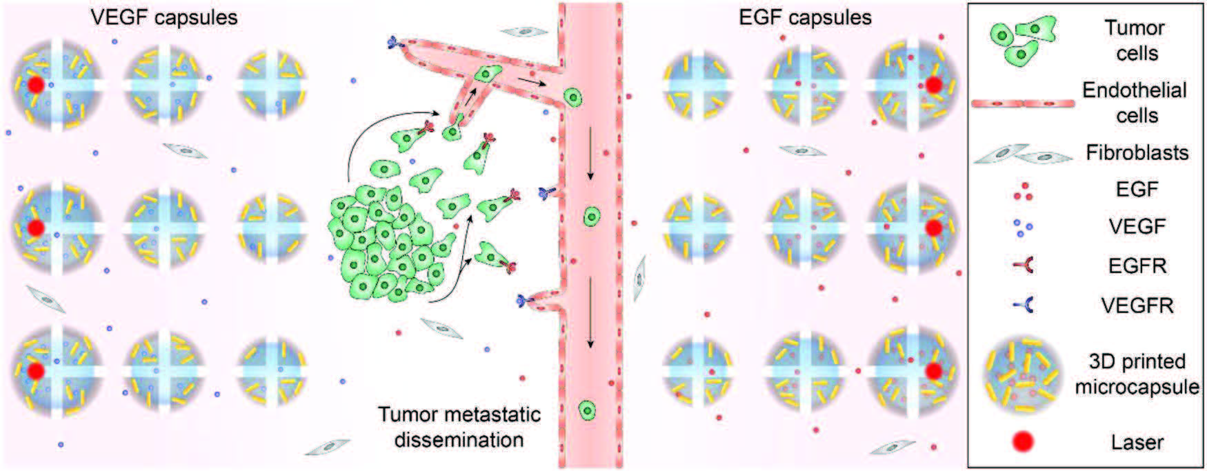
“This model is closer to the environment of the cells in the body. “Panoskaltsis-Mortari said. “so it’s more meaningful to study the effects of drugs on cells at this level than in 2D models, and it’s closer to the environment in vivo. “this 3D vascularized tumor tissue provides a new platform for identifying and screening potential anticancer drugs. More importantly, the model also provides a new way to study metastatic tumor cells.
“One of the reasons for the success of this model is that we can better control the environment. “Fanben Meng, a postdoctoral fellow at the School of Science and Engineering at the University of Minnesota, said. “We can control the release of chemicals more slowly, creating a chemical gradient that allows cells to react in a way that is closer to the body. All of these benefits from our personalized custom 3D printing technology, which allows us to accurately control the location of cell clusters and chemical libraries in the 3D environment. “
At present, researchers mainly focus on lung cancer and melanoma. They will then integrate more types of cells, especially immune cells, as well as cell therapy, and study these interactions.
“Detection of anticancer drugs and cell therapy are two of the world’s most famous concepts at the University of Minnesota, and using this model we can take these innovations to the next climax. “Dr. Daniel Vallera, a member of the Masonic Cancer Center at the University of Minnesota, said. “Studies like this can produce important results, such as revealing the relationship between blood vessels and drugs, because it’s a modular product that you can add other elements to make it more advanced. You can even use the patient’s own tumor cells in this model. “
Reference
1. Meng, Fanben, et al. “3D bioprinted in vitro metastatic models via reconstruction of tumor microenvironments.” Advanced Materials 31.10 (2019): 1806899.