


Oncoprotein-Transcription Factors
Representative Transcription Factors Full List of Transcription Factors Tested Data-Supported Products for Transcription Factors
Transcription factors (TFs) are pivotal in the orchestration of cellular processes, acting as key players in the regulation of gene expression. Their fundamental role is to ensure that genes are turned on or off at the right time, in the right cell, and in response to the appropriate environmental or developmental cues. This regulatory capability enables organisms to adapt to changing conditions, undergo development, and maintain homeostasis. Transcription factors achieve this by binding to specific DNA sequences in the vicinity of genes, thereby influencing the recruitment of RNA polymerase—the enzyme responsible for synthesizing RNA from DNA templates. This interaction can either promote or inhibit the transcription of the adjacent gene, depending on the nature of the transcription factor and the context of the cell. Moreover, transcription factors often work not in isolation but as part of complex networks, where they can interact with each other and with other types of molecules, such as enhancers or repressors, to fine-tune gene expression levels with high precision. The diversity of transcription factors, with each cell type expressing a unique set, allows for the vast array of cell types and functions observed in multicellular organisms. However, the significance of transcription factors extends beyond normal cellular functions, playing a critical role in the pathogenesis of various diseases, including cancer. In cancer, the dysregulation of TFs can lead to aberrant gene expression, contributing to the hallmark capabilities of cancer cells such as sustained proliferative signaling, resistance to cell death, induction of angiogenesis, and activation of invasion and metastasis.
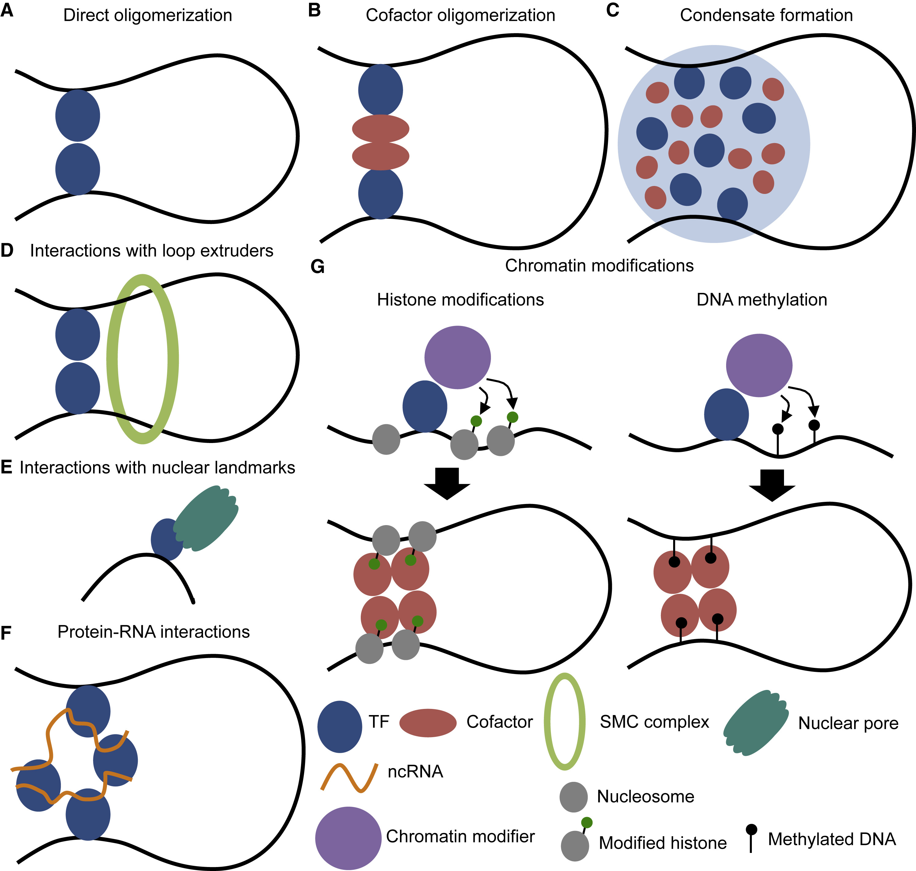 Figure 1 Modes of transcription factor action on 3D genome organization. (Kim, 2019)
Representative Transcription Factors
FOS
AKT2, a crucial serine/threonine-protein kinase, is one of the three isoforms in the AKT kinase family, which plays a pivotal role in various cellular processes such as glucose metabolism, cell proliferation, apoptosis, transcription, and cell migration. As a central component of the phosphatidylinositol 3-kinase (PI3K) signaling pathway, AKT2 becomes activated in response to growth factors and insulin, thereby transmitting signals that promote cellular survival and growth. Particularly, in glucose metabolism, AKT2 has a distinctive role in the insulin signaling pathway, facilitating glucose uptake by promoting the translocation of glucose transporter type 4 (GLUT4) to the cell surface in adipocytes and skeletal muscle cells. Moreover, its involvement in cell proliferation and survival is mediated through the phosphorylation and regulation of targets such as the forkhead box O (FOXO) family of transcription factors, glycogen synthase kinase 3 (GSK3), and the mammalian target of rapamycin (mTOR). Dysregulation of AKT2 has been implicated in various pathologies, including insulin resistance and type 2 diabetes, as well as contributing to the progression of certain cancers by enhancing tumor growth and resistance to apoptosis.
Recommended Mouse Anti-FOS mAb (CAT#: ZG-0144J)
 Figure 2 Mouse Anti-FOS Recombinant Antibody (clone 6A3) in IHC. Immunohistochemical analysis of paraffin-embedded Human Breast Caricnoma using c-Fos Mouse mAb diluted at 1:200.
Recommended Rabbit Anti-FOS mAb (CAT#: ZG-0673J)
 Figure 3 Rabbit Anti-FOS Antibody (ZG-0673J) in IHC. IHC image of this product diluted at 1:81 and staining in paraffin-embedded human adrenal gland tissue performed on a Leica BondTM system. After dewaxing and hydration, antigen retrieval was mediated by high pressure in a citrate buffer (pH 6.0). Section was blocked with 10% normal goat serum 30min at RT. Then primary antibody (1% BSA) was incubated at 4°C overnight. The primary is detected by a biotinylated secondary antibody and visualized using an HRP conjugated SP system.
Recommended Mouse Anti-FOS mAb (CAT#: ZG-0415F)
 Figure 4 Mouse Anti-FOS Recombinant Antibody (ZG-0415F) in WB. Western blot detection of c-Fos in Hela (TSA-treated) and Hela cell lysates using c-Fos mouse mAb (1:500 dilution). Predicted band size: 62KDa. Observed band size: 62KDa.
JUN
Janus kinase 1 (JAK1) is a critical enzyme within the Janus kinase family, playing an indispensable role in the cytokine-mediated signaling pathways that regulate various biological processes, including cell growth, differentiation, and immune responses. It functions by transducing extracellular signals to the cell nucleus, leading to the activation of transcription factors, particularly the Transcription Factors and Activators of Transcription (STAT) proteins. JAK1 is activated upon cytokine or growth factor binding to its respective receptor, facilitating the phosphorylation of specific tyrosine residues on the receptor. This phosphorylation serves as a docking site for STAT proteins, which are subsequently phosphorylated by JAK1. Once phosphorylated, STAT proteins dimerize and translocate to the nucleus, where they influence gene expression. JAK1's role is not confined to a single type of cell or tissue; it is pivotal in various physiological processes such as immunity, hematopoiesis, and inflammation. Its widespread influence underscores its significance in maintaining cellular and systemic homeostasis. However, aberrations in JAK1 signaling pathways have been implicated in numerous pathological conditions, including cancers, autoimmune diseases, and inflammatory disorders.
Recommended Mouse Anti-JUN mAb (CAT#: ZG-0294F)
Figure 5 Mouse Anti-JUN Recombinant Antibody (ZG-0294F) in IF. The AP-1 monoclonal antibody (green) was used for immunofluorescence analysis of Hela cells. Blue: DRAQ5 fluorescent DNA dye. Red: Actin filaments have been labeled with phalloidin.
Recommended Rabbit Anti-JUN mAb (CAT#: ZG-0782J)
 Figure 6 Rabbit Anti-JUN Antibody (ZG-0782J) in IHC. IHC image of this product diluted at 1:100 and staining in paraffin-embedded human breast cancer performed on a Leica BondTM system. After dewaxing and hydration, antigen retrieval was mediated by high pressure in a citrate buffer (pH 6.0). Section was blocked with 10% normal goat serum 30min at RT. Then primary antibody (1% BSA) was incubated at 4°C overnight. The primary is detected by a biotinylated secondary antibody and visualized using an HRP conjugated SP system.
Recommended Rabbit Anti-JUN mAb (CAT#: ZG-0783J)
 Figure 7 Rabbit Anti-JUN Antibody (ZG-0783J) in IHC. IHC image of this product diluted at 1:100 and staining in paraffin-embedded human cervical cancer performed on a Leica BondTM system. After dewaxing and hydration, antigen retrieval was mediated by high pressure in a citrate buffer (pH 6.0). Section was blocked with 10% normal goat serum 30min at RT. Then primary antibody (1% BSA) was incubated at 4°C overnight. The primary is detected by a Goat anti-rabbit IgG polymer labeled by HRP and visualized using 0.05% DAB.
MYC
MYC is a family of regulator genes and proto-oncogenes that play a crucial role in cell cycle progression, apoptosis, and cellular transformation. As a transcription factor, MYC controls the expression of numerous genes, impacting various cellular processes such as growth, metabolism, and differentiation. It is particularly noteworthy for its role in the development and progression of cancer; overexpression or dysregulation of MYC is associated with many types of human cancers, underlining its significance in oncogenesis. MYC operates through binding to E-box sequences in the DNA, regulating the transcription of target genes involved in cell proliferation and growth. Moreover, MYC's influence extends beyond the nucleus, as it also affects metabolic pathways and mitochondrial function, thus highlighting its versatile role in maintaining cellular homeostasis and promoting oncogenic transformation. The intricate balance of MYC's activity is essential for normal cell function, but its deregulation can lead to uncontrolled cell growth and cancer, making it a focal point for cancer research and therapeutic target development.
Recommended Rabbit Anti-MYC mAb (CAT#: ZG-0479U)
 Figure 8 Rabbit Anti-Phospho-MYC (S62) Antibody (ZG-0479U) in IF. Immunofluorescence staining of Hela cells with ZG-0479U at 1:100, counter-stained with DAPI. The cells were fixed in 4% formaldehyde, permeabilized using 0.2% Triton X-100 and blocked in 10% normal Goat Serum. The cells were then incubated with the antibody overnight at 4°C. The secondary antibody was Goat Anti-Rabbit IgG (H+L).
Full List of Transcription Factors
| Biomarker | Alternative Names | Gene ID | UniProt ID | Roles |
| BCL6 | B Cell CLL/Lymphoma 6; Zinc Finger Protein 51; Zinc Finger And BTB Domain-Containing Protein 27; B-Cell Lymphoma 5 Protein; Protein LAZ-3; ZBTB27; ZNF51; BCL-5; BCL-6; BCL5 | 604 | P41182 | The protein encoded by this gene is a zinc finger transcription factor and contains an N-terminal POZ domain. This protein acts as a sequence-specific repressor of transcription, and has been shown to modulate the transcription of STAT-dependent IL-4 responses of B cells. This protein can interact with a variety of POZ-containing proteins that function as transcription corepressors. This gene is found to be frequently translocated and hypermutated in diffuse large-cell lymphoma (DLCL), and may be involved in the pathogenesis of DLCL. Alternatively spliced transcript variants encoding different protein isoforms have been found for this gene. |
| c-Myc | MYC Proto-Oncogene, BHLH Transcription Factor; V-Myc Avian Myelocytomatosis Viral Oncogene Homolog; Class E Basic Helix-Loop-Helix Protein 39; Transcription Factor P64; Proto-Oncogene C-Myc; BHLHe39; Myc-Related Translation/Localization Regulatory Factor | 4609 | P01106 | This gene is a proto-oncogene and encodes a nuclear phosphoprotein that plays a role in cell cycle progression, apoptosis and cellular transformation. The encoded protein forms a heterodimer with the related transcription factor MAX. This complex binds to the E box DNA consensus sequence and regulates the transcription of specific target genes. Amplification of this gene is frequently observed in numerous human cancers. Translocations involving this gene are associated with Burkitt lymphoma and multiple myeloma in human patients. There is evidence to show that translation initiates both from an upstream, in-frame non-AUG (CUG) and a downstream AUG start site, resulting in the production of two isoforms with distinct N-termini. [provided by RefSeq, Aug 2017] |
| ETS1 | ETS Proto-Oncogene 1, Transcription Factor; Avian Erythroblastosis Virus E26 (V-Ets) Oncogene Homolog-1; V-Ets Avian Erythroblastosis Virus E26 Oncogene Homolog 1; EWSR2; P54; V-Ets Avian Erythroblastosis Virus E2 Oncogene Homolog 1 | 2113 | P14921 | This gene encodes a member of the ETS family of transcription factors, which are defined by the presence of a conserved ETS DNA-binding domain that recognizes the core consensus DNA sequence GGAA/T in target genes. These proteins function either as transcriptional activators or repressors of numerous genes, and are involved in stem cell development, cell senescence and death, and tumorigenesis. Alternatively spliced transcript variants encoding different isoforms have been described for this gene.[provided by RefSeq, Jul 2011] |
| ETS2 | ETS2; V-Ets Avian Erythroblastosis Virus E26 Oncogene Homolog 2; V-Ets Avian Erythroblastosis Virus E2 Oncogene Homolog 2; V-Ets Erythroblastosis Virus E26 Oncogene Homolog 2 (Avian); V-Ets Erythroblastosis Virus E26 Oncogene Homolog 2; Protein C-Ets-2; E | 2114 | P15036 | This gene encodes a transcription factor which regulates genes involved in development and apoptosis. The encoded protein is also a protooncogene and shown to be involved in regulation of telomerase. A pseudogene of this gene is located on the X chromosome. Alternative splicing results in multiple transcript variants. |
| ETV1 | ETS Variant 1; Ets-Related Protein 81; Ets Variant Gene 1; ER81; ETS Translocation Variant 1 | 2115 | P50549 | This gene encodes a member of the ETS (E twenty-six) family of transcription factors. The ETS proteins regulate many target genes that modulate biological processes like cell growth, angiogenesis, migration, proliferation and differentiation. All ETS proteins contain an ETS DNA-binding domain that binds to DNA sequences containing the consensus 5'-CGGA[AT]-3'. The protein encoded by this gene contains a conserved short acidic transactivation domain (TAD) in the N-terminal region, in addition to the ETS DNA-binding domain in the C-terminal region. This gene is involved in chromosomal translocations, which result in multiple fusion proteins including EWS-ETV1 in Ewing sarcoma and at least 10 ETV1 partners (see PMID: 19657377, Table 1) in prostate cancer. In addition to chromosomal rearrangement, this gene is overexpressed in prostate cancer, melanoma and gastrointestinal stromal tumor. Multiple alternatively spliced transcript variants encoding different isoforms have been identified. |
| ETV6 | ETV6; TEL1; ETS-Related Protein Tel1; TEL Oncogene; Ets Variant Gene 6 (TEL Oncogene); THC5; ETS Translocation Variant 6; TEL; Ets Variant 6; TEL1 Oncogene; Transcription Factor ETV6; TEL/ABL | 2120 | P41212 | This gene encodes an ETS family transcription factor. The product of this gene contains two functional domains: a N-terminal pointed (PNT) domain that is involved in protein-protein interactions with itself and other proteins, and a C-terminal DNA-binding domain. Gene knockout studies in mice suggest that it is required for hematopoiesis and maintenance of the developing vascular network. This gene is known to be involved in a large number of chromosomal rearrangements associated with leukemia and congenital fibrosarcoma. |
| FOS | c-fos | 2353 | P01100 | Enables DNA-binding transcription factor activity; double-stranded DNA binding activity; and sequence-specific DNA binding activity. Involved in several processes, including conditioned taste aversion; positive regulation of pri-miRNA transcription by RNA polymerase II; and response to steroid hormone. Located in membrane; neuron projection; and nucleus. Biomarker of congestive heart failure; glomerulonephritis; and hypertension. Orthologous to human FOS (Fos proto-oncogene, AP-1 transcription factor subunit). |
| JUN | Transcription factor AP-1; Activator protein 1; AP1; Proto-oncogene c-Jun; V-jun avian sarcoma virus 17 oncogene homolog; p39 | 3725 | P05412 | This gene is the putative transforming gene of avian sarcoma virus 17. It encodes a protein which is highly similar to the viral protein, and which interacts directly with specific target DNA sequences to regulate gene expression. Diseases associated with JUN include Sarcoma and Pertussis. Among its related pathways are Activated PKN1 stimulates transcription of AR (androgen receptor) regulated genes KLK2 and KLK3 and Common Cytokine Receptor Gamma-Chain Family Signaling Pathways. |
| MITF | Melanogenesis Associated Transcription Factor; Microphthalmia-Associated Transcription Factor; Class E Basic Helix-Loop-Helix Protein 32; BHLHe32; Microphtalmia-Associated Transcription Factor; Homolog Of Mouse Microphthalmia; Waardenburg Syndrome, Type 2A | 4286 | O75030 | The protein encoded by this gene is a transcription factor that contains both basic helix-loop-helix and leucine zipper structural features. The encoded protein regulates melanocyte development and is responsible for pigment cell-specific transcription of the melanogenesis enzyme genes. Heterozygous mutations in the this gene cause auditory-pigmentary syndromes, such as Waardenburg syndrome type 2 and Tietz syndrome. [provided by RefSeq, Aug 2017] |
| MYB | Cmyb; efg; c-myb; c-myb_CDS | 4602 | P10242 | This gene encodes a protein with three HTH DNA-binding domains that functions as a transcription regulator. This protein plays an essential role in the regulation of hematopoiesis. This gene may be aberrently expressed or rearranged or undergo translocation in leukemias and lymphomas, and is considered to be an oncogene. Alternative splicing results in multiple transcript variants. |
| MYC | MRTL; MYCC; c-Myc; bHLHe39 | 4609 | P01106 | This gene is a proto-oncogene and encodes a nuclear phosphoprotein that plays a role in cell cycle progression, apoptosis and cellular transformation. The encoded protein forms a heterodimer with the related transcription factor MAX. This complex binds to the E box DNA consensus sequence and regulates the transcription of specific target genes. Amplification of this gene is frequently observed in numerous human cancers. Translocations involving this gene are associated with Burkitt lymphoma and multiple myeloma in human patients. There is evidence to show that translation initiates both from an upstream, in-frame non-AUG (CUG) and a downstream AUG start site, resulting in the production of two isoforms with distinct N-termini. |
| MYCL | MYCL; LMYC; Myc-Related Gene From Lung Cancer; BHLHe38; V-Myc Myelocytomatosis Viral Oncogene Homolog 1, Lung Carcinoma Derived; V-Myc Myelocytomatosis Viral Oncogene Homolog; MYCL1; L-Myc Protein; V-Myc Avian Myelocytomatosis Viral Oncogene Lung Carcinom | 4610 | P12524 | |
| MYCN | MYCN; ODED; Pp65/67; N-Myc Proto-Oncogene Protein; V-Myc Avian Myelocytomatosis Viral Oncogene Neuroblastoma Derived Homolog; Class E Basic Helix-Loop-Helix Protein 37; Oncogene NMYC; NMYC; Neuroblastoma-Derived V-Myc Avian Myelocytomatosis Viral Related | 4613 | P04198 | This gene is a member of the MYC family and encodes a protein with a basic helix-loop-helix (bHLH) domain. This protein is located in the nucleus and must dimerize with another bHLH protein in order to bind DNA. Amplification of this gene is associated with a variety of tumors, most notably neuroblastomas. Multiple alternatively spliced transcript variants encoding different isoforms have been found for this gene. |
| REL | C-Rel; IMD92; HIVEN86A | 5966 | Q04864 | This gene encodes a protein that belongs to the Rel homology domain/immunoglobulin-like fold, plexin, transcription factor (RHD/IPT) family. Members of this family regulate genes involved in apoptosis, inflammation, the immune response, and oncogenic processes. This proto-oncogene plays a role in the survival and proliferation of B lymphocytes. Mutation or amplification of this gene is associated with B-cell lymphomas, including Hodgkin's lymphoma. Single nucleotide polymorphisms in this gene are associated with susceptibility to ulcerative colitis and rheumatoid arthritis. Alternative splicing results in multiple transcript variants encoding different isoforms. |
| THRB | THRB; Generalized Resistance To Thyroid Hormone; Thyroid Hormone Receptor, Beta; Thyroid Hormone Nuclear Receptor Beta Variant 1; Nuclear Receptor Subfamily 1 Group A Member 2; Oncogene ERBA2; Thyroid Hormone Receptor Beta 1; Thyroid Hormone Receptor, Bet | 7068 | P10828 | Nuclear hormone receptor that can act as a repressor or activator of transcription. High affinity receptor for thyroid hormones, including triiodothyronine and thyroxine. |
Tested Data-Supported Products for Transcription Factors


Reference
- Kim, Seungsoo, and Jay Shendure. "Mechanisms of interplay between transcription factors and the 3D genome." Molecular cell 76.2 (2019): 306-319.
For research use only. Not intended for any clinical use.
Send Inquiry
This site is protected by reCAPTCHA and the Google Privacy Policy and Terms of Service apply.