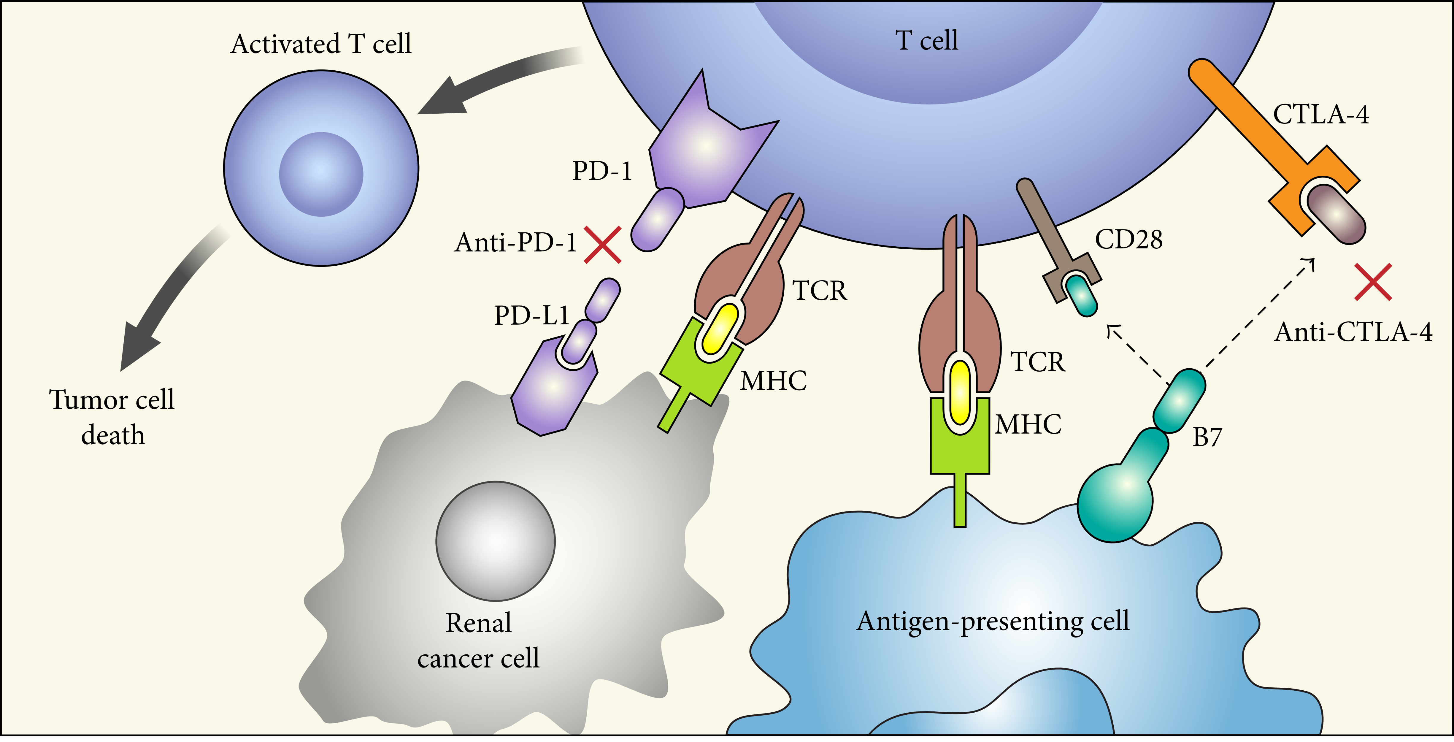

Tumor Antigens
Tumor antigens are unique or overexpressed proteins on the surface of cancer cells, distinguishing them from normal cells and making them prime targets for cancer immunotherapy. These antigens arise due to mutations in cancer cells, abnormal gene expressions, or the presence of viral antigens in virus-induced cancers, playing a pivotal role in the development and progression of tumors. They are classified into several types, including tumor-specific antigens (TSAs), which are unique to cancer cells, and tumor-associated antigens (TAAs), which are present on both cancerous and normal cells but are overexpressed in cancer cells. The discovery and characterization of tumor antigens have revolutionized cancer treatment by enabling the development of targeted therapies, such as cancer vaccines, monoclonal antibodies, and adoptive cell therapies. These treatments aim to stimulate the patient's immune system to recognize and destroy cancer cells specifically, offering a more personalized and less toxic alternative to traditional cancer treatments like chemotherapy and radiation. Furthermore, the identification of tumor antigens has facilitated the development of diagnostic and prognostic biomarkers, providing valuable tools for early cancer detection, monitoring treatment response, and predicting patient outcomes.
 Figure 1 Structural and Functional Basis of chimeric antigen receptor (CAR).
Representative Tumor Antigen Molecules
CA9
Carbonic Anhydrase IX (CA9) is a transmembrane enzyme belonging to the carbonic anhydrase family, primarily recognized for its role in regulating cellular pH. CA9 is distinct for its expression primarily in response to hypoxic conditions, a characteristic environment of various solid tumors, making it a marker of tumor hypoxia and an attractive target for cancer therapy. The enzyme catalyzes the reversible hydration of carbon dioxide to bicarbonate and protons, a fundamental process in maintaining acid-base balance and facilitating CO2 transport. In the tumor microenvironment, CA9 expression contributes to the acidification of the extracellular space, enabling tumor cells to survive and proliferate under hypoxic conditions, thus promoting tumor growth and metastasis. Additionally, CA9 plays a crucial role in cell adhesion and migration, indicating its involvement in cancer progression beyond its enzymatic function. The overexpression of CA9 in a wide range of solid tumors, coupled with its limited expression in normal tissues, underscores its potential as a biomarker for cancer diagnosis and prognosis, as well as a therapeutic target.


CD63
CD63, a member of the tetraspanin family, is a transmembrane protein that plays critical roles in various cellular processes, including cell adhesion, migration, and the regulation of signal transduction pathways. This protein is ubiquitously expressed in many cell types and is notably involved in the biogenesis and function of multivesicular bodies (MVBs) and exosomes, small vesicles involved in intercellular communication. CD63 is often used as a marker for these vesicles due to its enrichment in their membranes. Through its interactions with other membrane proteins, lipids, and cytosolic factors, CD63 influences various physiological and pathological processes. It has been implicated in the immune response by modulating the activity of immune cells, such as T cells and dendritic cells, thereby affecting antigen presentation and the inflammatory response. Additionally, CD63 plays a significant role in cancer, where its expression levels and distribution have been associated with tumor progression, metastasis, and the modulation of cancer cell signaling pathways.


CTNNB1
CTNNB1 (catenin beta 1) is a crucial protein encoded by the CTNNB1 gene in humans, playing a pivotal role in the regulation of cell-cell adhesion and gene transcription. It is a central component of the Wnt signaling pathway, a complex network that influences cell fate determination, migration, and organogenesis during embryonic development and contributes to the maintenance of adult tissue homeostasis. CTNNB1 functions by coordinating with cadherins in cell adhesion complexes, thereby stabilizing cell structure and facilitating intercellular communication. In the nucleus, it acts as a transcriptional coactivator for T-cell factor/lymphoid enhancer-binding factor (TCF/LEF) family members, turning on genes essential for cell proliferation and differentiation. The dysregulation of CTNNB1 is linked to a variety of diseases, including cancer, where mutations can lead to its accumulation and the inappropriate activation of target genes, promoting uncontrolled cell growth and resistance to cell death. Additionally, its role in the Wnt pathway makes it a target for therapeutic intervention in conditions such as colorectal cancer, where aberrant Wnt signaling is a hallmark.


Full List of Tumor Antigen Molecules
| Biomarker | Alternative Names | Gene ID | UniProt ID | Roles |
| ABL1 | ABL Proto-Oncogene 1, Non-Receptor Tyrosine Kinase; V-Abl Abelson Murine Leukemia Viral Oncogene Homolog 1; C-Abl Oncogene 1, Receptor Tyrosine Kinase; Abelson Tyrosine-Protein Kinase 1; Proto-Oncogene C-Abl; EC 2.7.10.2; JTK7; P150; ABL; Abelson Murine Leukemia Viral Oncogene Homolog 1; C-Abl Oncogene 1, Non-Receptor Tyrosine Kinase | 25 | A0A024R8E2 | This gene is a protooncogene that encodes a protein tyrosine kinase involved in a variety of cellular processes, including cell division, adhesion, differentiation, and response to stress. The activity of the protein is negatively regulated by its SH3 domain, whereby deletion of the region encoding this domain results in an oncogene. The ubiquitously expressed protein has DNA-binding activity that is regulated by CDC2-mediated phosphorylation, suggesting a cell cycle function. This gene has been found fused to a variety of translocation partner genes in various leukemias, most notably the t(9;22) translocation that results in a fusion with the 5' end of the breakpoint cluster region gene. Alternative splicing of this gene results in two transcript variants, which contain alternative first exons that are spliced to the remaining common exons. |
| AFP | Alpha Fetoprotein; Alpha-1-Fetoprotein; Alpha-Fetoglobulin; HPAFP; Alpha-Fetoprotein; AFPD; FETA | 174 | P02771 | This gene encodes alpha-fetoprotein, a major plasma protein produced by the yolk sac and the liver during fetal life. Alpha-fetoprotein expression in adults is often associated with hepatoma or teratoma. However, hereditary persistance of alpha-fetoprotein may also be found in individuals with no obvious pathology. The protein is thought to be the fetal counterpart of serum albumin, and the alpha-fetoprotein and albumin genes are present in tandem in the same transcriptional orientation on chromosome 4. Alpha-fetoprotein is found in monomeric as well as dimeric and trimeric forms, and binds copper, nickel, fatty acids and bilirubin. The level of alpha-fetoprotein in amniotic fluid is used to measure renal loss of protein to screen for spina bifida and anencephaly. [provided by RefSeq, Jul 2008] |
| BRCA1 | IRIS; PSCP; BRCAI; BRCC1; FANCS; PNCA4; RNF53; BROVCA1; PPP1R53 | 672 | P38398 | This gene encodes a 190 kD nuclear phosphoprotein that plays a role in maintaining genomic stability, and it also acts as a tumor suppressor. The BRCA1 gene contains 22 exons spanning about 110 kb of DNA. The encoded protein combines with other tumor suppressors, DNA damage sensors, and signal transducers to form a large multi-subunit protein complex known as the BRCA1-associated genome surveillance complex (BASC). This gene product associates with RNA polymerase II, and through the C-terminal domain, also interacts with histone deacetylase complexes. This protein thus plays a role in transcription, DNA repair of double-stranded breaks, and recombination. Mutations in this gene are responsible for approximately 40% of inherited breast cancers and more than 80% of inherited breast and ovarian cancers. Alternative splicing plays a role in modulating the subcellular localization and physiological function of this gene. Many alternatively spliced transcript variants, some of which are disease-associated mutations, have been described for this gene, but the full-length natures of only some of these variants has been described. A related pseudogene, which is also located on chromosome 17, has been identified. |
| CA9 | CAIX; MN | 768 | Q16790 | Carbonic anhydrases (CAs) are a large family of zinc metalloenzymes that catalyze the reversible hydration of carbon dioxide. They participate in a variety of biological processes, including respiration, calcification, acid-base balance, bone resorption, and the formation of aqueous humor, cerebrospinal fluid, saliva, and gastric acid. They show extensive diversity in tissue distribution and in their subcellular localization. CA IX is a transmembrane protein and is one of only two tumor-associated carbonic anhydrase isoenzymes known. It is expressed in all clear-cell renal cell carcinoma, but is not detected in normal kidney or most other normal tissues. It may be involved in cell proliferation and transformation. This gene was mapped to 17q21.2 by fluorescence in situ hybridization, however, radiation hybrid mapping localized it to 9p13-p12. |
| CCNB1 | Cyclin B1; CCNB; G2/Mitotic-Specific Cyclin B1; G2/Mitotic-Specific Cyclin-B1; | 891 | P14635 | CCNB1 (Cyclin B1) is a Protein Coding gene. Diseases associated with CCNB1 include Thyroid Lymphoma and Adrenal Carcinoma. Among its related pathways are Mitotic Prometaphase and Mitotic Prophase. Gene Ontology (GO) annotations related to this gene include protein kinase binding and patched binding. An important paralog of this gene is CCNB2. |
| CD19 | CD19; B4; CVID3; MGC12802 | 930 | P15391 | Lymphocytes proliferate and differentiate in response to various concentrations of different antigens. The ability of the B cell to respond in a specific, yet sensitive manner to the various antigens is achieved with the use of low-affinity antigen receptors. This gene encodes a cell surface molecule which assembles with the antigen receptor of B lymphocytes in order to decrease the threshold for antigen receptor-dependent stimulation. |
| CD22 | SIGLEC2; SIGLEC-2 | 933 | P20273 | Predicted to enable CD4 receptor binding activity; protein phosphatase binding activity; and sialic acid binding activity. Involved in B cell activation; negative regulation of B cell receptor signaling pathway; and regulation of endocytosis. Located in early endosome and recycling endosome. |
| CD24 | CD24A | 100133941 | P25063 | This gene encodes a sialoglycoprotein that is expressed on mature granulocytes and B cells and modulates growth and differentiation signals to these cells. The precursor protein is cleaved to a short 32 amino acid mature peptide which is anchored via a glycosyl phosphatidylinositol (GPI) link to the cell surface. This gene was missing from previous genome assemblies, but is properly located on chromosome 6. Non-transcribed pseudogenes have been designated on chromosomes 1, 15, 20, and Y. Alternative splicing results in multiple transcript variants. |
| CD5 | CD5; LEU1 | 921 | P06127 | This gene encodes a member of the scavenger receptor cysteine-rich (SRCR) superfamily. Members of this family are secreted or membrane-anchored proteins mainly found in cells associated with the immune system. This protein is a type-I transmembrane glycoprotein found on the surface of thymocytes, T lymphocytes and a subset of B lymphocytes. The encoded protein contains three SRCR domains and may act as a receptor to regulate T-cell proliferation. Alternative splicing results in multiple transcript variants encoding different isoforms. |
| CD63 | CD63 antigen; Granulophysin; LAMP-3; Limp1; Melanoma-associated antigen ME491; OMA81H; Ocular melanoma-associated antigen; Tetraspanin-30; Tspan-30 | 967 | P08962 | The protein encoded by this gene is a member of the transmembrane 4 superfamily, also known as the tetraspanin family. Most of these members are cell-surface proteins that are characterized by the presence of four hydrophobic domains. The proteins mediate signal transduction events that play a role in the regulation of cell development, activation, growth and motility. The encoded protein is a cell surface glycoprotein that is known to complex with integrins. It may function as a blood platelet activation marker. Deficiency of this protein is associated with Hermansky-Pudlak syndrome. Also this gene has been associated with tumor progression. Alternative splicing results in multiple transcript variants encoding different protein isoforms. |
| CDK4 | CMM3; PSK-J3 | 1019 | P11802 | The protein encoded by this gene is a member of the Ser/Thr protein kinase family. This protein is highly similar to the gene products of S. cerevisiae cdc28 and S. pombe cdc2. It is a catalytic subunit of the protein kinase complex that is important for cell cycle G1 phase progression. The activity of this kinase is restricted to the G1-S phase, which is controlled by the regulatory subunits D-type cyclins and CDK inhibitor p16(INK4a). This kinase was shown to be responsible for the phosphorylation of retinoblastoma gene product (Rb). Mutations in this gene as well as in its related proteins including D-type cyclins, p16(INK4a) and Rb were all found to be associated with tumorigenesis of a variety of cancers. Multiple polyadenylation sites of this gene have been reported. |
| CEACAM1 | CEACAM1; carcinoembryonic antigen-related cell adhesion molecule 1 (biliary glycoprotein); BGP; BGP1; BGPI; carcinoembryonic antigen-related cell adhesion molecule 1; antigen CD66; CD66a antigen | 634 | P13688 | This gene encodes a member of the carcinoembryonic antigen (CEA) gene family, which belongs to the immunoglobulin superfamily. Two subgroups of the CEA family, the CEA cell adhesion molecules and the pregnancy-specific glycoproteins, are located within a 1.2 Mb cluster on the long arm of chromosome 19. Eleven pseudogenes of the CEA cell adhesion molecule subgroup are also found in the cluster. The encoded protein was originally described in bile ducts of liver as biliary glycoprotein. Subsequently, it was found to be a cell-cell adhesion molecule detected on leukocytes, epithelia, and endothelia. The encoded protein mediates cell adhesion via homophilic as well as heterophilic binding to other proteins of the subgroup. Multiple cellular activities have been attributed to the encoded protein, including roles in the differentiation and arrangement of tissue three-dimensional structure, angiogenesis, apoptosis, tumor suppression, metastasis, and the modulation of innate and adaptive immune responses. Multiple transcript variants encoding different isoforms have been reported, but the full-length nature of all variants has not been defined. |
| CEACAM3 | CEA; CGM1; W264; W282; CD66D | 1084 | P40198 | This gene encodes a member of the family of carcinoembryonic antigen-related cell adhesion molecules (CEACAMs), which are used by several bacterial pathogens to bind and invade host cells. The encoded transmembrane protein directs phagocytosis of several bacterial species that is dependent on the small GTPase Rac. It is thought to serve an important role in controlling human-specific pathogens by the innate immune system. Alternatively spliced transcript variants have been described. |
| CEACAM5 | CEACAM-5; CD66e; CEA; Meconium antigen 100 | 1048 | P06731 | This gene encodes a cell surface glycoprotein that represents the founding member of the carcinoembryonic antigen (CEA) family of proteins. The encoded protein is used as a clinical biomarker for gastrointestinal cancers and may promote tumor development through its role as a cell adhesion molecule. Additionally, the encoded protein may regulate differentiation, apoptosis, and cell polarity. This gene is present in a CEA family gene cluster on chromosome 19. Alternative splicing results in multiple transcript variants. |
| CEACAM6 | CD66c; CEAL; NCA | 4680 | P40199 | This gene encodes a protein that belongs to the carcinoembryonic antigen (CEA) family whose members are glycosyl phosphatidyl inositol (GPI) anchored cell surface glycoproteins. Members of this family play a role in cell adhesion and are widely used as tumor markers in serum immunoassay determinations of carcinoma. This gene affects the sensitivity of tumor cells to adenovirus infection. The protein encoded by this gene acts as a receptor for adherent-invasive E. coli adhesion to the surface of ileal epithelial cells in patients with Crohns disease. This gene is clustered with genes and pseudogenes of the cell adhesion molecules subgroup of the CEA family on chromosome 19. |
| CLCA2 | CLCA2; Human CLCA2 | 9635 | Q9UQC9 | |
| CTAG2 | CTAG2; Cancer/testis antigen 2; CT2; ESO2; CAMEL; CT6.2; CT6.2a; CT6.2b; LAGE-1; LAGE2B | 30848 | O75638 | This gene encodes an autoimmunogenic tumor antigen that belongs to the ESO/LAGE family of cancer-testis antigens. This protein is expressed in a wide array of cancers including melanoma, breast cancer, bladder cancer and prostate cancer. This protein is also expressed in normal testis tissue. An alternative open reading frame product of this gene has been described in PMID:10399963. This alternate protein, termed CAMEL, is a tumor antigen that is recognized by melanoma-specific cytotoxic T-lymphocytes. Alternate splicing results in multiple transcript variants. |
| CTNNB1 | Catenin Beta 1; Catenin (Cadherin-Associated Protein), Beta 1, 88kDa; CTNNB; Catenin (Cadherin-Associated Protein), Beta 1 (88kD); Catenin (Cadherin-Associated Protein), Beta 1; Catenin Beta-1; | 12387 | Q02248 | Catenin beta-1, also known as beta-catenin (β-catenin), is a protein that in humans is encoded by the CTNNB1 gene. |
| EGFR | Epidermal Growth Factor Receptor; Receptor Tyrosine-Protein Kinase ErbB-1; Erb-B2 Receptor Tyrosine Kinase 1; Proto-Oncogene C-ErbB-1; EC 2.7.10.1; ERBB1; ERBB; HER1; Epidermal Growth Factor Receptor (Avian Erythroblastic Leukemia Viral (V-Erb-B) Oncogene Homolog); Erythroblastic Leukemia Viral (V-Erb-B) Oncogene Homolog (Avian) | 1956 | P00533 | The protein encoded by this gene is a transmembrane glycoprotein that is a member of the protein kinase superfamily. This protein is a receptor for members of the epidermal growth factor family. EGFR is a cell surface protein that binds to epidermal growth factor. Binding of the protein to a ligand induces receptor dimerization and tyrosine autophosphorylation and leads to cell proliferation. Mutations in this gene are associated with lung cancer. [provided by RefSeq, Jun 2016] |
| ERBIN | Erbb2 Interacting Protein; Densin-180-Like Protein; ERBB2-Interacting Protein; Protein LAP2; ERBB2IP; LAP2 | 55914 | Q96RT1 | This gene is a member of the leucine-rich repeat and PDZ domain (LAP) family. The encoded protein contains 17 leucine-rich repeats and one PDZ domain. It binds to the unphosphorylated form of the ERBB2 protein and regulates ERBB2 function and localization. It has also been shown to affect the Ras signaling pathway by disrupting Ras-Raf interaction. Alternatively spliced transcript variants encoding multiple isoforms have been observed for this gene. |
| fibronectin | FN; CIG; FNZ; MSF; ED-B; FINC; GFND; LETS; GFND2 | 2335 | P02751 | Fibronectin (FN) is a glycoprotein component of the extracellular matrix of the extracellular matrix (ECM) with roles in embryogenesis, development, and wound healing. More recently, FN has emerged as player in platelet thrombus formation and diseases associated with thrombosis including vascular remodeling, atherosclerosis, and cardiac repair following a myocardial infarct. Each monomer of FN consists of three types of homologous repeating units, that is 12 type I repeats, two type II repeats and 15-17 type III repeats. The occurrence of multiple isoforms results from alternative mRNA splicing of the ED-A, ED-B and III-CS regions, and subsequent post-translational modification. As an ECM component and one of the primary cell adhesion molecules, Fibronectin can be a ligand for fibrin, heparin, chondroitin sulfate, collagen/gelatin, as well as many integrin receptors through which FN mediates the variety of cellular signaling pathways. The study of solid human tumors showed among the early signs of malignant transformation the fragmentation of pericellular FN, concommitent with the increase of its production by the peritumoral stroma. These results should encourage further investigations concerning the potential importance of Fn production and breakdown during cancer progression. FN1 expression has been described to increase significantly from the morula towards the early blastocyst stage, suggesting that FN1 may also be involved in early blastocyst formation. The fragment 2 of FN comprises the first 7 FN type III repeats and is suggested to be important for self association during fibril growth via the key module III2. |
| gp100 | YLE-8A; YLEPGPVTL | 6490 | P40967 | This gene encodes a melanocyte-specific type I transmembrane glycoprotein. The encoded protein is enriched in melanosomes, which are the melanin-producing organelles in melanocytes, and plays an essential role in the structural organization of premelanosomes. This protein is involved in generating internal matrix fibers that define the transition from Stage I to Stage II melanosomes. This protein undergoes a complex pattern of prosttranslational processing and modification that is essential to the proper functioning of the protein. A secreted form of this protein that is released by proteolytic ectodomain shedding may be used as a melanoma-specific serum marker. Alternate splicing results in multiple transcript variants. |
| HER2 | NEU; NGL; HER2; TKR1; CD340; HER-2; MLN 19; HER-2/neu | 2064 | P04626 | This gene encodes a member of the epidermal growth factor (EGF) receptor family of receptor tyrosine kinases. This protein has no ligand binding domain of its own and therefore cannot bind growth factors. However, it does bind tightly to other ligand-bound EGF receptor family members to form a heterodimer, stabilizing ligand binding and enhancing kinase-mediated activation of downstream signalling pathways, such as those involving mitogen-activated protein kinase and phosphatidylinositol-3 kinase. Allelic variations at amino acid positions 654 and 655 of isoform a (positions 624 and 625 of isoform b) have been reported, with the most common allele, Ile654/Ile655, shown here. Amplification and/or overexpression of this gene has been reported in numerous cancers, including breast and ovarian tumors. Alternative splicing results in several additional transcript variants, some encoding different isoforms and others that have not been fully characterized. |
| HMMR | Hyaluronan Mediated Motility Receptor; Intracellular Hyaluronic Acid-Binding Protein; Receptor For Hyaluronan-Mediated Motility; IHABP; RHAMM; Hyaluronan-Mediated Motility Receptor (RHAMM); Hyaluronan-Mediated Motility Receptor; CD168 Antigen; CD168 | 3161 | O75330 | The protein encoded by this gene is involved in cell motility. It is expressed in breast tissue and together with other proteins, it forms a complex with BRCA1 and BRCA2, thus is potentially associated with higher risk of breast cancer. Alternatively spliced transcript variants encoding different isoforms have been noted for this gene. |
| KLK3 | Kallikrein Related Peptidase 3; Gamma-Seminoprotein; P-30 Antigen; Kallikrein-3; Semenogelase; Seminin; APS; PSA | 354 | P07288 | Kallikreins are a subgroup of serine proteases having diverse physiological functions. Growing evidence suggests that many kallikreins are implicated in carcinogenesis and some have potential as novel cancer and other disease biomarkers. This gene is one of the fifteen kallikrein subfamily members located in a cluster on chromosome 19. Its protein product is a protease present in seminal plasma. It is thought to function normally in the liquefaction of seminal coagulum, presumably by hydrolysis of the high molecular mass seminal vesicle protein. Serum level of this protein, called PSA in the clinical setting, is useful in the diagnosis and monitoring of prostatic carcinoma. Alternate splicing of this gene generates several transcript variants encoding different isoforms. [provided by RefSeq, Jul 2008] |
| KRAS | NS; NS3; CFC2; RALD; K-Ras; KRAS1; KRAS2; RASK2; KI-RAS; C-K-RAS; K-RAS2A; K-RAS2B; K-RAS4A; K-RAS4B; c-Ki-ras2 | 3845 | P01116 | This gene, a Kirsten ras oncogene homolog from the mammalian ras gene family, encodes a protein that is a member of the small GTPase superfamily. A single amino acid substitution is responsible for an activating mutation. The transforming protein that results is implicated in various malignancies, including lung adenocarcinoma, mucinous adenoma, ductal carcinoma of the pancreas and colorectal carcinoma. Alternative splicing leads to variants encoding two isoforms that differ in the C-terminal region. [provided by RefSeq, Jul 2008] |
| MLANA | Melan-A; Antigen LB39-AA; Antigen SK29-AA; Protein Melan-A; MART-1; MART1; Melanoma Antigen Recognized By T-Cells 1 | 2315 | Q16655 | MLANA (Melan-A) is a Protein Coding gene. Diseases associated with MLANA include Melanoma and Microphthalmia. Among its related pathways are NF-kappaB Signaling. |
| MSLN | Mesothelin; Pre-Pro-Megakaryocyte-Potentiating Factor; CAK1 Antigen; MPF; Soluble MPF Mesothelin Related Protein; Megakaryocyte Potentiating Factor; SMRP; BAY 94-9343; unconjugated:BAY 86-1903; MSLN | 10232 | Q13421 | This gene encodes a preproprotein that is proteolytically processed to generate two protein products, megakaryocyte potentiating factor and mesothelin. Megakaryocyte potentiating factor functions as a cytokine that can stimulate colony formation of bone marrow megakaryocytes. Mesothelin is a glycosylphosphatidylinositol-anchored cell-surface protein that may function as a cell adhesion protein. This protein is overexpressed in epithelial mesotheliomas, ovarian cancers and in specific squamous cell carcinomas. Alternative splicing results in multiple transcript variants, at least one of which encodes an isoform that is proteolytically processed. [provided by RefSeq, Feb 2016] |
| MUC1 | ADMCKD; ADMCKD1; CA 15-3; CD227; EMA; H23AG; KL-6; MAM6; MCD; MCKD; MCKD1; MUC-1; MUC-1/SEC; MUC-1/X; MUC1/ZD; PEM; PEMT; PUM | 4582 | P15941 | This gene encodes a membrane-bound protein that is a member of the mucin family. Mucins are O-glycosylated proteins that play an essential role in forming protective mucous barriers on epithelial surfaces. These proteins also play a role in intracellular signaling. This protein is expressed on the apical surface of epithelial cells that line the mucosal surfaces of many different tissues including lung, breast stomach and pancreas. This protein is proteolytically cleaved into alpha and beta subunits that form a heterodimeric complex. The N-terminal alpha subunit functions in cell-adhesion and the C-terminal beta subunit is involved in cell signaling. Overexpression, aberrant intracellular localization, and changes in glycosylation of this protein have been associated with carcinomas. This gene is known to contain a highly polymorphic variable number tandem repeats (VNTR) domain. Alternate splicing results in multiple transcript variants. |
| MUM1 | EXPAND1; HSPC211; MUM-1 | 84939 | Q2TAK8 | MUM1 (Melanoma Associated Antigen (Mutated) 1) is a Protein Coding gene. Diseases associated with MUM1 include Melanoma. Among its related pathways are DNA Damage. Gene Ontology (GO) annotations related to this gene include nucleosome binding. An important paralog of this gene is MUM1L1. |
| NY-ESO-1 | NY-ESO-1 | 1485 | P78358 | The protein encoded by this gene is an antigen that is overexpressed in many cancers but that is also expressed in normal testis. This gene is found in a duplicated region of the X-chromosome and therefore has a neighboring gene of identical sequence. |
| p53 | 7157 | K7PPA8 | ||
| PCNA | Proliferating Cell Nuclear Antigen; Cyclin; DNA Polymerase Delta Auxiliary Protein; ATLD2 | 5111 | P12004 | The protein encoded by this gene is found in the nucleus and is a cofactor of DNA polymerase delta. The encoded protein acts as a homotrimer and helps increase the processivity of leading strand synthesis during DNA replication. In response to DNA damage, this protein is ubiquitinated and is involved in the RAD6-dependent DNA repair pathway. Two transcript variants encoding the same protein have been found for this gene. Pseudogenes of this gene have been described on chromosome 4 and on the X chromosome. |
| PRAME | MAPE; OIP4; CT130; OIP-4 | 23532 | P78395 | This gene encodes an antigen that is preferentially expressed in human melanomas and that is recognized by cytolytic T lymphocytes. It is not expressed in normal tissues, except testis. The encoded protein acts as a repressor of retinoic acid receptor, and likely confers a growth advantage to cancer cells via this function. Alternative splicing results in multiple transcript variants. |
| PSA | Psa | 354 | P07288 | |
| RAGE | Receptor for advanced glycation endproducts; RAGE | 11596 | Q62151 | RAGE (receptor for advanced glycation endproducts), also called AGER, is a 35 kilodalton transmembrane receptor of the immunoglobulin super family which was first characterized in 1992 by Neeper et al. |
| SSX2 | SSX2; SSX; HD21; CT5.2; CT5.2A; HOM-MEL-40 | 6757 | Q16385 | The product of this gene belongs to the family of highly homologous synovial sarcoma X (SSX) breakpoint proteins. These proteins may function as transcriptional repressors. They are also capable of eliciting spontaneous humoral and cellular immune responses in cancer patients, and are potentially useful targets in cancer vaccine-based immunotherapy. This gene, and also the SSX1 and SSX4 family members, have been involved in t(X;18)(p11.2;q11.2) translocations that are characteristically found in all synovial sarcomas. This translocation results in the fusion of the synovial sarcoma translocation gene on chromosome 18 to one of the SSX genes on chromosome X. The encoded hybrid proteins are likely responsible for transforming activity. Alternative splicing of this gene results in multiple transcript variants. This gene also has an identical duplicate, GeneID: 727837, located about 45 kb downstream in the opposite orientation on chromosome X. |
| Survivin | Survivin; baculoviral inhibitor of apoptosis repeat-containing 5; BIRC5 | 332 | O15392 | This gene is a member of the inhibitor of apoptosis (IAP) gene family, which encode negative regulatory proteins that prevent apoptotic cell death. IAP family members usually contain multiple baculovirus IAP repeat (BIR) domains, but this gene encodes proteins with only a single BIR domain. The encoded proteins also lack a C-terminus RING finger domain. Gene expression is high during fetal development and in most tumors, yet low in adult tissues. Alternatively spliced transcript variants encoding distinct isoforms have been found for this gene. |
| TERT | TP2; TRT; CMM9; EST2; TCS1; hTRT; DKCA2; DKCB4; hEST2; PFBMFT1 | 7015 | O14746 | Telomerase is a ribonucleoprotein polymerase that maintains telomere ends by addition of the telomere repeat TTAGGG. The enzyme consists of a protein component with reverse transcriptase activity, encoded by this gene, and an RNA component which serves as a template for the telomere repeat. Telomerase expression plays a role in cellular senescence, as it is normally repressed in postnatal somatic cells resulting in progressive shortening of telomeres. Deregulation of telomerase expression in somatic cells may be involved in oncogenesis. Studies in mouse suggest that telomerase also participates in chromosomal repair, since de novo synthesis of telomere repeats may occur at double-stranded breaks. Alternatively spliced variants encoding different isoforms of telomerase reverse transcriptase have been identified; the full-length sequence of some variants has not been determined. Alternative splicing at this locus is thought to be one mechanism of regulation of telomerase activity. |
| TGFBR2 | AAT3; FAA3; LDS1B; LDS2; LDS2B; MFS2; RIIC; TAAD2; TBR-ii; TBRII; TGFbeta-RII; TGFR-2 | 7048 | P37173 | The protein encoded by this gene is a transmembrane protein that has a protein kinase domain, forms a heterodimeric complex with TGF-beta receptor type-1, and binds TGF-beta. This receptor/ligand complex phosphorylates proteins, which then enter the nucleus and regulate the transcription of genes related to cell proliferation, cell cycle arrest, wound healing, immunosuppression, and tumorigenesis. Mutations in this gene have been associated with Marfan Syndrome, Loeys-Deitz Aortic Aneurysm Syndrome, and the development of various types of tumors. Alternatively spliced transcript variants encoding different isoforms have been characterized. |
| TYRP1 | TYRP1; TRP; CAS2; CATB; GP75; OCA3; TRP1; TYRP; b-PROTEIN | 7306 | P17643 | This gene encodes a melanosomal enzyme that belongs to the tyrosinase family and plays an important role in the melanin biosynthetic pathway. Defects in this gene are the cause of rufous oculocutaneous albinism and oculocutaneous albinism type III. |
| WDR46 | WD Repeat Domain 46; WD Repeat-Containing Protein BING4; C6orf11; BING4; Chromosome 6 Open Reading Frame 11; WD Repeat-Containing Protein 46; FP221; UTP7 | 9277 | O15213 | WDR46 (WD Repeat Domain 46) is a Protein Coding gene. Among its related pathways are Gene Expression and rRNA processing in the nucleus and cytosol. |
| WT1 | Wilms Tumor 1; WT33; Wilms Tumor Protein; NPHS4; WIT-2; WAGR; AWT1; GUD | 7490 | P19544 | This gene encodes a transcription factor that contains four zinc-finger motifs at the C-terminus and a proline/glutamine-rich DNA-binding domain at the N-terminus. It has an essential role in the normal development of the urogenital system, and it is mutated in a small subset of patients with Wilms tumor. This gene exhibits complex tissue-specific and polymorphic imprinting pattern, with biallelic, and monoallelic expression from the maternal and paternal alleles in different tissues. Multiple transcript variants have been described. In several variants, there is evidence for the use of a non-AUG (CUG) translation initiation codon upstream of, and in-frame with the first AUG. Authors of PMID:7926762 also provide evidence that WT1 mRNA undergoes RNA editing in human and rat, and that this process is tissue-restricted and developmentally regulated. [provided by RefSeq, Mar 2015] |
Tested Data-Supported Products Targeting Tumor Antigen Molecules
- Lopez-Beltran, Antonio, et al. "The identification of immunological biomarkers in kidney cancers." Frontiers in Oncology 8 (2018): 456.
For research use only. Not intended for any clinical use.
This site is protected by reCAPTCHA and the Google Privacy Policy and Terms of Service apply.


-1.png)


-1.png)
-1.png)


-1.png)


-1-1.png)


-1.png)


