


Glycolysis
Representative Targets in Glycolysis Full List of Targets in Glycolysis Tested Data-Supported Products for Targeting Glycolysis
Glycolysis is a central metabolic pathway that plays a crucial role in energy production in cells, converting glucose into pyruvate while generating ATP (adenosine triphosphate) and NADH (reduced nicotinamide adenine dinucleotide). This process occurs in the cytoplasm of both aerobic and anaerobic organisms and is the first step in the breakdown of carbohydrates to extract energy for cellular metabolism. Glycolysis is a sequence of ten enzyme-catalyzed reactions, beginning with the phosphorylation of glucose to glucose-6-phosphate and culminating in the production of pyruvate. Each step is precisely regulated to ensure efficiency and responsiveness to cellular energy demands. The pathway is divided into two phases: the energy investment phase, where ATP is consumed to activate glucose, and the energy payoff phase, where ATP and NADH are produced. The key regulatory steps in glycolysis are catalyzed by the enzymes hexokinase, phosphofructokinase-1, and pyruvate kinase, each of which is tightly regulated through feedback mechanisms involving ATP and other metabolic intermediates. This regulation ensures that glycolysis responds dynamically to changes in cellular energy status and metabolic needs.
Glycolysis is not only important for energy production but also provides critical intermediates for other metabolic pathways, including the pentose phosphate pathway and fatty acid synthesis. In anaerobic conditions, pyruvate is further metabolized to lactate, facilitating a continuous flux through glycolysis by regenerating NAD+ needed for glycolysis to proceed. This adaptability makes glycolysis essential for energy production in environments with varying oxygen availability. In medical contexts, alterations in glycolysis have been linked to various diseases, including cancer, where cancer cells often exhibit increased glycolysis to support rapid growth and proliferation, known as the Warburg effect.
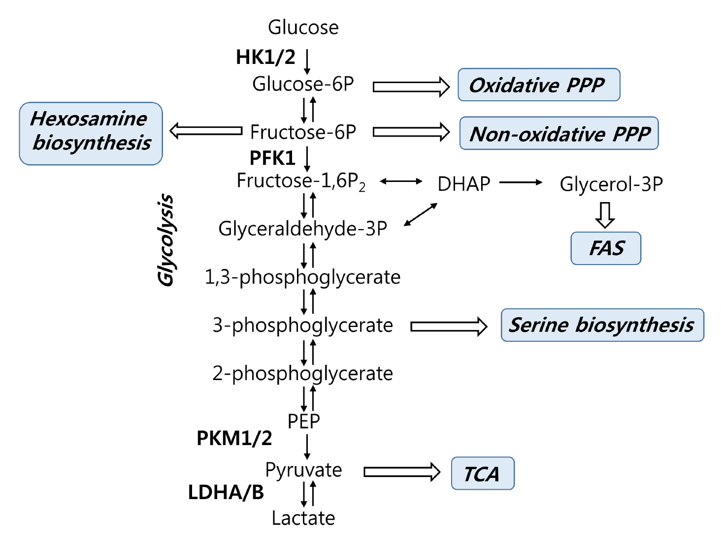 Figure 1 Glycolysis and its branching pathways. (Kim, 2018)
Representative Targets in Glycolysis
PDHA1
PDHA1, or Pyruvate Dehydrogenase E1 Alpha 1 subunit, is a critical enzyme component of the pyruvate dehydrogenase complex (PDC), which plays a pivotal role in cellular energy metabolism. Specifically, PDHA1 is involved in the conversion of pyruvate, derived from glucose through glycolysis, into acetyl-CoA, which enters the citric acid cycle to generate ATP, the cell's main energy currency. This conversion is a key regulatory step linking glycolysis and the Krebs cycle, and it occurs in the mitochondria of cells. The PDHA1 subunit is essential for the proper function of the PDC, and mutations in the PDHA1 gene can lead to a variety of metabolic disorders, most notably pyruvate dehydrogenase deficiency. This condition is characterized by a buildup of pyruvate and a corresponding deficiency in acetyl-CoA, leading to severe neurological dysfunction and metabolic abnormalities, manifesting early in life. The symptoms and severity can vary widely but often include developmental delay, chronic neurological dysfunction, and, in some cases, lactic acidosis. Given the central role of PDHA1 in energy metabolism, its activity is tightly regulated by multiple mechanisms. These include phosphorylation by specific kinases (PDKs) that deactivate the enzyme, and dephosphorylation by phosphatases (PDPs) that reactivate it. This regulation ensures that energy production is closely matched to cellular needs. The clinical importance of PDHA1 extends beyond metabolic disorders; it also has implications in cancer biology. Tumors often exhibit altered metabolic pathways, including enhanced glycolysis and reduced oxidative phosphorylation (a phenomenon known as the Warburg effect), and PDHA1's role in these pathways makes it a potential target for cancer therapy.
Recommended Mouse Anti-PDHA1 mAb (CAT#: ZG-041R)
 Figure 2 Mouse Anti-PDHA1 Antibody (ZG-041R) in ICC. Immunocytochemistry staining of HeLa cells fixed with 4% Paraformaldehyde and using anti-pyruvate dehydrogenase (lipoamide) alpha1 mouse mAb (dilution 1:100).
Recommended Rabbit Anti-PDHA1 mAb (CAT#: VS3-FY1118)
 Figure 3 Recombinant Rabbit Anti-PDHA1 Antibody (clone R06-1J4) in ICC. Immunocytochemical analysis of pyruvate dehydrogenase E1alpha subunit (green) in Hela using pyruvate dehydrogenase E1alpha subunit antibody and DAPI (blue).
Recommended Rabbit Anti-PDHA1 mAb (CAT#: ZG-0364C)
 Figure 4 Mouse Anti-PDHA1 Antibody (ZG-0364C) in IF. Immunofluorescence analysis of HeLa cells using PDH-E1α monoclonal antibody.
PKM2
PKM2, or Pyruvate Kinase M2, is an enzyme that catalyzes the final step in glycolysis, the conversion of phosphoenolpyruvate (PEP) to pyruvate, with the concurrent production of ATP. This enzyme exists in two forms, M1 and M2, which are products of alternative splicing of the PKM gene. PKM2 is distinctively expressed in embryonic and proliferative tissues, including cancer cells, where it supports the metabolic needs of rapid cell growth and division. The functional versatility of PKM2 extends beyond its enzymatic role in glycolysis. Unlike PKM1, which is always highly active, PKM2 can exist in both a highly active tetrameric form and a less active dimeric form. This unique feature allows cells expressing PKM2 to modulate their pyruvate kinase activity in response to different cellular needs and conditions. In its less active form, PKM2 facilitates the accumulation of upstream glycolytic intermediates that are essential for anabolic processes, such as nucleotide, amino acid, and lipid synthesis, which are crucial for cell proliferation and growth. In cancer, the expression of PKM2 is particularly significant. The enzyme's ability to regulate the balance between energy production and the synthesis of biosynthetic precursors helps cancer cells adapt their metabolism to support both their survival and rapid proliferation, a phenomenon central to cancer progression known as the Warburg effect. Therefore, PKM2 is not just a facilitator of glycolysis but also a regulator of cellular metabolism that contributes to tumorigenesis.
Recommended Mouse Anti-PKM2 mAb (CAT#: ZG-0378C)
 Figure 5 Mouse Anti-PKM Antibody (ZG-0378C) in ICC. Immunocytochemistry staining of Hela cells fixed with 4% Paraformaldehyde and using anti-PKM2 mouse mAb (dilution 1:400).
Recommended Mouse Anti-PKM2 mAb (CAT#: ZG-0379C)
 Figure 6 Mouse Anti-PKM Antibody (ZG-0379C) in ICC. Immunocytochemistry staining of Hela cells fixed with 4% Paraformaldehyde and using anti-PKM2 mouse mAb (dilution 1:400).
SLC2A1
SLC2A1, also known as GLUT1 (Glucose Transporter Type 1), is a key protein in the facilitation of glucose transport across the plasma membranes of mammalian cells. It is part of the larger solute carrier family 2 (SLC2), which is responsible for the transport of glucose and other hexose sugars. GLUT1 is ubiquitously expressed but is particularly abundant in tissues with high basal glucose requirements, such as the brain, erythrocytes, and the endothelial cells forming the blood-brain barrier. The function of GLUT1 is to ensure a steady supply of glucose to cells, independent of fluctuating external glucose levels, thus providing a fundamental energy source necessary for cellular processes. In the brain, GLUT1 is critical as neurons have high and constant energy demands that are primarily met through glucose metabolism. Consequently, dysfunction or deficiency of GLUT1 can have severe neurological implications. GLUT1 deficiency syndrome, for instance, is a genetic disorder characterized by a reduced ability of GLUT1 to transport glucose into the brain, leading to developmental delays, seizures, and other neurological impairments. In addition to its physiological roles, GLUT1 is notably involved in pathological conditions. In cancer, many types of tumor cells exhibit upregulated GLUT1 expression, enhancing their glucose uptake to support rapid proliferation and growth (the Warburg effect).
Recommended Mouse Anti-SLC2A1 mAb (CAT#: ZG-0078C)
 Figure 7 Mouse Anti-SLC2A1 Antibody (clone ABT-GLUT1) in IHC. Immunohistochemical analysis of paraffin-embedded Colon Carcinoma. 1, Antibody was diluted at 1:200 (4° overnight). 2, TRIS-EDTA of pH 8.0 was used for antigen retrieval. 3, Secondary antibody was diluted at 1:200 (room temperature, 30 min).
Recommended Rabbit Anti-SLC2A1 mAb (CAT#: ZG-0564U)
 Figure 8 Rabbit Anti-SLC2A1 Antibody (ZG-0564U) in IHC. IHC image of ZG-0564U diluted at 1:100 and staining in paraffin-embedded human lung cancer performed on a Leica Bond™ system. After dewaxing and hydration, antigen retrieval was mediated by high pressure in a citrate buffer (pH 6.0). Section was blocked with 10% normal goat serum 30min at RT. Then primary antibody (1% BSA) was incubated at 4°C overnight. The primary is detected by a Goat anti-rabbit IgG polymer labeled by HRP and visualized using 0.05% DAB.
Recommended Mouse Anti-SLC2A1 mAb (CAT#: VS3-FY1346)
 Figure 9 Recombinant Mouse Anti-SLC2A1 Antibody (clone 1G5-8F2-7D10) in IHC-P. Immunohistochemical analysis of paraffin-embedded human placenta using an antibody to the glucose transporter GLUT1. High pressure and high temperature sodium citrate pH 6.0 for antigen retrieval.
Full List of Targets in Glycolysis
| Biomarker | Alternative Names | Gene ID | UniProt ID | Roles |
| GPI | Glucose-6-Phosphate Isomerase; Autocrine Motility Factor; Phosphoglucose Isomerase; Phosphohexose Isomerase; Neuroleukin; EC 5.3.1.9; SA-36; PGI; AMF; NLK; PHI | 2821 | P06744 | This gene encodes a member of the glucose phosphate isomerase protein family. The encoded protein has been identified as a moonlighting protein based on its ability to perform mechanistically distinct functions. In the cytoplasm, the gene product functions as a glycolytic enzyme (glucose-6-phosphate isomerase) that interconverts glucose-6-phosphate and fructose-6-phosphate. Extracellularly, the encoded protein (also referred to as neuroleukin) functions as a neurotrophic factor that promotes survival of skeletal motor neurons and sensory neurons, and as a lymphokine that induces immunoglobulin secretion. The encoded protein is also referred to as autocrine motility factor based on an additional function as a tumor-secreted cytokine and angiogenic factor. Defects in this gene are the cause of nonspherocytic hemolytic anemia and a severe enzyme deficiency can be associated with hydrops fetalis, immediate neonatal death and neurological impairment. Alternative splicing results in multiple transcript variants. |
| HK2 | Hexokinase 2; Muscle Form Hexokinase; Hexokinase Type II; EC 2.7.1.1; HK II; Hexokinase-2, Muscle; | 3099 | P52789 | Hexokinases phosphorylate glucose to produce glucose-6-phosphate, the first step in most glucose metabolism pathways. This gene encodes hexokinase 2, the predominant form found in skeletal muscle. It localizes to the outer membrane of mitochondria. Expression of this gene is insulin-responsive, and studies in rat suggest that it is involved in the increased rate of glycolysis seen in rapidly growing cancer cells. |
| PDHA1 | Pyruvate Dehydrogenase E1 Alpha 1 Subunit; Pyruvate Dehydrogenase E1 Component Subunit Alpha, Somatic Form, Mitochondrial; Pyruvate Dehydrogenase (Lipoamide) Alpha 1; Pyruvate Dehydrogenase Alpha 1; PDHE1-A Type I; EC 1.2.4.1; PHE1A | 5160 | P08559 | The pyruvate dehydrogenase (PDH) complex is a nuclear-encoded mitochondrial multienzyme complex that catalyzes the overall conversion of pyruvate to acetyl-CoA and CO(2), and provides the primary link between glycolysis and the tricarboxylic acid (TCA) cycle. The PDH complex is composed of multiple copies of three enzymatic components: pyruvate dehydrogenase (E1), dihydrolipoamide acetyltransferase (E2) and lipoamide dehydrogenase (E3). The E1 enzyme is a heterotetramer of two alpha and two beta subunits. This gene encodes the E1 alpha 1 subunit containing the E1 active site, and plays a key role in the function of the PDH complex. Mutations in this gene are associated with pyruvate dehydrogenase E1-alpha deficiency and X-linked Leigh syndrome. Alternatively spliced transcript variants encoding different isoforms have been found for this gene.[provided by RefSeq, Mar 2010] |
| PDHB | PDHB; Pyruvate Dehydrogenase E1 Component Subunit Beta, Mitochondrial; PDHE1-B; PDHBD; EC 1.2.4.1; PHE1B | 5162 | P11177 | The pyruvate dehydrogenase (PDH) complex is a nuclear-encoded mitochondrial multienzyme complex that catalyzes the overall conversion of pyruvate to acetyl-CoA and carbon dioxide, and provides the primary link between glycolysis and the tricarboxylic acid (TCA) cycle. The PDH complex is composed of multiple copies of three enzymatic components: pyruvate dehydrogenase (E1), dihydrolipoamide acetyltransferase (E2) and lipoamide dehydrogenase (E3). The E1 enzyme is a heterotetramer of two alpha and two beta subunits. This gene encodes the E1 beta subunit. Mutations in this gene are associated with pyruvate dehydrogenase E1-beta deficiency. Alternatively spliced transcript variants have been found for this gene. |
| PDK1 | PDK1; PDPK1 | 5163 | Q15118 | Pyruvate dehydrogenase (PDH) is a mitochondrial multienzyme complex that catalyzes the oxidative decarboxylation of pyruvate and is one of the major enzymes responsible for the regulation of homeostasis of carbohydrate fuels in mammals. The enzymatic activity is regulated by a phosphorylation/dephosphorylation cycle. Phosphorylation of PDH by a specific pyruvate dehydrogenase kinase (PDK) results in inactivation. Multiple alternatively spliced transcript variants have been found for this gene. |
| PDK2 | Pyruvate Dehydrogenase Kinase 2; Pyruvate Dehydrogenase Kinase, Isoenzyme 2; Pyruvate Dehydrogenase Kinase, Isozyme 2; PDH Kinase 2; EC 2.7.11.2; PDHK2 | 5164 | Q15119 | This gene encodes a member of the pyruvate dehydrogenase kinase family. The encoded protein phosphorylates pyruvate dehydrogenase, down-regulating the activity of the mitochondrial pyruvate dehydrogenase complex. Overexpression of this gene may play a role in both cancer and diabetes. Alternatively spliced transcript variants encoding multiple isoforms have been observed for this gene. [provided by RefSeq, Dec 2010] |
| PDK3 | CMTX6; GS1-358P8.4 | 5165 | Q15120 | The pyruvate dehydrogenase (PDH) complex is a nuclear-encoded mitochondrial multienzyme complex that catalyzes the overall conversion of pyruvate to acetyl-CoA and CO(2). It provides the primary link between glycolysis and the tricarboxylic acid (TCA) cycle, and thus is one of the major enzymes responsible for the regulation of glucose metabolism. The enzymatic activity of PDH is regulated by a phosphorylation/dephosphorylation cycle, and phosphorylation results in inactivation of PDH. The protein encoded by this gene is one of the three pyruvate dehydrogenase kinases that inhibits the PDH complex by phosphorylation of the E1 alpha subunit. This gene is predominantly expressed in the heart and skeletal muscles. Alternatively spliced transcript variants encoding different isoforms have been found for this gene. |
| PDK4 | Pyruvate Dehydrogenase Kinase 4; EC 2.7.11.2; Pyruvate Dehydrogenase Kinase; Isoenzyme 4; Pyruvate Dehydrogenase Kinase; Isozyme 4; Pyruvate Dehydrogenase Kinase Isoform 4; EC 2.7.11; PDHK4 | 5166 | Q16654 | This gene is a member of the PDK/BCKDK protein kinase family and encodes a mitochondrial protein with a histidine kinase domain. This protein is located in the matrix of the mitrochondria and inhibits the pyruvate dehydrogenase complex by phosphorylating one of its subunits, thereby contributing to the regulation of glucose metabolism. Expression of this gene is regulated by glucocorticoids, retinoic acid and insulin. |
| PKM2 | Pyruvate kinase isoform M2 | 5315 | P14618 | Pyruvate kise isoenzyme type M2 (PKM2 or M2-PK) is one of the two isozymes encoded by the human PKM gene. The two isozymes M1 and M2 are altertive splicing products and differ in one of eleven exons. Pyruvate kise is a critical enzyme in glycolysis as it dephosphorylates phosphoenolpyruvate (PEP) to pyruvate resulting in the net production ATP. |
| SLC16A1 | Monocarboxylate transporter 1; MCT 1; Solute carrier family 16 member 1; SLC16A1; MCT1 | 6566 | P53985 | Proton-coupled monocarboxylate transporter. Catalyzes the rapid transport across the plasma membrane of many monocarboxylates such as lactate, pyruvate, branched-chain oxo acids derived from leucine, valine and isoleucine, and the ketone bodies acetoacetate, beta-hydroxybutyrate and acetate. Depending on the tissue and on cicumstances, mediates the import or export of lactic acid and ketone bodies. Required for normal nutrient assimilation, increase of white adipose tissue and body weight gain when on a high-fat diet. Plays a role in cellular responses to a high-fat diet by modulating the cellular levels of lactate and pyruvate, small molecules that contribute to the regulation of central metabolic pathways and insulin secretion, with concomitant effects on plasma insulin levels and blood glucose homeostasis. |
| SLC16A4 | SLC16A4; MCT5; Solute Carrier Family 16 Member 4; Solute Carrier Family 16 (Monocarboxylic Acid Transporters), Member 4; MCT 4; Solute Carrier Family 16, Member 4 (Monocarboxylic Acid Transporter 5); Solute Carrier Family 16, Member 4; MCT 5; Monocarboxylate Transporter 4; Monocarboxylate Transporter 5; MCT4 | 9122 | O15374 | Proton-linked monocarboxylate transporter. Catalyzes the rapid transport across the plasma membrane of many monocarboxylates such as lactate, pyruvate, branched-chain oxo acids derived from leucine, valine and isoleucine, and the ketone bodies acetoacetate, beta-hydroxybutyrate and acetate (By similarity). |
| SLC2A1 | Solute Carrier Family 2 Member 1; Choreoathetosis/Spasticity, Episodic (Paroxysmal Choreoathetosis/Spasticity); Solute Carrier Family 2 (Facilitated Glucose Transporter), Member 1; Human T-Cell Leukemia Virus (I And II) Receptor; Glucose Transporter Type 1, Erythrocyte/Brain; HepG2 Glucose Transporter; GLUT-1; GLUT1; Solute Carrier Family 2, Facilitated Glucose Transporter Member 1; Receptor For HTLV-1 And HTLV-2; GLUT1DS | 6513 | P11166 | This gene encodes a major glucose transporter in the mammalian blood-brain barrier. The encoded protein is found primarily in the cell membrane and on the cell surface, where it can also function as a receptor for human T-cell leukemia virus (HTLV) I and II. Mutations in this gene have been found in a family with paroxysmal exertion-induced dyskinesia. [provided by RefSeq, Apr 2013] |
| SLC2A3 | Solute Carrier Family 2 Member 3; Solute Carrier Family 2 (Facilitated Glucose Transporter), Member 3; Glucose Transporter Type 3, Brain; GLUT-3; GLUT3; Solute Carrier Family 2, Facilitated Glucose Transporter Member 3 | 6515 | P11169 | Glucose transporter 3 (or GLUT3), also known as solute carrier family 2, facilitated glucose transporter member 3 (SLC2A3) is a protein that in humans is encoded by the SLC2A3 gene. |
Tested Data-Supported Products for Targeting Glycolysis


Reference
- Kim, Jung-Ae, and Young Il Yeom. "Metabolic signaling to epigenetic alterations in cancer." Biomolecules & therapeutics 26.1 (2018): 69.
For research use only. Not intended for any clinical use.
Send Inquiry
This site is protected by reCAPTCHA and the Google Privacy Policy and Terms of Service apply.