


Liver Cancer Biomarkers
Representative Biomarkers Full List of Biomarkers Tested Data-Supported Products
Liver cancer, a pressing global health issue, is projected to affect over 1 million individuals annually by 2025. Hepatocellular carcinoma (HCC), constituting approximately 90% of liver cancer cases, is significantly influenced by hepatitis B and C virus infections. The rising prevalence of non-alcoholic steatohepatitis (NASH), associated with metabolic syndrome or diabetes, is becoming a prominent risk factor, particularly in Western populations. About 25% of HCC cases harbor potentially actionable mutations, yet the translation of these discoveries into clinical practice remains limited. The diagnosis of HCC often relies on non-invasive criteria, challenged by the need for molecular profiling. Recent advances have led to the approval of six systemic therapies based on phase III trials, significantly impacting the management of advanced HCC. Ongoing research into combination therapies, including checkpoint inhibitors and tyrosine kinase inhibitors, promises to revolutionize HCC treatment across all stages of the disea.
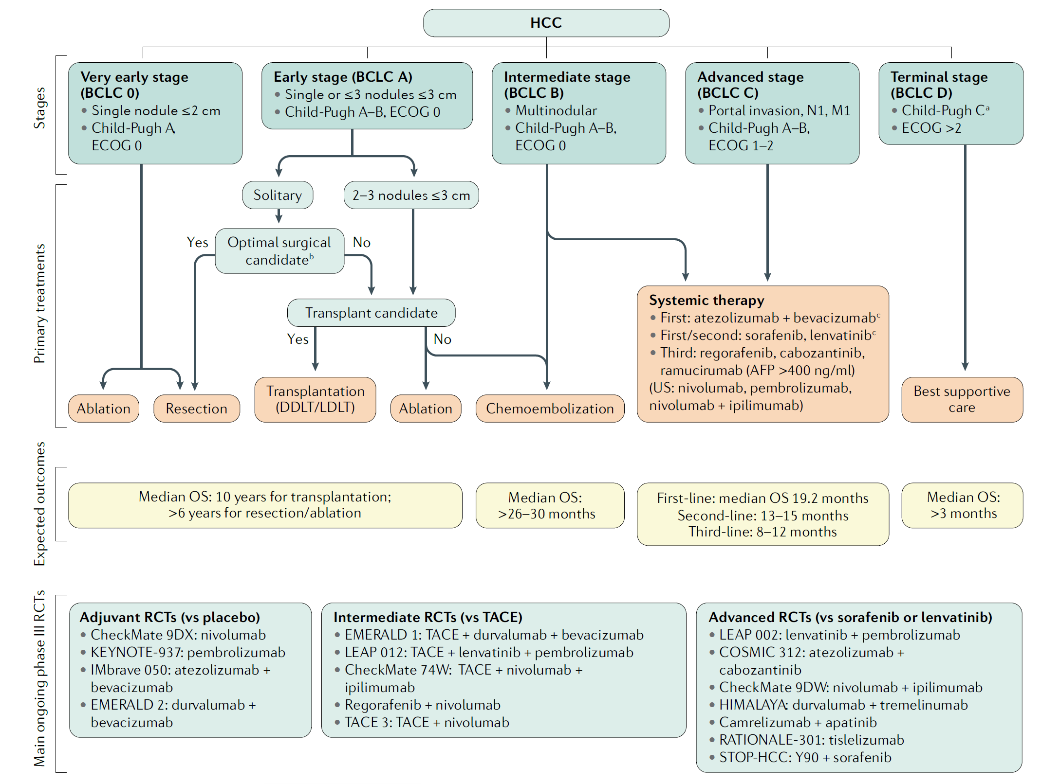 Figure 1 Treatment strategy in the management of hepatocellular carcinoma. (Llovet, 2021)
Representative Biomarkers of Liver Cancer
AFP
Glypican-3 (GPC3) is a heparan sulfate proteoglycan attached to the cell membrane, playing a crucial role in the regulation of cell growth and division. Particularly in hepatocellular carcinoma (HCC), GPC3 is significantly upregulated, contrasting its minimal or non-existent expression in normal liver tissues, which highlights its potential as a biomarker for diagnosis and a target for therapy. GPC3's involvement in HCC is multifaceted; it modulates various signaling pathways, including the Wnt/β-catenin and YAP/TAZ pathways, crucial for cell proliferation, differentiation, and apoptosis. This regulatory capacity influences tumor growth, angiogenesis, and metastasis, underlying the aggressive nature of HCC.
Recommended Humanized Anti-GPC3 mAb (Codrituzumab) (CAT#: TAB-H14)
 Figure 2 ELISA analysis of TAB-H14 was performed by coating with Human GPC3 Protein (His Tag). Then blocked with BSA and incubated with TAB-H14. The HRP-conjugated goat anti-human IgG as a secondary antibody (1: 5000). Detection was performed using TMB substrate and stopped with sulfuric acid. The absorbances were read on a spectrophotometer at 450 nm.
Recommended Human Anti-GPC3 mAb (CAT#: HPAB-0563-CN)
 Figure 3 HPLC analysis of HPAB-0563-CN shows the purity is 96.8%.
Recommended Rabbit Anti-GPC3 mAb (CAT#: ZG-0690J)
 Figure 4 Immunofluorescence staining of HepG2 cells with this product at 1:30, counter-stained with DAPI. The cells were fixed in 4% formaldehyde, permeabilized using 0.2% Triton X-100 and blocked in 10% normal Goat Serum. The cells were then incubated with the antibody overnight at 4°C. The secondary antibody was Alexa Fluor 488-congugated Goat Anti-Rabbit IgG (H+L).
GPC3
Alpha-fetoprotein (AFP) is a glycoprotein primarily produced by the fetal liver and yolk sac, with its levels declining rapidly post-birth. It re-emerges as a significant biomarker in hepatocellular carcinoma (HCC), where its gene regulation becomes complex, often resulting in overexpression associated with HCC's aggressiveness. AFP functions as a pro-proliferative protein, modulating apoptotic regulation and cytoplasmic signaling, thereby promoting tumour cell growth. Its levels correlate with molecular HCC subclasses, reflecting the tumor's heterogeneity on macroscopic, histopathological, and molecular levels. High AFP serum levels are linked to poorer prognosis in HCC patients, making it a valuable prognostic marker. Additionally, AFP's role extends to predicting treatment response and monitoring disease progression, emphasizing its importance in the clinical management of HCC.
Recommended Mouse Anti-AFP mAb (CAT#: ZG-0018J)
 Figure 5 Immunohistochemical analysis of paraffin-embedded Human gallbladder. 1. Antibody was diluted at 1:100 (4°C overnight). 2, High-pressure and temperature EDTA, pH8.0 was used for antigen retrieval. 3, Secondary Antibody was diluted at 1:200 (room temperature, 30min)
LMNB1
Lamin B1 (LMNB1) is a pivotal structural protein of the nuclear lamina, a fibrous network located underneath the inner nuclear membrane, playing essential roles in maintaining nuclear structure, organizing chromatin, and regulating gene expression. In the context of hepatocellular carcinoma (HCC), the most common type of liver cancer, LMNB1 has garnered attention for its multifaceted roles. Aberrant expression of LMNB1 has been associated with the progression and prognosis of HCC. It influences various cellular mechanisms critical for cancer development, including cell cycle progression, apoptosis, and DNA damage response. Studies have shown that overexpression of LMNB1 in HCC can lead to alterations in nuclear morphology and function, promoting cellular proliferation and inhibiting apoptosis, thereby facilitating tumor growth and metastasis. Additionally, LMNB1's involvement in chromatin organization and gene regulation can significantly impact the expression of oncogenes and tumor suppressor genes, further contributing to HCC pathogenesis.
Recommended Mouse Anti-LMNB1 mAb (CAT#: MOB-0073F)
 Figure 6 Immunohistochemical analysis of paraffin-embedded human uterine tissue. 1. The antibody is diluted 1:200 (overnight at 4°C). 2. Sodium citrate pH 6.0 is used for antibody repair (> 98°C, 20 minutes). 3. Dilute the secondary antibody at 1:200 (room temperature, 30min). The negative control is only used by the secondary antibody.
Recommended Mouse Anti-LMNB1 mAb (CAT#: ZG-0953F)
 Figure 7 Immunohistochemistry on frozen sections of swine liver showing nuclear lamina staining in hepatocytes.
Recommended Mouse Anti-AFP mAb (CAT#: ZG-0264C)
 Figure 8 Immunocytochemistry staining of Hela cells fixed with 4% Paraformaldehyde and using anti-Lamin B1 mouse mAb (dilution 1:100).
Full List of Liver Cancer Biomarkers
| Biomarker | Alternative Names | Gene ID | UniProt ID | Roles |
| AFP | Alpha Fetoprotein; Alpha-1-Fetoprotein; Alpha-Fetoglobulin; HPAFP; Alpha-Fetoprotein; AFPD; FETA | 174 | P02771 | This gene encodes alpha-fetoprotein, a major plasma protein produced by the yolk sac and the liver during fetal life. Alpha-fetoprotein expression in adults is often associated with hepatoma or teratoma. However, hereditary persistance of alpha-fetoprotein may also be found in individuals with no obvious pathology. The protein is thought to be the fetal counterpart of serum albumin, and the alpha-fetoprotein and albumin genes are present in tandem in the same transcriptional orientation on chromosome 4. Alpha-fetoprotein is found in monomeric as well as dimeric and trimeric forms, and binds copper, nickel, fatty acids and bilirubin. The level of alpha-fetoprotein in amniotic fluid is used to measure renal loss of protein to screen for spina bifida and anencephaly. [provided by RefSeq, Jul 2008] |
| ATAD2 | ATPase Family, AAA Domain Containing 2; AAA Nuclear Coregulator Cancer-Associated Protein; ANCCA; EC 3.6.1.3; PRO2000; CT137 | 29028 | A0A024R9G7 | A large family of ATPases has been described, whose key feature is that they share a conserved region of about 220 amino acids that contains an ATP-binding site. The proteins that belong to this family either contain one or two AAA (ATPases Associated with diverse cellular Activities) domains. AAA family proteins often perform chaperone-like functions that assist in the assembly, operation, or disassembly of protein complexes. The protein encoded by this gene contains two AAA domains, as well as a bromodomain. |
| DKK1 | DKK-1; SK | 22943 | O94907 | This gene encodes a member of the dickkopf family of proteins. Members of this family are secreted proteins characterized by two cysteine-rich domains that mediate protein-protein interactions. The encoded protein binds to the LRP6 co-receptor and inhibits beta-catenin-dependent Wnt signaling. This gene plays a role in embryonic development and may be important in bone formation in adults. Elevated expression of this gene has been observed in numerous human cancers and this protein may promote proliferation, invasion and growth in cancer cell lines. |
| GPC3 | DGSX; GTR2-2; MXR7; OCI-5; SDYS; SGB; SGBS; SGBS1 | 2719 | P51654 | Cell surface heparan sulfate proteoglycans are composed of a membrane-associated protein core substituted with a variable number of heparan sulfate chains. Members of the glypican-related integral membrane proteoglycan family (GRIPS) contain a core protein anchored to the cytoplasmic membrane via a glycosyl phosphatidylinositol linkage. These proteins may play a role in the control of cell division and growth regulation. The protein encoded by this gene can bind to and inhibit the dipeptidyl peptidase activity of CD26, and it can induce apoptosis in certain cell types. Deletion mutations in this gene are associated with Simpson-Golabi-Behmel syndrome, also known as Simpson dysmorphia syndrome. Alternative splicing results in multiple transcript variants. References: Fu Ying,Urban Daniel J,Nani Roger R et al. Glypican-3-Specific Antibody Drug Conjugates Targeting Hepatocellular Carcinoma..Hepatology, 2019, 70: 563-576. Zhang Yi-Fan,Ho Mitchell,Humanization of high-affinity antibodies targeting glypican-3 in hepatocellular carcinoma. |
| GPC4 | Glypican 4; Glypican Proteoglycan 4; K-Glypican; DJ900E8.1 (Glypican 4) | 2239 | O75487 | Cell surface heparan sulfate proteoglycans are composed of a membrane-associated protein core substituted with a variable number of heparan sulfate chains. Members of the glypican-related integral membrane proteoglycan family (GRIPS) contain a core protein anchored to the cytoplasmic membrane via a glycosyl phosphatidylinositol linkage. These proteins may play a role in the control of cell division and growth regulation. The GPC4 gene is adjacent to the 3' end of GPC3 and may also play a role in Simpson-Golabi-Behmel syndrome. |
| LMNB1 | LMN; ADLD; LMN2; LMNB | 4001 | P20700 | The nuclear lamina consists of a two-dimensional matrix of proteins located next to the inner nuclear membrane. The lamin family of proteins make up the matrix and are highly conserved in evolution. |
| PDGFRL | PDGFRL; Platelet-Derived Growth Factor Receptor-Like Protein; PDGRL; Platelet-Derived Growth Factor Receptor-Like; Platelet-Derived Growth Factor-Beta-Like Tumor Suppressor; PDGFR-Like Protein | 5157 | Q15198 | This gene encodes a protein with significant sequence similarity to the ligand binding domain of platelet-derived growth factor receptor beta. Mutations in this gene, or deletion of a chromosomal segment containing this gene, are associated with sporadic hepatocellular carcinomas, colorectal cancers, and non-small cell lung cancers. This suggests this gene product may function as a tumor suppressor. |
Tested Data-Supported Products Targeting Liver Cancer Biomarkers


-1.png)
-1.png)


-1.png)


References
- Llovet, Josep M et al. “Hepatocellular carcinoma.” Nature reviews. Disease primers vol. 7,1 6. 21 Jan. 2021, doi:10.1038/s41572-020-00240-3
For research use only. Not intended for any clinical use.
Send Inquiry
This site is protected by reCAPTCHA and the Google Privacy Policy and Terms of Service apply.