


PI3K Pathway
Representative Targets in PI3K Pathway Full List of Targets in PI3K Pathway Tested Data-Supported Products for Targeting PI3K Pathway
The phosphoinositide 3-kinases (PI3Ks) are a family of enzymes involved in complex intracellular signaling pathways, playing pivotal roles in regulating cell functions such as growth, proliferation, differentiation, motility, survival, and intracellular trafficking. These enzymes are activated by various stimuli, including hormones, growth factors, and insulin, catalyzing the phosphorylation of the 3' hydroxyl group of the inositol ring of phosphatidylinositol lipids. The PI3K pathway is integral to cellular signaling, with its activation leading to the production of lipid second messengers that initiate a cascade of downstream effects, involving the serine/threonine kinase AKT (also known as Protein Kinase B) and other effectors. This signaling cascade influences numerous cellular processes and is tightly regulated by various mechanisms, including the action of phosphatase and tensin homolog (PTEN), a tumor suppressor gene that acts as a PI3K antagonist. Dysregulation of the PI3K pathway has been implicated in many human diseases, particularly cancer, where mutations in PI3K or PTEN lead to unchecked cellular proliferation and survival.
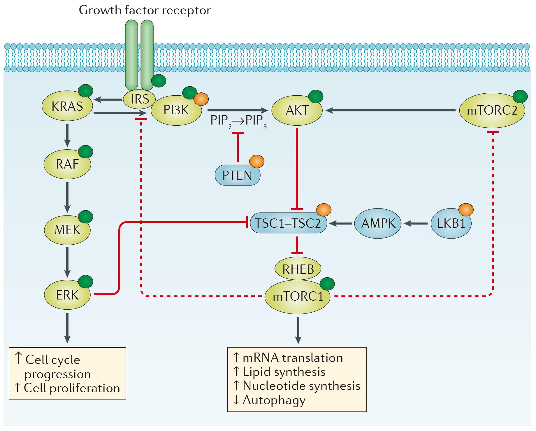 Figure 1 Intracellular signaling via the PI3K–AKT–mTOR pathway. (Janku, 2018)
Representative Targets in PI3K Pathway
AKT3
AKT3, belonging to the AKT serine/threonine kinase family, plays a critical role in regulating biological processes that are essential for maintaining cellular homeostasis. This kinase is particularly significant in the context of neuronal development and function, as well as in the modulation of cellular growth, proliferation, and survival pathways. AKT3 is activated in response to growth factors and nutrients through phosphoinositide 3-kinase (PI3K)-dependent signaling pathways, thereby influencing the activity of downstream targets involved in the control of cell cycle progression and apoptosis. Its distinct expression patterns, especially in the brain, suggest a unique role in the development and maintenance of neural tissues, including the regulation of brain size and function. Furthermore, alterations in AKT3 have been implicated in various pathological conditions, including cancer and neurological disorders, highlighting its importance not only in normal cellular physiology but also in disease states.
Recommended Rabbit Anti-AKT3 mAb (CAT#: ZG-0437U)
 Figure 2 Rabbit Anti-AKT3 Antibody (ZG-0437U) in FC. Profile of Akt3 expression in human blood lymphocytes analyzed by the BD FACSCanto II. Blood cells of patient with chronic lymphocytic leukemia were fixed, permeabilized and stained with anti-human Akt3 FITC (red, used 10 μl per test) or with an isotype control (black).
Recommended Rabbit Anti-AKT3 mAb (CAT#: ZG-0385U)
 Figure 3 Rabbit Anti-AKT3 Antibody (ZG-0385U) in IF. Representative picture of Akt3 expression in HEK293 cells, visualized with rabbit anti-Akt3 antibody. Primary antibody dilution 1:500.
mTOR
The mechanistic Target of Rapamycin (mTOR) is a central cell-signaling kinase integral to a wide array of cellular processes, including protein synthesis, nutrient sensing, cell growth, and autophagy. This highly conserved serine/threonine kinase operates within two distinct complexes, mTOR Complex 1 (mTORC1) and mTOR Complex 2 (mTORC2), each orchestrating different cellular responses to environmental cues. mTORC1 is sensitive to rapamycin and primarily regulates cell growth by controlling protein synthesis through the phosphorylation of S6K1 and 4EBP1, pivotal in the translation initiation process. It also responds to nutrient availability, energy levels, and growth factors. On the other hand, mTORC2, which is rapamycin-insensitive under acute treatment, plays a critical role in cytoskeletal organization, cell survival, and lipid metabolism, through the direct phosphorylation of AKT, SGK1, and PKCα. The regulation of mTOR activity is complex, involving inputs from upstream pathways such as PI3K/AKT and AMPK, which sense changes in the cellular environment and adjust mTOR signaling accordingly. Dysregulation of mTOR signaling is implicated in numerous diseases, including cancer, metabolic disorders, and neurodegeneration.
Recommended Rabbit Anti-mTOR mAb (CAT#: ZG-0473U)
 Figure 4 Rabbit Anti-Phospho-MTOR (S2481) Antibody (ZG-0473U) in IF. Immunofluorescence staining of Hela cells with ZG-0473U at 1:100, counter-stained with DAPI. The cells were fixed in 4% formaldehyde, permeabilized using 0.2% Triton X-100 and blocked in 10% normal Goat Serum. The cells were then incubated with the antibody overnight at 4°C. The secondary antibody was Alexa Fluor 488-congugated Goat Anti-Rabbit IgG (H+L).
Recommended Rabbit Anti-mTOR mAb (CAT#: ZG-0474U)
 Figure 5 Rabbit Anti-Phospho-MTOR (S2448) Antibody (ZG-0474U) in IHC. IHC image of ZG-0474U diluted at 1:100 and staining in paraffin-embedded human breast cancer performed on a Leica BondTM system. After dewaxing and hydration, antigen retrieval was mediated by high pressure in a citrate buffer (pH 6.0). Section was blocked with 10% normal goat serum 30min at RT. Then primary antibody (1% BSA) was incubated at 4°C overnight. The primary is detected by a biotinylated secondary antibody and visualized using an HRP conjugated SP system.
Recommended Rabbit Anti-mTOR mAb (CAT#: VS3-FY973)
 Figure 6 Recombinant Rabbit Anti-MTOR (phospho Ser2448) Antibody (clone R08-7G7) in IHC-P. Immunohistochemical analysis of paraffin-embedded human lung carcinoma using Phospho-mTOR (Ser2448) Antibody. High pressure and high temperature sodium citrate pH 6.0 for antigen retrieval.
PTEN
PTEN (Phosphatase and tensin homolog) is a crucial tumor suppressor gene that plays a significant role in cell growth, division, and apoptosis. This dual-specificity phosphatase acts by dephosphorylating phosphatidylinositol (3,4,5)-trisphosphate (PIP3), a key lipid second messenger involved in the PI3K/AKT/mTOR signaling pathway, thereby negatively regulating cell survival and proliferation signals. Mutations or deletions in the PTEN gene are associated with a variety of cancers, including breast, thyroid, and prostate cancer, highlighting its pivotal role in cancer biology. Beyond its tumor-suppressive functions, PTEN is involved in several other physiological processes, such as DNA repair, apoptosis, and cell migration.
Recommended Mouse Anti-PTEN mAb (CAT#: MOB-0232F)
 Figure 7 Mouse Anti-PTEN Recombinant Antibody (clone 9E8) (MOB-0232F) in IHC. PTEN Mouse mAb diluted 1:200 was used for immunohistochemical analysis of paraffin-embedded human lung cancer tissues.
Recommended Mouse Anti-PTEN mAb (CAT#: MOB-0231F)
 Figure 8 Mouse Anti-PTEN Recombinant Antibody (clone 2C10) (MOB-0231F) in IHC. PTEN Mouse mAb diluted 1:200 was used for immunohistochemical analysis of paraffin-embedded human colon cancer tissue.
Full List of Transcription Factors
| Biomarker | Alternative Names | Gene ID | UniProt ID | Roles |
| AKT1 | AKT Serine/Threonine Kinase 1; V-Akt Murine Thymoma Viral Oncogene Homolog 1; Protein Kinase B Alpha; Proto-Oncogene C-Akt; RAC-PK-Alpha; EC 2.7.11.1; PKB Alpha; PKB; RAC; V-Akt Murine Thymoma Viral Oncogene-Like Protein 1; RAC-Alpha Serine/Threonine-Protein Kinase | 207 | B0LPE5 | The serine-threonine protein kinase encoded by the AKT1 gene is catalytically inactive in serum-starved primary and immortalized fibroblasts. AKT1 and the related AKT2 are activated by platelet-derived growth factor. The activation is rapid and specific, and it is abrogated by mutations in the pleckstrin homology domain of AKT1. It was shown that the activation occurs through phosphatidylinositol 3-kinase. In the developing nervous system AKT is a critical mediator of growth factor-induced neuronal survival. Survival factors can suppress apoptosis in a transcription-independent manner by activating the serine/threonine kinase AKT1, which then phosphorylates and inactivates components of the apoptotic machinery. Mutations in this gene have been associated with the Proteus syndrome. Multiple alternatively spliced transcript variants have been found for this gene. [provided by RefSeq, Jul 2011] |
| AKT1S1 | AKT1 Substrate 1; 40 KDa Proline-Rich AKT Substrate; PRAS40; Proline-Rich Akt Substrate; 40 KDa; AKT1 Substrate 1 (Proline-Rich); AKT1 Substrate 1 (Proline Rich); Proline-Rich AKT1 Substrate 1; Lobe | 84335 | Q96B36 | AKT1S1 is a proline-rich substrate of AKT (MIM 164730) that binds 14-3-3 protein (see YWHAH, MIM 113508) when phosphorylated (Kovacina et al., 2003 ). |
| AKT2 | AKT Serine/Threonine Kinase 2; V-Akt Murine Thymoma Viral Oncogene Homolog 2; Protein Kinase B Beta; Protein Kinase Akt-2; RAC-PK-Beta; EC 2.7.11.1; PKB Beta; Putative V-Akt Murine Thymoma Viral Oncoprotein 2; RAC-Beta Serine/Threonine-Protein Kinase | 208 | B4DG79 | This gene is a putative oncogene encoding a protein belonging to a subfamily of serine/threonine kinases containing SH2-like (Src homology 2-like) domains. The gene was shown to be amplified and overexpressed in 2 of 8 ovarian carcinoma cell lines and 2 of 15 primary ovarian tumors. Overexpression contributes to the malignant phenotype of a subset of human ductal pancreatic cancers. The encoded protein is a general protein kinase capable of phophorylating several known proteins. |
| AKT3 | AKT Serine/Threonine Kinase 3; RAC-PK-Gamma; EC 2.7.11.1; PKB Gamma; STK-2; PKBG; V-Akt Murine Thymoma Viral Oncogene Homolog 3 (Protein Kinase B, Gamma); V-Akt Murine Thymoma Viral Oncogene Homolog 3; RAC-Gamma Serine/Threonine Protein Kinase; RAC-Gamma Serine/Threonine-Protein Kinase | 10000 | Q9Y243 | The protein encoded by this gene is a member of the AKT, also called PKB, serine/threonine protein kinase family. AKT kinases are known to be regulators of cell signaling in response to insulin and growth factors. They are involved in a wide variety of biological processes including cell proliferation, differentiation, apoptosis, tumorigenesis, as well as glycogen synthesis and glucose uptake. This kinase has been shown to be stimulated by platelet-derived growth factor (PDGF), insulin, and insulin-like growth factor 1 (IGF1). Alternatively splice transcript variants encoding distinct isoforms have been described. |
| MLST8 | GBL; LST8; POP3; WAT1; GbetaL | 64223 | Q9BVC4 | |
| MTOR | Mechanistic Target Of Rapamycin Kinase; Rapamycin And FKBP12 Target 1; Mammalian Target Of Rapamycin; FK506-Binding Protein 12-Rapamycin Complex-Associated Protein 1; Mechanistic Target Of Rapamycin (Serine/Threonine Kinase); FK506 Binding Protein 12-Rapamycin Associated Protein 2; FKBP12-Rapamycin Complex-Associated Protein 1; Rapamycin Associated Protein FRAP2; FKBP-Rapamycin Associated Protein; Mechanistic Target Of Rapamycin; Rapamycin Target Protein 1; FRAP1; FRAP2 | 2475 | P42345 | The protein encoded by this gene belongs to a family of phosphatidylinositol kinase-related kinases. These kinases mediate cellular responses to stresses such as DNA damage and nutrient deprivation. This protein acts as the target for the cell-cycle arrest and immunosuppressive effects of the FKBP12-rapamycin complex. The ANGPTL7 gene is located in an intron of this gene. [provided by RefSeq, Sep 2008] |
| PDK1 | PDK1; PDPK1 | 5163 | Q15118 | Pyruvate dehydrogenase (PDH) is a mitochondrial multienzyme complex that catalyzes the oxidative decarboxylation of pyruvate and is one of the major enzymes responsible for the regulation of homeostasis of carbohydrate fuels in mammals. The enzymatic activity is regulated by a phosphorylation/dephosphorylation cycle. Phosphorylation of PDH by a specific pyruvate dehydrogenase kinase (PDK) results in inactivation. Multiple alternatively spliced transcript variants have been found for this gene. |
| PIK3CA | Phosphatidylinositol-4,5-Bisphosphate 3-Kinase Catalytic Subunit Alpha; Phosphoinositide-3-Kinase, Catalytic, Alpha Polypeptide; Serine/Threonine Protein Kinase PIK3CA; PtdIns-3-Kinase Subunit P110-Alpha; PI3K-Alpha; Phosphatidylinositol-4,5-Bisphosphate 3-Kinase Catalytic Subunit, Alpha Isoform; Phosphatidylinositol 4,5-Bisphosphate 3-Kinase Catalytic Subunit Alpha Isoform; Phosphatidylinositol-4,5-Bisphosphate 3-Kinase 110 KDa Catalytic Subunit Alpha; Phosphatidylinositol 4,5-Bisphosphate 3-Kinase 110 KDa Catalytic Subunit Alpha; Phosphatidylinositol-4,5-Bisphosphate 3-Kinase, Catalytic Subunit Alpha; Phosphatidylinositol 3-Kinase, Catalytic, Alpha Polypeptide; Phosphatidylinositol 3-Kinase, Catalytic, 110-KD, Alpha; Phosphoinositide-3-Kinase Catalytic Alpha Polypeptide; PI3-Kinase P110 Subunit Alpha; PtdIns-3-Kinase Subunit Alpha | 5290 | P42336 | Phosphatidylinositol 3-kinase is composed of an 85 kDa regulatory subunit and a 110 kDa catalytic subunit. The protein encoded by this gene represents the catalytic subunit, which uses ATP to phosphorylate PtdIns, PtdIns4P and PtdIns(4,5)P2. This gene has been found to be oncogenic and has been implicated in cervical cancers. A pseudogene of this gene has been defined on chromosome 22. [provided by RefSeq, Apr 2016] |
| PIK3CB | Phosphatidylinositol-4,5-Bisphosphate 3-Kinase Catalytic Subunit Beta; Phosphatidylinositol 4,5-Bisphosphate 3-Kinase 110 KDa Catalytic Subunit Beta; Phosphoinositide-3-Kinase, Catalytic, Beta Polypeptide; PtdIns-3-Kinase Subunit P110-Beta; PtdIns-3-Kinase Subunit Beta; PI3-Kinase Subunit Beta; EC 2.7.1.153; PI3K-Beta; P110BETA | 5291 | P42338 | This gene encodes an isoform of the catalytic subunit of phosphoinositide 3-kinase (PI3K). These kinases are important in signaling pathways involving receptors on the outer membrane of eukaryotic cells and are named for their catalytic subunit. The encoded protein is the catalytic subunit for PI3Kbeta (PI3KB). PI3KB has been shown to be part of the activation pathway in neutrophils which have bound immune complexes at sites of injury or infection. Alternative splicing results in multiple transcript variants. [provided by RefSeq, Dec 2011] |
| PIK3CD | APDS; PI3K; IMD14; p110D; IMD14A; IMD14B; ROCHIS; P110DELTA | 5293 | O00329 | Phosphoinositide 3-kinases (PI3Ks) phosphorylate inositol lipids and are involved in the immune response. The protein encoded by this gene is a class I PI3K found primarily in leukocytes. Like other class I PI3Ks (p110-alpha p110-beta, and p110-gamma), the encoded protein binds p85 adapter proteins and GTP-bound RAS. However, unlike the other class I PI3Ks, this protein phosphorylates itself, not p85 protein. |
| PIK3R1 | Phosphoinositide-3-Kinase Regulatory Subunit 1; Phosphatidylinositol 3-Kinase 85 KDa Regulatory Subunit Alpha; Phosphoinositide-3-Kinase, Regulatory Subunit 1 (Alpha); Phosphoinositide-3-Kinase Regulatory Subunit Alpha; PtdIns-3-Kinase Regulatory Subunit Alpha; PI3K Regulatory Subunit Alpha; PI3-Kinase Subunit P85-Alpha; GRB1; Phosphatidylinositol 3-Kinase, Regulatory Subunit, Polypeptide 1 (P85 Alpha) | 5295 | P27986 | Phosphatidylinositol 3-kinase phosphorylates the inositol ring of phosphatidylinositol at the 3-prime position. The enzyme comprises a 110 kD catalytic subunit and a regulatory subunit of either 85, 55, or 50 kD. This gene encodes the 85 kD regulatory subunit. Phosphatidylinositol 3-kinase plays an important role in the metabolic actions of insulin, and a mutation in this gene has been associated with insulin resistance. Alternative splicing of this gene results in four transcript variants encoding different isoforms. [provided by RefSeq, Jun 2011] |
| PTEN | BZS; DEC; CWS1; GLM2; MHAM; TEP1; MMAC1; PTEN1; 10q23del; PTENbeta | 5728 | P60484 | This gene was identified as a tumor suppressor that is mutated in a large number of cancers at high frequency. The protein encoded by this gene is a phosphatidylinositol-3,4,5-trisphosphate 3-phosphatase. |
| RICTOR | RPTOR Independent Companion Of MTOR Complex 2; Rapamycin-Insensitive Companion Of MTOR; AVO3 Homolog; Pianissimo; HAVO3; RPTOR Independent Companion Of MTOR, Complex 2 | 253260 | Q6R327 | RICTOR and MTOR (FRAP1; MIM 601231) are components of a protein complex that integrates nutrient- and growth factor-derived signals to regulate cell growth. |
| RPS6KB1 | Ribosomal Protein S6 Kinase B1; Serine/Threonine-Protein Kinase 14A; Ribosomal Protein S6 Kinase I; EC 2.7.11.1; S6K-Beta-1; P70 S6KA; STK14A; S6K1; Ribosomal Protein S6 Kinase, 70kDa, Polypeptide 1; Ribosomal Protein S6 Kinase, 70kD, Polypeptide 1; 70 KDa Ribosomal Protein S6 Kinase 1; Ribosomal Protein S6 Kinase Beta-1; Serine/Threonine Kinase 14 Alpha | 6198 | P23443 | This gene encodes a member of the ribosomal S6 kinase family of serine/threonine kinases. The encoded protein responds to mTOR (mammalian target of rapamycin) signaling to promote protein synthesis, cell growth, and cell proliferation. Activity of this gene has been associated with human cancer. Alternatively spliced transcript variants have been observed. The use of alternative translation start sites results in isoforms with longer or shorter N-termini which may differ in their subcellular localizations. There are two pseudogenes for this gene on chromosome 17. [provided by RefSeq, Jan 2013] |
| RPTOR | Regulatory Associated Protein Of MTOR Complex 1; Raptor; P150 Target Of Rapamycin (TOR)-Scaffold Protein Containing WD-Repeats; Regulatory Associated Protein Of MTOR; Complex 1; P150 Target Of Rapamycin (TOR)-Scaffold Protein; Regulatory Associated Protein Of MTOR | 57521 | Q8N122 | This gene encodes a component of a signaling pathway that regulates cell growth in response to nutrient and insulin levels. The encoded protein forms a stoichiometric complex with the mTOR kinase, and also associates with eukaryotic initiation factor 4E-binding protein-1 and ribosomal protein S6 kinase. The protein positively regulates the downstream effector ribosomal protein S6 kinase, and negatively regulates the mTOR kinase. Multiple transcript variants encoding different isoforms have been found for this gene. |
Tested Data-Supported Products for Transcription Factors


Reference
- Janku, Filip, Timothy A. Yap, and Funda Meric-Bernstam. "Targeting the PI3K pathway in cancer: are we making headway?." Nature reviews Clinical oncology 15.5 (2018): 273-291.
For research use only. Not intended for any clinical use.
Send Inquiry
This site is protected by reCAPTCHA and the Google Privacy Policy and Terms of Service apply.