


Immune Checkpoints
Representative Immune Checkpoints Full List of Immune Checkpoints Tested Data-Supported Products
Immune checkpoints are critical regulators of the immune system, acting as complex molecular switches that can either stimulate or inhibit immune responses. This regulatory mechanism is essential for maintaining self-tolerance and minimizing collateral damage during immune responses to infection. However, cancer cells cleverly exploit these pathways by upregulating immune checkpoint proteins, effectively dampening the immune system's ability to recognize and destroy them. The discovery and understanding of immune checkpoints have paved the way for revolutionary cancer treatments, including checkpoint inhibitors, which block these proteins and reinvigorate the immune system against cancer cells. Among the most well-known checkpoints are CTLA-4 (Cytotoxic T-Lymphocyte-Associated protein 4) and PD-1 (Programmed Death-1), along with their respective ligands, CD80/CD86 and PD-L1, which play pivotal roles in immune regulation. The clinical success of antibodies targeting CTLA-4, PD-1, and PD-L1 has underscored the potential of immune checkpoint blockade as a therapeutic strategy, offering new hope to patients with various types of cancer. This burgeoning field continues to expand as research delves deeper into the intricate web of immune regulation, seeking to identify new targets for cancer immunotherapy and improve patient outcomes.
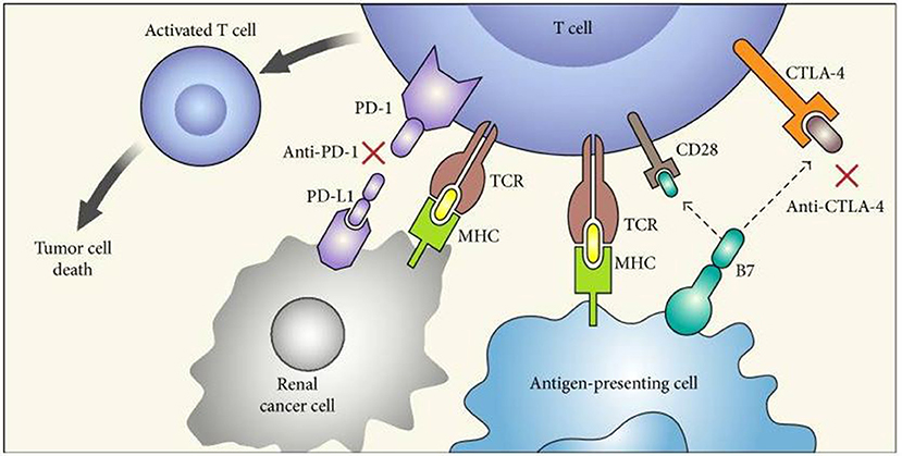 Figure 1 Mechanism of action of immune checkpoint inhibitors. (Lopez-Beltran, 2018)
Representative Immune Checkpoint Molecules
PD-1
Programmed cell death protein 1 (PD-1; encoded by the PDCD1 gene) is a cell surface receptor belonging to the CD28 family, crucial in regulating the immune system's response by maintaining the balance between T cell activation, tolerance, and immunopathology. Expressed on T cells, B cells, and myeloid cells, PD-1 plays a pivotal role in the inhibition of immune responses, acting as a checkpoint that prevents autoimmune reactions when bound to its ligands, PD-L1 and PD-L2, which are found on various cells, including some cancer cells. This interaction leads to the downregulation of T cell effector functions, promoting self-tolerance and minimizing tissue damage during inflammatory responses. However, the PD-1 pathway is also exploited by tumors to evade the immune system by expressing PD-L1, thereby inhibiting the cytotoxic T cell activity against cancer cells. Consequently, targeting the PD-1/PD-L1 axis has emerged as a revolutionary approach in cancer immunotherapy, aiming to enhance the immune system's ability to recognize and destroy cancer cells. By blocking PD-1 or PD-L1, these therapies can restore the immune system's ability to target cancer cells, leading to significant clinical benefits across a variety of cancer types.
Recommended Human Anti-PD-1 mAb (CAT#: TAB-H55)
 Figure 2 Sensorgram fit of human PD-1 analyte-binding anti-PD-1 antibody ligand (Cat# TAB-H55). Dilute the antibody ligand (5 μg/mL) and antigen analyte with running buffer. The ligand was injected to sample channel (Fc2) at a flow rate of 10 μL/min to reach a capture level of about 400 RU. The analyte was injected to Fc1-Fc2 of the channel at a flow rate of 30 μL/min for an association phase of the corresponding time. The association and dissociation process were all handling in the running buffer. Repeat 6 cycles of analyte according to analyte concentrations in ascending order.
Recommended Human Anti-CD19 mAb (CAT#: HPAB-0268-CN)
 Figure 3 Immunohistochemical analysis of paraffin-embedded tonsils. 1. The antibody is diluted 1:200 (overnight at 4°C). 2. Citric acid, pH 6.0 is used for antigen retrieval. 3. Dilute the secondary antibody at 1:200 (room temperature, 30min).
Recommended Human Anti-PD-1 mAb (CAT#: TAB-770)
 Figure 4 ELISA analysis of anti-PD-1 antibody (Cat# TAB-770) was performed by coating with recombinant human PD-1 protein (His tag). Then blocked with BSA and incubated with anti-PD-1 antibodies. The HRP-conjugated goat anti-human IgG was used as a secondary antibody. Detection was performed using TMB substrate and stopped with sulfuric acid. The absorbances were read on a spectrophotometer at 450 nm..
CD40
CD40 is a crucial costimulatory protein found on the surface of antigen-presenting cells (APCs) such as B cells, dendritic cells, and macrophages, playing a vital role in the immune system's regulation. It is a member of the tumor necrosis factor (TNF) receptor superfamily, recognized for its significant function in mediating B-cell proliferation, differentiation, and immunoglobulin class switching. Upon binding with its ligand, CD154 (CD40L), primarily expressed on activated T-helper cells, CD40 triggers a series of intracellular signaling cascades that enhance the immune response. This interaction is essential for the formation of memory B cells and the generation of a high-affinity, isotype-switched antibody response, pivotal for effective humoral immunity. Moreover, CD40-CD40L engagement is crucial for the activation of APCs, leading to the upregulation of co-stimulatory molecules and cytokine production, thereby promoting T-cell activation and differentiation. Dysregulation of CD40 signaling pathways can contribute to various autoimmune diseases, chronic inflammatory conditions, and cancer, highlighting its importance in maintaining immune homeostasis and serving as a potential therapeutic target in immunotherapy. Through its multifaceted roles, CD40 acts as a bridge between innate and adaptive immunity, orchestrating a coordinated and robust immune defense.
SIRPA
Signal regulatory protein alpha (SIRPA) is a transmembrane glycoprotein that plays a pivotal role in the immune system's regulation, particularly in the modulation of immune responses and maintenance of immune homeostasis. It is primarily expressed in neurons, dendritic cells, macrophages, and other immune cells. SIRPA functions by binding to its ligand, CD47, a ubiquitously expressed protein known as the "don't eat me" signal, which inhibits phagocytosis and thus prevents the immune system from attacking self-cells. This interaction is crucial for protecting healthy cells during immune surveillance and for the clearance of aged or damaged cells without inducing an inflammatory response. Furthermore, SIRPA signaling is involved in the negative regulation of receptor tyrosine kinases, which impacts cell adhesion, migration, and the cytoskeletal organization. Its role extends beyond immunological activities to include contributions to neuronal development, synaptic plasticity, and the regulation of inflammation. The dysregulation of SIRPA signaling pathways has been implicated in various diseases, including cancer, where tumor cells overexpress CD47 to evade immune detection, as well as in neurodegenerative disorders and inflammatory conditions.
Full List of Immune Checkpoint Molecules
| Biomarker | Alternative Names | Gene ID | UniProt ID | Roles |
| ADORA2A | Adenosine A2a Receptor; ADORA2; Adenosine Receptor Subtype A2a; Adenosine Receptor A2a; A2aR; RDC8 | 135 | P29274 | Adenosine A2a Receptor is a member of the guanine nucleotide-binding protein (G protein)-coupled receptor (GPCR) superfamily, which is subdivided into classes and subtypes. The receptors are seven-pass transmembrane proteins that respond to extracellular cues and activate intracellular signal transduction pathways. This protein, an adenosine receptor of A2A subtype, uses adenosine as the preferred endogenous agonist and preferentially interacts with the G(s) and G(olf) family of G proteins to increase intracellular cAMP levels. It plays an important role in many biological functions, such as cardiac rhythm and circulation, cerebral and renal blood flow, immune function, pain regulation, and sleep. It has been implicated in pathophysiological conditions such as inflammatory diseases and neurodegenerative disorders. Alternative splicing results in multiple transcript variants. |
| B7-H3 | B7-H3; CD276; B7 homolog 3; B7H3 | 80381 | Q5ZPR3 | The protein encoded by this gene belongs to the immunoglobulin superfamily, and thought to participate in the regulation of T-cell-mediated immune response. Studies show that while the transcript of this gene is ubiquitously expressed in normal tissues and solid tumors, the protein is preferentially expressed only in tumor tissues. Additionally, it was observed that the 3 UTR of this transcript contains a target site for miR29 microRNA, and there is an inverse correlation between the expression of this protein and miR29 levels, suggesting regulation of expression of this gene product by miR29. Alternatively spliced transcript variants encoding different isoforms have been found for this gene. |
| B7-H4 | B7-H4; B7h.5; B7H4; B7S1; B7X; PRO1291; VCTN1 | 79679 | Q7Z7D3 | This gene encodes a protein belonging to the B7 costimulatory protein family. Proteins in this family are present on the surface of antigen-presenting cells and interact with ligand bound to receptors on the surface of T cells. Studies have shown that high levels of the encoded protein has been correlated with tumor progression. A pseudogene of this gene is located on chromosome 20. Multiple transcript variants encoding different isoforms have been found for this gene. |
| BTLA | B And T Lymphocyte Associated; B- And T-Lymphocyte-Associated Protein; B- And T-Lymphocyte Attenuator; CD272 Antigen; BTLA1; CD272 | 151888 | Q7Z6A9 | This gene encodes a member of the immunoglobulin superfamily. The encoded protein contains a single immunoglobulin (Ig) domain and is a receptor that relays inhibitory signals to suppress the immune response. Alternative splicing results in multiple transcript variants. Polymorphisms in this gene have been associated with an increased risk of rheumatoid arthritis. [provided by RefSeq, Aug 2011] |
| CD155 | PVR; FLJ25946; PVS; CD155; TAGE4; HVED; NECL5 | 5817 | Q8K094 | The protein encoded by this gene is a transmembrane glycoprotein belonging to the immunoglobulin superfamily. The external domain mediates cell attachment to the extracellular matrix molecule vitronectin, while its intracellular domain interacts with the dynein light chain Tctex-1/DYNLT1. The gene is specific to the primate lineage, and serves as a cellular receptor for poliovirus in the first step of poliovirus replication. Multiple transcript variants encoding different isoforms have been found for this gene. |
| CD160 | BY55; NK1; NK28 | 11126 | O95971 | CD160 is an 27 kDa glycoprotein which was initially identified with the monoclonal antibody BY55. Its expression is tightly associated with peripheral blood NK cells and CD8 T lymphocytes with cytolytic effector activity. The cDNA sequence of CD160 predicts a cysteine-rich, glycosylphosphatidylinositol-anchored protein of 181 amino acids with a single Ig-like domain weakly homologous to KIR2DL4 molecule. CD160 is expressed at the cell surface as a tightly disulfide-linked multimer. RNA blot analysis revealed CD160 mRNAs of 1.5 and 1.6 kb whose expression was highly restricted to circulating NK and T cells, spleen and small intestine. Within NK cells CD160 is expressed by CD56dimCD16+ cells whereas among circulating T cells its expression is mainly restricted to TCRgd bearing cells and to TCRab+CD8brightCD95+CD56+CD28-CD27-cells. In tissues, CD160 is expressed on all intestinal intraepithelial lymphocytes. CD160 shows a broad specificity for binding to both classical and nonclassical MHC class I molecules. |
| CD200 | CD200; MOX1; MOX2; MRC; OX-2; My033 | 4345 | P41217 | This gene encodes a type I membrane glycoprotein containing two extracellular immunoglobulin domains, a transmembrane and a cytoplasmic domain. This gene is expressed by various cell types, including B cells, a subset of T cells, thymocytes, endothelial cells, and neurons. The encoded protein plays an important role in immunosuppression and regulation of anti-tumor activity. Alternative splicing results in multiple transcript variants encoding different isoforms. |
| Cd226 | CD226 Molecule; CD226 Antigen; DNAM-1; DNAM1; T Lineage-Specific Activation Antigen 1 Antigen; Platelet And T Cell Activation Antigen 1 | 10666 | Q15762 | CD226 (CD226 Molecule) is a Protein Coding gene. Diseases associated with CD226 include Ovarian Cystic Teratoma and Mature Teratoma Of The Ovary. Among its related pathways are Hematopoietic Stem Cell Differentiation Pathways and Lineage-specific Markers and Innate Immune System. Gene Ontology (GO) annotations related to this gene include protein kinase binding and cell adhesion molecule binding. |
| CD244 | CD244; 2B4; SLAMF4; NKR2B4; NAIL; h2B4 | 51744 | Q9BZW8 | This gene encodes a cell surface receptor expressed on natural killer (NK) cells (and some T cells) that mediate non-major histocompatibility complex (MHC) restricted killing. The interaction between NK-cell and target cells via this receptor is thought to modulate NK-cell cytolytic activity. Alternatively spliced transcript variants encoding different isoforms have been found for this gene. |
| CD27 | CD27; TNFRSF7; S152; T14; Tp55 | 939 | P26842 | The protein encoded by this gene is a member of the TNF-receptor superfamily. This receptor is required for generation and long-term maintenance of T cell immunity. It binds to ligand CD70, and plays a key role in regulating B-cell activation and immunoglobulin synthesis. This receptor transduces signals that lead to the activation of NF-kappaB and MAPK8/JNK. Adaptor proteins TRAF2 and TRAF5 have been shown to mediate the signaling process of this receptor. CD27-binding protein (SIVA), a proapoptotic protein, can bind to this receptor and is thought to play an important role in the apoptosis induced by this receptor. |
| Cd274 | CD274 Molecule; CD274 Antigen; B7 Homolog 1; Programmed Cell Death 1 Ligand 1; PDCD1 Ligand 1; PDCD1LG1; PDCD1L1 | 29126 | Q9NZQ7 | CD274 is a gene also known as PD-L1 (Programmed Death-Ligand 1). It is an immune checkpoint protein that is involved in regulating the function of the immune system. CD274/PD-L1 inhibits the activation of T cells by binding to its receptor PD-1, thereby preventing excessive immune responses. |
| CD28 | CD28; Tp44 | 940 | P31041 | The protein encoded by this gene is essential for T-cell proliferation and survival, cytokine production, and T-helper type-2 development. Several alternatively spliced transcript variants encoding different isoforms have been found for this gene. |
| CD40 | CD40; Bp50; CDW40; MGC9013; TNFRSF5; p50 | 958 | P25942 | This gene is a member of the TNF-receptor superfamily. The encoded protein is a receptor on antigen-presenting cells of the immune system and is essential for mediating a broad variety of immune and inflammatory responses including T cell-dependent immunoglobulin class switching, memory B cell development, and germinal center formation. AT-hook transcription factor AKNA is reported to coordinately regulate the expression of this receptor and its ligand, which may be important for homotypic cell interactions. Adaptor protein TNFR2 interacts with this receptor and serves as a mediator of the signal transduction. The interaction of this receptor and its ligand is found to be necessary for amyloid-beta-induced microglial activation, and thus is thought to be an early event in Alzheimer disease pathogenesis. Mutations affecting this gene are the cause of autosomal recessive hyper-IgM immunodeficiency type 3 (HIGM3). Multiple alternatively spliced transcript variants of this gene encoding distinct isoforms have been reported. |
| CD40L | CD40 Ligand; CD40L; CD154; T-BAM; TRAP; HIGM1 | CD40LG (CD40 Ligand) is a Protein Coding gene. Diseases associated with CD40LG include Immunodeficiency With Hyper-Igm, Type 1 and Toxoplasmosis. Among its related pathways are Photodynamic therapy-induced NF-kB survival signaling and B Cell Development Pathways. Gene Ontology (GO) annotations related to this gene include cytokine activity and CD40 receptor binding. An important paralog of this gene is TNFSF15. | ||
| CD47 | CD47; MER6; IAP; OA3 | 961 | Q08722 | Leukocyte surface antigen CD47 is also known as Antigenic surface determinant protein OA3, Integrin-associated protein (IAP) and Protein MER6. CD47 contains 1 Ig-like V-type (immunoglobulin-like) domain. CD47 is very broadly distributed on normal adult tissues. CD47 has a role in both cell adhesion by acting as an adhesion receptor for THBS1 on platelets, and in the modulation of integrins and plays an important role in memory formation and synaptic plasticity in the hippocampus by similarity. CD47 is the receptor for SIRPA, binding to which prevents maturation of immature dendritic cells and inhibits cytokine production by mature dendritic cells. CD47 Interaction with SIRPG mediates cell-cell adhesion, enhances superantigen-dependent T-cell-mediated proliferation and costimulates T-cell activation. |
| CD48 | CD48; BCM1; SLAMF2; BLAST; BLAST1; MEM-102; TCT.1; BCM-1; SLAMF-2; BLAST-1 | 962 | P09326 | This gene encodes a member of the CD2 subfamily of immunoglobulin-like receptors which includes SLAM (signaling lymphocyte activation molecules) proteins. The encoded protein is found on the surface of lymphocytes and other immune cells, dendritic cells and endothelial cells, and participates in activation and differentiation pathways in these cells. The encoded protein does not have a transmembrane domain, however, but is held at the cell surface by a GPI anchor via a C-terminal domain which maybe cleaved to yield a soluble form of the receptor. Multiple transcript variants encoding different isoforms have been found for this gene. |
| CD70 | CD70; CD27LG; TNFSF7; TNFSF7G; CD27L | 970 | P32970 | The protein encoded by this gene is a cytokine that belongs to the tumor necrosis factor (TNF) ligand family. This cytokine is a ligand for TNFRSF27/CD27. It is a surface antigen on activated, but not on resting, T and B lymphocytes. It induces proliferation of costimulated T cells, enhances the generation of cytolytic T cells, and contributes to T cell activation. This cytokine is also reported to play a role in regulating B-cell activation, cytotoxic function of natural killer cells, and immunoglobulin sythesis. |
| CD80 | B7; BB1; B7-1; B7.1; LAB7; CD28LG; CD28LG1 | 941 | P33681 | The protein encoded by this gene is a membrane receptor that is activated by the binding of CD28 or CTLA-4. The activated protein induces T-cell proliferation and cytokine production. |
| CD86 | B70; B7-2; B7.2; LAB72; CD28LG2 | 942 | P42081 | This gene encodes a type I membrane protein that is a member of the immunoglobulin superfamily. This protein is expressed by antigen-presenting cells, and it is the ligand for two proteins at the cell surface of T cells, CD28 antigen and cytotoxic T-lymphocyte-associated protein 4. Binding of this protein with CD28 antigen is a costimulatory signal for activation of the T-cell. Binding of this protein with cytotoxic T-lymphocyte-associated protein 4 negatively regulates T-cell activation and diminishes the immune response. Alternative splicing results in several transcript variants encoding different isoforms. |
| CD96 | TACTILE | 10225 | P40200 | The protein encoded by this gene belongs to the immunoglobulin superfamily. It is a type I membrane protein. The protein may play a role in the adhesive interactions of activated T and NK cells during the late phase of the immune response. It may also function in antigen presentation. Alternative splicing generates multiple transcript variants encoding distinct isoforms. |
| CEACAM1 | CEACAM1; carcinoembryonic antigen-related cell adhesion molecule 1 (biliary glycoprotein); BGP; BGP1; BGPI; carcinoembryonic antigen-related cell adhesion molecule 1; antigen CD66; CD66a antigen | 634 | P13688 | This gene encodes a member of the carcinoembryonic antigen (CEA) gene family, which belongs to the immunoglobulin superfamily. Two subgroups of the CEA family, the CEA cell adhesion molecules and the pregnancy-specific glycoproteins, are located within a 1.2 Mb cluster on the long arm of chromosome 19. Eleven pseudogenes of the CEA cell adhesion molecule subgroup are also found in the cluster. The encoded protein was originally described in bile ducts of liver as biliary glycoprotein. Subsequently, it was found to be a cell-cell adhesion molecule detected on leukocytes, epithelia, and endothelia. The encoded protein mediates cell adhesion via homophilic as well as heterophilic binding to other proteins of the subgroup. Multiple cellular activities have been attributed to the encoded protein, including roles in the differentiation and arrangement of tissue three-dimensional structure, angiogenesis, apoptosis, tumor suppression, metastasis, and the modulation of innate and adaptive immune responses. Multiple transcript variants encoding different isoforms have been reported, but the full-length nature of all variants has not been defined. |
| HHLA2 | HHLA2; HERV-H LTR-associating 2; B7H7; B7-H7 | 11148 | Q9UM44 | The antibodies capable of immunospecifically binding to the B7-H7 counter-receptor, H7CR are used in enhancing immune responses and the treatment and diagnosis of cancer and other diseases. |
| HVEM | ATAR; CD270; HVEA; HVEM; LIGHTR; TR2 | 8764 | Q71F55 | This gene encodes a member of the TNF (tumor necrosis factor) receptor superfamily. The encoded protein functions in signal transduction pathways that activate inflammatory and inhibitory T-cell immune response. It binds herpes simplex virus (HSV) viral envelope glycoprotein D (gD), mediating its entry into cells. Alternative splicing results in multiple transcript variants. |
| ICOS | AILIM; CD278; CVID1 | 29851 | Q9Y6W8 | The protein encoded by this gene belongs to the CD28 and CTLA-4 cell-surface receptor family. It forms homodimers and plays an important role in cell-cell signaling, immune responses, and regulation of cell proliferation. |
| Icosl | Icosl; icos ligand; B7h; GI50; GL50; B7-H2; LICOS; B7RP-1; GL50-B; ICOS-L; Icoslg; Ly115l; AU044799; BG071784; mKIAA0653 | 50723 | Q9JHJ8 | Ligand for the T-cell-specific cell surface receptor ICOS. Acts as a costimulatory signal for T-cell proliferation and cytokine secretion; induces also B-cell proliferation and differentiation into plasma cells. Could play an important role in mediating local tissue responses to inflammatory conditions, as well as in modulating the secondary immune response by co-stimulating memory T-cell function. |
| LAG3 | LAG3, CD223, lymphocyte activating 3; REGN3767 | 3902 | P18627 | The LAG3 gene contains 8 exons. The sequence data, exon/intron organization, and chromosomal localization all indicate a close relationship of LAG3 to CD4. The gene for LAG-3 lies adjacent to the gene for CD4 on human chromosome 12 (12p13) and is approximately 20% identical to the CD4 gene. |
| LGALS9 | Galectin-9; LGALS9; Ecalectin; Gal-9 | 3965 | O00182 | The galectins are a family of beta-galactoside-binding proteins implicated in modulating cell-cell and cell-matrix interactions. The protein encoded by this gene is an S-type lectin. It is overexpressed in Hodgkins disease tissue and might participate in the interaction between the H&RS cells with their surrounding cells and might thus play a role in the pathogenesis of this disease and/or its associated immunodeficiency. Multiple alternatively spliced transcript variants have been found for this gene. |
| LIGHT | TNFSF14; CD258; HVEML; LIGHT; LTg | 8740 | O43557 | The protein encoded by this gene is a member of the tumor necrosis factor (TNF) ligand family. This protein is a ligand for TNFRSF14, which is a member of the tumor necrosis factor receptor superfamily, and which is also known as a herpesvirus entry mediator (HVEM). This protein may function as a costimulatory factor for the activation of lymphoid cells and as a deterrent to infection by herpesvirus. This protein has been shown to stimulate the proliferation of T cells, and trigger apoptosis of various tumor cells. This protein is also reported to prevent tumor necrosis factor alpha mediated apoptosis in primary hepatocyte. Two alternatively spliced transcript variant encoding distinct isoforms have been reported. |
| OX40 | OX40L; TNFSF4; CD252; Glycoprotein Gp34; TXGP1; CD134 ligand; CD134L | 7293 | P43489 | This gene encodes a cytokine of the tumor necrosis factor (TNF) ligand family. The encoded protein functions in T cell antigen-presenting cell (APC) interactions and mediates adhesion of activated T cells to endothelial cells. Polymorphisms in this gene have been associated with Sjogrens syndrome and systemic lupus erythematosus. Alternative splicing results in multiple transcript variants |
| PDCD1 | PDCD1; PD1; CD279; SLEB2 | 5133 | Q15116 | This protein is expressed in pro-B-cells and is thought to play a role in their differentiation. In mice, expression of this gene is induced in the thymus when anti-CD3 antibodies are injected and large numbers of thymocytes undergo apoptosis. Mice deficient for this gene bred on a BALB/c background developed dilated cardiomyopathy and died from congestive heart failure. These studies suggest that this gene product may also be important in T cell function and contribute to the prevention of autoimmune diseases. |
| Pdcd1lg2 | B7DC; bA574F11.2; Btdc; CD273; PD-L2; PDCD1L2; PDL2 | 58205 | Q3U304 | Involved in the costimulatory signal, essential for T-cell proliferation and IFNG production in a PDCD1-independent manner. Interaction with PDCD1 inhibits T-cell proliferation by blocking cell cycle progression and cytokine production (By similarity). |
| PVR | PVR; PVS; Poliovirus receptor; Nectin-like protein 5; NECL-5; CD155 | 5817 | P15151 | CD155 (cluster of differentiation 155), also known as the poliovirus receptor, is a protein that in humans is encoded by the PVR gene. |
| Sirpa | Signal Regulatory Protein Alpha; CD172 Antigen-Like Family Member A; Inhibitory Receptor SHPS-1; Macrophage Fusion Receptor; PTPNS1; SHPS1; SIRP; P84; BIT; MFR; Brain-Immunoglobulin-Like Molecule With Tyrosine-Based Activation Motifs; Brain Ig-Like Molecule With Tyrosine-Based Activation Motifs; Protein Tyrosine Phosphatase, Non-Receptor Type Substrate 1; Tyrosine-Protein Phosphatase Non-Receptor Type Substrate 1; Tyrosine Phosphatase SHP Substrate 1; Signal-Regulatory Protein Alpha-1 | 19261 | P97797 | The protein encoded by this gene is a member of the signal-regulatory-protein (SIRP) family, and also belongs to the immunoglobulin superfamily. SIRP family members are receptor-type transmembrane glycoproteins known to be involved in the negative regulation of receptor tyrosine kinase-coupled signaling processes. This protein can be phosphorylated by tyrosine kinases. The phospho-tyrosine residues of this PTP have been shown to recruit SH2 domain containing tyrosine phosphatases (PTP), and serve as substrates of PTPs. This protein was found to participate in signal transduction mediated by various growth factor receptors. CD47 has been demonstrated to be a ligand for this receptor protein. This gene and its product share very high similarity with several other members of the SIRP family. These related genes are located in close proximity to each other on chromosome 20p13. Multiple alternatively spliced transcript variants have been determined for this gene. [provided by RefSeq, Jul 2008] |
| TIGIT | TIGIT; VSIG9; VSTM3 | 100043314 | P86176 | This gene encodes a member of the PVR (poliovirus receptor) family of immunoglobin proteins. The product of this gene is expressed on several classes of T cells including follicular B helper T cells (TFH). The protein has been shown to bind PVR with high affinity; this binding is thought to assist interactions between TFH and dendritic cells to regulate T cell dependent B cell responses. |
| Tim-3 | T-cell immunoglobulin and mucin domain containing molecule-3; TIM3 | Q8VIM0 | T cell immunoglobulin and mucin-domain-containing molecule-3 (Tim-3) has been shown to influence autoimmune diseases; however, its function in viral infection has not been well-defined. | |
| TMIGD2 | TMIGD2; Immunoglobulin-Containing And Proline-Rich Receptor 1; CD28 Homolog; Transmembrane And Immunoglobulin Domain Containing 2; Transmembrane And Immunoglobulin Domain-Containing Protein 2 Variant 3; IGPR-1; CD28H; Transmembrane And Immunoglobulin Doma | 126259 | Q96BF3 | Plays a role in cell-cell interaction, cell migration, and angiogenesis. Through interaction with HHLA2, costimulates T-cells in the context of TCR-mediated activation. Enhances T-cell proliferation and cytokine production via an AKT-dependent signaling cascade. |
| TNFRSF18 | AITR; GITR; TNFRSF18; CD357 | 8784 | Q9Y5U5 | This gene encodes a member of the TNF-receptor superfamily. The encoded receptor has been shown to have increased expression upon T-cell activation, and it is thought to play a key role in dominant immunological self-tolerance maintained by CD25(+)CD4(+) regulatory T cells. Knockout studies in mice also suggest the role of this receptor is in the regulation of CD3-driven T-cell activation and programmed cell death. Three alternatively spliced transcript variants of this gene encoding distinct isoforms have been reported. |
| TNFRSF25 | TNF Receptor Superfamily Member 25; Tumor Necrosis Factor Receptor Superfamily, Member 12 (Translocating Chain-Association Membrane Protein); Lymphocyte-Associated Receptor Of Death; Apoptosis-Mediating Receptor TRAMP; Apoptosis-Mediating Receptor DR3; Apoptosis-Inducing Receptor AIR; Protein WSL-1; TNFRSF12; APO-3; DDR3; LARD; DR3; Tumor Necrosis Factor Receptor Superfamily, Member 25; Tumor Necrosis Factor Receptor Superfamily Member 25 | 8718 | Q93038 | The protein encoded by this gene is a member of the TNF-receptor superfamily. This receptor is expressed preferentially in the tissues enriched in lymphocytes, and it may play a role in regulating lymphocyte homeostasis. This receptor has been shown to stimulate NF-kappa B activity and regulate cell apoptosis. The signal transduction of this receptor is mediated by various death domain containing adaptor proteins. Knockout studies in mice suggested the role of this gene in the removal of self-reactive T cells in the thymus. Multiple alternatively spliced transcript variants of this gene encoding distinct isoforms have been reported, most of which are potentially secreted molecules. The alternative splicing of this gene in B and T cells encounters a programmed change upon T-cell activation, which predominantly produces full-length, membrane bound isoforms, and is thought to be involved in controlling lymphocyte proliferation induced by T-cell activation. |
| TNFRSF9 | TNFRSF9; 4-1BB; CD137; CDw137; ILA | 3604 | Q07011 | The protein encoded by this gene is a member of the TNF-receptor superfamily. This receptor contributes to the clonal expansion, survival, and development of T cells. It can also induce proliferation in peripheral monocytes, enhance T cell apoptosis induced by TCR/CD3 triggered activation, and regulate CD28 co-stimulation to promote Th1 cell responses. The expression of this receptor is induced by lymphocyte activation. TRAF adaptor proteins have been shown to bind to this receptor and transduce the signals leading to activation of NF-kappaB. |
| TNFSF18 | TNFSF18; AITRL; TL6; hGITRL; GITR Ligand | 8995 | Q9UNG2 | The protein encoded by this gene is a cytokine that belongs to the tumor necrosis factor (TNF) ligand family. This cytokine is a ligand for receptor TNFRSF18/AITR/GITR. It has been shown to modulate T lymphocyte survival in peripheral tissues. This cytokine is also found to be expressed in endothelial cells, and is thought to be important for interaction between T lymphocytes and endothelial cells. |
| TNFSF4 | CD134L; CD252; GP34; OX-40L; OX4OL; TNLG2B; TXGP1 | 7292 | P23510 | This gene encodes a cytokine of the tumor necrosis factor (TNF) ligand family. The encoded protein functions in T cell antigen-presenting cell (APC) interactions and mediates adhesion of activated T cells to endothelial cells. Polymorphisms in this gene have been associated with Sjogren's syndrome and systemic lupus erythematosus. Alternative splicing results in multiple transcript variants. [provided by RefSeq, Jul 2014] |
| TNFSF9 | 4-1BB Ligand; TNFSF9; CD137L | 8744 | P41273 | The protein encoded by this gene is a cytokine that belongs to the tumor necrosis factor (TNF) ligand family. This transmembrane cytokine is a bidirectional signal transducer that acts as a ligand for TNFRSF9/4-1BB, which is a costimulatory receptor molecule in T lymphocytes. This cytokine and its receptor are involved in the antigen presentation process and in the generation of cytotoxic T cells. The receptor TNFRSF9/4-1BB is absent from resting T lymphocytes but rapidly expressed upon antigenic stimulation. The ligand encoded by this gene, TNFSF9/4-1BBL, has been shown to reactivate anergic T lymphocytes in addition to promoting T lymphocyte proliferation. This cytokine has also been shown to be required for the optimal CD8 responses in CD8 T cells. This cytokine is expressed in carcinoma cell lines, and is thought to be involved in T cell-tumor cell interaction. |
Tested Data-Supported Products Targeting Immune Checkpoint Molecules


-YJ1.png)


-1.png)


-1.png)


Reference
- Lopez-Beltran, Antonio, et al. "The identification of immunological biomarkers in kidney cancers." Frontiers in Oncology 8 (2018): 456.
For research use only. Not intended for any clinical use.
Send Inquiry
This site is protected by reCAPTCHA and the Google Privacy Policy and Terms of Service apply.