


Oncoprotein-Apoptosis Regulation
Representative Oncoproteins in Apoptosis Regulation Full List of Oncoproteins in Apoptosis Regulation Tested Data-Supported Products for Oncoproteins in Apoptosis Regulation
Apoptosis, often described as programmed cell death, is a fundamental process vital for the maintenance of cellular homeostasis and development in multicellular organisms. It is characterized by a series of biochemical events leading to cell morphological changes and death, playing a critical role in removing damaged, diseased, or unwanted cells without causing harm to the surrounding tissue. On the other hand, oncoproteins are a class of proteins that, when dysregulated, can drive the transformation of a normal cell into a cancerous one, largely by interfering with normal cellular processes including apoptosis. The regulation of apoptosis by oncoproteins is a complex interplay that involves inhibiting apoptotic pathways to promote cellular survival and proliferation, which is a hallmark of cancer progression.
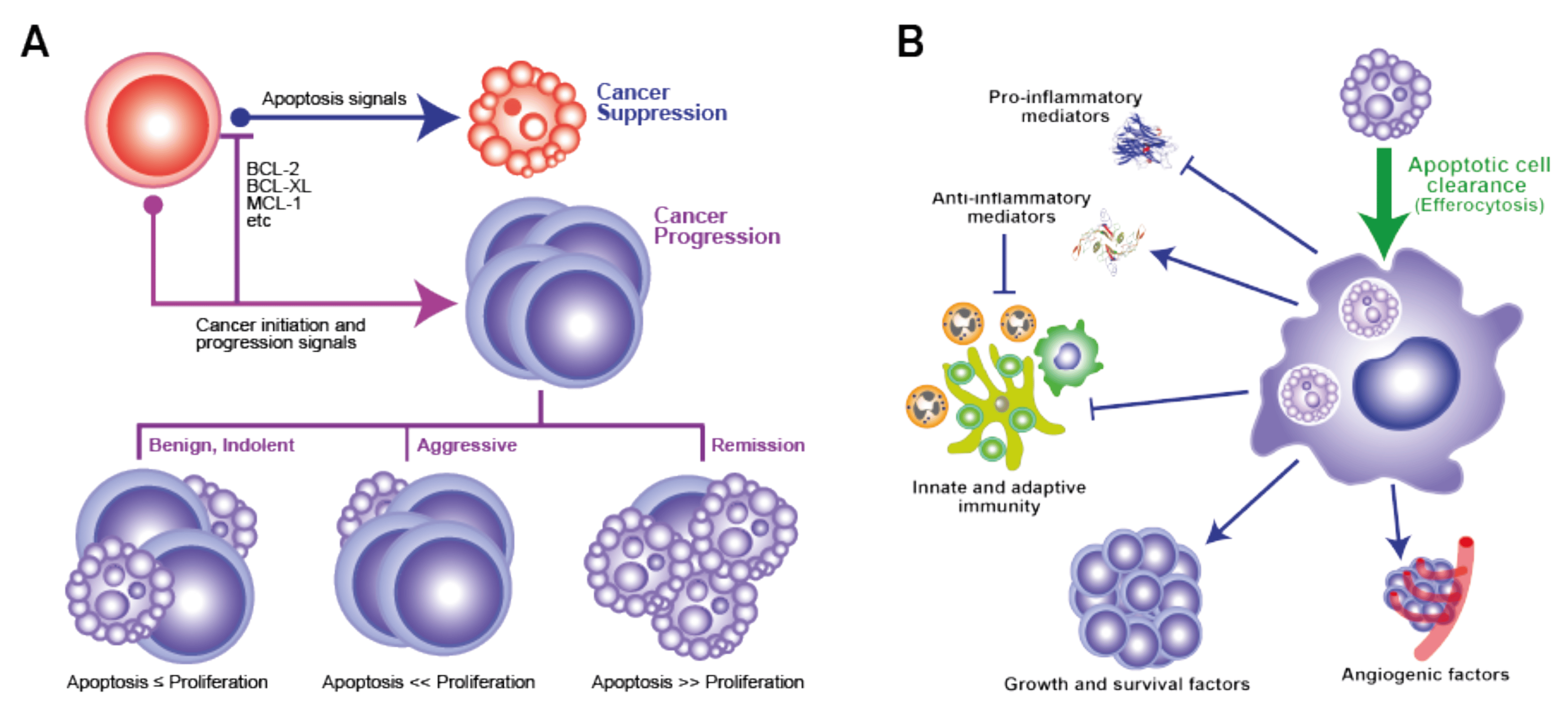 Figure 1 The ‘apoptosis paradox’ in malignant disease (Morana, 2022)
Representative Oncoproteins in Apoptosis Regulation
BCL-2
BCL-2 (B-cell lymphoma 2) is a pivotal protein in the regulation of apoptosis, the process of programmed cell death, which is essential for the maintenance of tissue homeostasis and the elimination of harmful cells in multicellular organisms. Originating from the BCL-2 gene, this protein is best known for its role as an anti-apoptotic factor, acting to inhibit the cell death pathway and thereby contributing to cell survival. Its function is intricately balanced with pro-apoptotic proteins, such as those from the BAX and BAK families, in a regulatory network that determines cell fate. The significance of BCL-2 extends beyond normal physiological processes; its overexpression has been implicated in the development of various cancers, particularly in the evasion of cancer cells from the immune system's surveillance mechanism, leading to uncontrolled cell proliferation. Therapeutically, targeting the BCL-2 protein has become a focal point in the development of novel cancer treatments, aimed at inducing apoptosis in tumor cells. This has led to the emergence of BCL-2 inhibitors as a class of drugs with promising potential in cancer therapy. Through its dual roles in maintaining cellular health and contributing to disease pathology, BCL-2 sits at the crossroads of vital pathways, making it a key subject of research in understanding and treating disease.
MDM2
The human MDM2 (Mouse Double Minute 2 homolog) protein plays a pivotal role in the regulation of the cell cycle and acts as a major negative regulator of the tumor suppressor p53, thus serving a critical function in the control of cell proliferation and apoptosis. Structurally, MDM2 is an E3 ubiquitin ligase that targets p53 for proteasomal degradation, effectively controlling the levels of this protein in cells under normal and stress conditions. Beyond its interaction with p53, MDM2 also engages with various other proteins and signaling pathways, influencing cellular processes such as DNA repair, transcription, and cell fate decisions. Its overexpression has been observed in a variety of human cancers, where it contributes to tumorigenesis by inhibiting p53 activity and thus promoting cell survival and proliferation. Conversely, the modulation of MDM2-p53 interaction offers a therapeutic avenue for cancer treatment, highlighting the importance of understanding MDM2's biology. Additionally, MDM2's roles extend to the regulation of apoptosis, senescence, and metabolism, underscoring its multifaceted contribution to cellular homeostasis and the complexity of its potential as a target in cancer therapy.
Recommended Rabbit Anti-MDM2 mAb (CAT#: VS3-FY919)
 Figure 2 Western Blot analysis of Phospho-MDM2 (Ser166) in Jurkat, C6, Hela lysates using Phospho-MDM2 (Ser166) Antibody.
Recommended Rabbit Anti-MDM2 mAb (CAT#: ZG-0396U)
 Figure 3 Immunohistochemical staining patterns of formalin fixed and paraffin embedded human liposarcoma tissue (4 μm sections) with anti-MDM2 antibody using Ventana BenchMark. The liposarcoma tissues show strong nuclear MDM2 expression.
MDM4
MDM4 (Mouse Double Minute 4), also known as MDMX or HDMX in humans, is a critical regulatory protein implicated in cell cycle control and apoptosis, functioning primarily through its interaction with the p53 tumor suppressor pathway. Structurally similar to MDM2, MDM4 does not possess intrinsic E3 ubiquitin ligase activity but significantly contributes to the inhibition of p53-mediated transcriptional activation and apoptosis by directly binding to p53. This interaction prevents p53 from activating its target genes involved in cell cycle arrest and apoptosis, effectively regulating cell proliferation and survival. Moreover, MDM4 plays a pivotal role in embryonic development and the response to DNA damage, underscoring its importance in maintaining genomic integrity. Research has elucidated that the overexpression of MDM4 can lead to enhanced tumor formation and progression, making it a potential target for cancer therapy. By modulating the p53 pathway, MDM4 influences various cellular processes, including DNA repair, cell cycle progression, and apoptosis, highlighting its crucial role in the balance between cell survival and death. Its complex interplay with MDM2 and p53 positions MDM4 at the heart of cellular growth control and the development of malignancies, demonstrating its significance in cancer biology and as a therapeutic target.
Recommended Mouse Anti-MDM4 mAb (CAT#: ZG-0275C)
 Figure 4 Confocal immunofluorescence analysis of Hela (left) and L-02 (right) cells using MDMX monoclonal antibody (green). Red: Actin filaments have been labeled with DY-554 phalloidin.
Full List of Oncoproteins in Apoptosis Regulation
| Biomarker | Alternative Names | Gene ID | UniProt ID | Roles |
| BCL-2 | BCL2, Apoptosis Regulator; Protein Phosphatase 1, Regulatory Subunit 50; B-Cell CLL/Lymphoma 2; Apoptosis Regulator Bcl-2; PPP1R50; Bcl-2 | 596 | P10415 | This gene encodes an integral outer mitochondrial membrane protein that blocks the apoptotic death of some cells such as lymphocytes. Constitutive expression of BCL2, such as in the case of translocation of BCL2 to Ig heavy chain locus, is thought to be the cause of follicular lymphoma. Alternative splicing results in multiple transcript variants. |
| MDM2 | MDM2 Proto-Oncogene; MDM2 Proto-Oncogene, E3 Ubiquitin Protein Ligase; Oncoprotein Mdm2; Hdm2; Mdm2, Transformed 3T3 Cell Double Minute 2, P53 Binding Protein (Mouse); Mdm2, Transformed 3T3 Cell Double Minute 2, P53 Binding Protein; Mouse Double Minute 2, Human Homolog Of; P53-Binding Protein; Double Minute 2, Human Homolog Of; P53-Binding Protein; Mdm2, P53 E3 Ubiquitin Protein Ligase Homolog; MDM2 Oncogene, E3 Ubiquitin Protein Ligase | 4193 | A7UKX8 | This gene encodes a nuclear-localized E3 ubiquitin ligase. The encoded protein can promote tumor formation by targeting tumor suppressor proteins, such as p53, for proteasomal degradation. This gene is itself transcriptionally-regulated by p53. Overexpression or amplification of this locus is detected in a variety of different cancers. There is a pseudogene for this gene on chromosome 2. Alternative splicing results in a multitude of transcript variants, many of which may be expressed only in tumor cells. [provided by RefSeq, Jun 2013] |
| MDM4 | HDMX; MDMX; MRP1 | 4194 | O15151 | This gene encodes a nuclear protein that contains a p53 binding domain at the N-terminus and a RING finger domain at the C-terminus, and shows structural similarity to p53-binding protein MDM2. Both proteins bind the p53 tumor suppressor protein and inhibit its activity, and have been shown to be overexpressed in a variety of human cancers. However, unlike MDM2 which degrades p53, this protein inhibits p53 by binding its transcriptional activation domain. This protein also interacts with MDM2 protein via the RING finger domain, and inhibits the latter's degradation. So this protein can reverse MDM2-targeted degradation of p53, while maintaining suppression of p53 transactivation and apoptotic functions. Alternatively spliced transcript variants encoding different isoforms have been noted for this gene. [provided by RefSeq, Feb 2011] |
Tested Data-Supported Products for Oncoproteins in Apoptosis Regulation
| CAT | Product Name | Biomarker | Assay | Image |
| ZG-0275C | Mouse Anti-MDM4 Recombinant Antibody (ZG-0275C) | MDM4 | WB |

|
| ZG-0396U | Rabbit Anti-MDM2 Recombinant Antibody (clone E22-L) | MDM2 | IHC-P |

|
| VS3-FY919 | Recombinant Rabbit Anti-MDM2 (phospho Ser166) Antibody (clone R08-8C7) | MDM2 | WB |

|
Reference
- Morana, Ornella, Will Wood, and Christopher D. Gregory. "The apoptosis paradox in cancer." International Journal of Molecular Sciences 23.3 (2022): 1328.
For research use only. Not intended for any clinical use.
Send Inquiry
This site is protected by reCAPTCHA and the Google Privacy Policy and Terms of Service apply.