Human Anti-IGF1R Recombinant Antibody (clone m590)
CAT#: FAMAB-0608WJ
This product is a chimeric (mouse/human) antibody targeting human IGF-1R. This antibody blocked the binding of IGF-I and IGF-II to IGF-IR, and inhibited both IGF-I and IGF-II induced phosphorylation of IGF-IR in MCF-7 cells. This antibody could be an useful antibody in diagnosis and treatment of cancer, as well as a research tool.
Published Data
Gene Expression

FC
Figure 1 Flow cytometry of breast cancer MCF-7 and ovarian cancer SKOV-3 cells stained with m590 and trastuzumab at 10 μg/mL.
Chen, C., Zhang, Y., Zhang, Y., Li, J., Tsao, S. W., & Zhang, M. Y. (2014). Superior Antitumor Activity of a Novel Bispecific Antibody Cotargeting Human Epidermal Growth Factor Receptor 2 and Type I Insulin-like Growth Factor ReceptorBispecific Antibody Cotargeting HER2 and IGF-IR. Molecular cancer therapeutics, 13(1), 90-100.

ELISA
Figure 2 Simultaneous binding of Bi-Ab to recombinant IGF-IR and HER2 ectodomains by indirect ELISA.
Recombinant IGF-IR ectodomain was coated in microplates. Three-fold serially diluted Bi-Ab, trastuzumab, and m590 were added to the wells followed by addition of 100 ng per well of biotinylated recombinant HER2 ectodomain. Bound HER2 ectodomain was detected by HRP conjugated to streptavidin and TMB.
Chen, C., Zhang, Y., Zhang, Y., Li, J., Tsao, S. W., & Zhang, M. Y. (2014). Superior Antitumor Activity of a Novel Bispecific Antibody Cotargeting Human Epidermal Growth Factor Receptor 2 and Type I Insulin-like Growth Factor ReceptorBispecific Antibody Cotargeting HER2 and IGF-IR. Molecular cancer therapeutics, 13(1), 90-100.

FC
Figure 3 Histogram overlay of SKOV-3 cells stained with m590, trastuzumab, and Bi-Ab at 2 and 10 μg/mL.
Chen, C., Zhang, Y., Zhang, Y., Li, J., Tsao, S. W., & Zhang, M. Y. (2014). Superior Antitumor Activity of a Novel Bispecific Antibody Cotargeting Human Epidermal Growth Factor Receptor 2 and Type I Insulin-like Growth Factor ReceptorBispecific Antibody Cotargeting HER2 and IGF-IR. Molecular cancer therapeutics, 13(1), 90-100.

FC
Figure 4 Binding kinetics of Bi-Ab, m590, and trastuzumab to membrane-associated IGF-IR and HER2 on SKOV-3 cells.
Mean fluorescence intensity (MFI) of SKOV-3 cells stained with the antibodies at different concentrations was measured.
Chen, C., Zhang, Y., Zhang, Y., Li, J., Tsao, S. W., & Zhang, M. Y. (2014). Superior Antitumor Activity of a Novel Bispecific Antibody Cotargeting Human Epidermal Growth Factor Receptor 2 and Type I Insulin-like Growth Factor ReceptorBispecific Antibody Cotargeting HER2 and IGF-IR. Molecular cancer therapeutics, 13(1), 90-100.

Inhib
Figure 5 A and B, inhibition of IGF-I (1.5 nmol/L) induced phosphorylation of Akt and ERK by m590 (40 nmol/L) alone (A) or in combination with trastuzumab (6.7 nmol/L; B) in MCF-7 cells.
Chen, C., Zhang, Y., Zhang, Y., Li, J., Tsao, S. W., & Zhang, M. Y. (2014). Superior Antitumor Activity of a Novel Bispecific Antibody Cotargeting Human Epidermal Growth Factor Receptor 2 and Type I Insulin-like Growth Factor ReceptorBispecific Antibody Cotargeting HER2 and IGF-IR. Molecular cancer therapeutics, 13(1), 90-100.

Inhib
Figure 6 Inhibition of IGF-I (1.5 nmol/L) induced phosphorylation of IGF-IR in SKOV-3 cells.
All antibodies were tested at 100 μg/mL and incubated with cells for 24 hours.
Chen, C., Zhang, Y., Zhang, Y., Li, J., Tsao, S. W., & Zhang, M. Y. (2014). Superior Antitumor Activity of a Novel Bispecific Antibody Cotargeting Human Epidermal Growth Factor Receptor 2 and Type I Insulin-like Growth Factor ReceptorBispecific Antibody Cotargeting HER2 and IGF-IR. Molecular cancer therapeutics, 13(1), 90-100.

Inhib
Figure 7 D and E, inhibition of IGF-I (1.5 nmol/L) induced phosphorylation of Akt and ERK in MCF-7 (D) and SKOV-3 (E) cells.
All antibodies were tested at 100 μg/mL and incubated with cells for 24 hours.
Chen, C., Zhang, Y., Zhang, Y., Li, J., Tsao, S. W., & Zhang, M. Y. (2014). Superior Antitumor Activity of a Novel Bispecific Antibody Cotargeting Human Epidermal Growth Factor Receptor 2 and Type I Insulin-like Growth Factor ReceptorBispecific Antibody Cotargeting HER2 and IGF-IR. Molecular cancer therapeutics, 13(1), 90-100.

Inhib
Figure 8 Inhibition of phosphorylation of Akt and ERK by the antibodies in SKOV-3 cells in the absence of IGF-I.
Chen, C., Zhang, Y., Zhang, Y., Li, J., Tsao, S. W., & Zhang, M. Y. (2014). Superior Antitumor Activity of a Novel Bispecific Antibody Cotargeting Human Epidermal Growth Factor Receptor 2 and Type I Insulin-like Growth Factor ReceptorBispecific Antibody Cotargeting HER2 and IGF-IR. Molecular cancer therapeutics, 13(1), 90-100.

Inhib
Figure 9 Dose-dependent inhibition of ERK phosphorylation, but not Akt phosphorylation by Bi-Ab in SKOV-3 cells in the absence of IGF-I. Bi-Ab: 200, 40, 8, 1.6, 0.32, and 0 μg/mL.
Chen, C., Zhang, Y., Zhang, Y., Li, J., Tsao, S. W., & Zhang, M. Y. (2014). Superior Antitumor Activity of a Novel Bispecific Antibody Cotargeting Human Epidermal Growth Factor Receptor 2 and Type I Insulin-like Growth Factor ReceptorBispecific Antibody Cotargeting HER2 and IGF-IR. Molecular cancer therapeutics, 13(1), 90-100.

Inhib
Figure 10 Inhibition of SKOV-3 cell proliferation in MTT assay.
Percentage of inhibition at each antibody concentration was used in one-way ANOVA statistical analysis to test whether there was significant difference between any two antibodies at the same concentration. Two paired antibodies with significant difference (*, P < 0.001) in percentage of inhibition are indicated.
Chen, C., Zhang, Y., Zhang, Y., Li, J., Tsao, S. W., & Zhang, M. Y. (2014). Superior Antitumor Activity of a Novel Bispecific Antibody Cotargeting Human Epidermal Growth Factor Receptor 2 and Type I Insulin-like Growth Factor ReceptorBispecific Antibody Cotargeting HER2 and IGF-IR. Molecular cancer therapeutics, 13(1), 90-100.

ADCC
Figure 11 Percentage of ADCC of the antibodies at 1 and 5 μg/mL.
The same ANOVA statistical analysis was done to test whether there was significant difference in percentage of ADCC between any two antibodies at the same concentration. Percentage of ADCC between Bi-Ab and Comb at 1 μg/mL showed significant difference (*, P < 0.05), which is indicated. Each antibody dilution was tested in triplicate and the assays were repeated once.
Chen, C., Zhang, Y., Zhang, Y., Li, J., Tsao, S. W., & Zhang, M. Y. (2014). Superior Antitumor Activity of a Novel Bispecific Antibody Cotargeting Human Epidermal Growth Factor Receptor 2 and Type I Insulin-like Growth Factor ReceptorBispecific Antibody Cotargeting HER2 and IGF-IR. Molecular cancer therapeutics, 13(1), 90-100.

FuncS
Figure 12 Diagram of the mouse study and tumor growth kinetics in each group of mice treated with or without antibodies (control).
A, diagram of the mouse study. Three million of SKOV-3-Luc cells were injected subcutaneously into each nude mouse on day 0. 100 μg of Bi-Ab, or m590, or trastuzumab, or Comb were injected intraperitoneally on day 1, 4, 6, and 8 postinoculations. Mouse imaging was performed on day 1, 4, 6, and 8 before antibody injections, and on day 11, 15, 25, and 35 postinoculations. Seven mice were included in each antibody-treated group, but only five mice were in the control group. B, average luminescence intensity in each group of mice at different time point. Logarithmic values of the average luminescence intensities and standard variations were shown.
Chen, C., Zhang, Y., Zhang, Y., Li, J., Tsao, S. W., & Zhang, M. Y. (2014). Superior Antitumor Activity of a Novel Bispecific Antibody Cotargeting Human Epidermal Growth Factor Receptor 2 and Type I Insulin-like Growth Factor ReceptorBispecific Antibody Cotargeting HER2 and IGF-IR. Molecular cancer therapeutics, 13(1), 90-100.

FuncS
Figure 13 Inhibition of cancer growth by Bi-Ab in SKOV-3 HER2- and IGF-IR-overexpressing xenograft mouse model in comparison with m590 and trastuzumab alone, or in combination (Comb).
A, average luminescence intensity in each group of mice at different time point. Logarithmic values of the average luminescence intensity were used in ANOVA statistical analysis to test whether there was significant difference between any two groups at the same time point. Two paired groups with significant difference (*, P < 0.001) are indicated. B, number of mice in each group with luminescence intensity 2-fold higher than the baseline level.
Chen, C., Zhang, Y., Zhang, Y., Li, J., Tsao, S. W., & Zhang, M. Y. (2014). Superior Antitumor Activity of a Novel Bispecific Antibody Cotargeting Human Epidermal Growth Factor Receptor 2 and Type I Insulin-like Growth Factor ReceptorBispecific Antibody Cotargeting HER2 and IGF-IR. Molecular cancer therapeutics, 13(1), 90-100.

ELISA
Figure 14 Binding of mouse IgG2b 4G11 and chimeric IgG1 m590 to non-denatured and denatured IGF-IR and IR ectodomains.
Both ectodomains were coated at 200 ng/well on Maxisorp plates. Denatured ectodomains were prepared as described in Materials and Methods. After blocking the plates with 3% BSA in PBS, three-fold serially diluted antibodies with a starting concentration of 20 µg/ml were added to the plates and bound antibodies were detected with anti-mouse IgG conjugated to HRP for 4G11 and anti-human Fc conjugated to HRP for m590.
Zhang, M. Y., Feng, Y., Wang, Y., & Dimitrov, D. S. (2009, September). Characterization of a chimeric monoclonal antibody against the insulin-like growth factor-I receptor. In MAbs (Vol. 1, No. 5, pp. 475-480). Taylor & Francis.

FC
Figure 15 Binding of human-mouse chimeric IgG1 m590, mouse IgG2b 4G11, anti-IGF-IR b subunit antibody GRII and control antibody anti-HIV-1 IgG1 X5 to NWT C43 cells and MCF-7 cells.
Ten 10 mg/ml of each antibody were incubated with stably transfected NWT C43 cells or breast cancer cell line MCF-7. Bound chimeric IgG1 m590 and control antibody IgG1 X5 were revealed by PE labeled anti-human IgG, F(ab')2, mouse IgG2b 4G11 and GRII by PE labeled anti-mouse polyclonal antibody.
Zhang, M. Y., Feng, Y., Wang, Y., & Dimitrov, D. S. (2009, September). Characterization of a chimeric monoclonal antibody against the insulin-like growth factor-I receptor. In MAbs (Vol. 1, No. 5, pp. 475-480). Taylor & Francis.

Inhib
Figure 16 Inhibition of IGF-I and IGF-II induced phosphorylation of IGF-IR by anti-IGF-IR human-mouse chimeric antibody IgG1 m590 and parental murine antibody IgG2b 4G11 in MCF-7 cells.
MCF-7 cells starved in serum-free medium were pre-incubated with indicated concentrations (in nM) of IgG1 m590 (Lanes 3 to 6) and IgG2b 4G11 (Lanes 7 to 9) for 30 min followed by addition of 1.5 nM IGF-I (left) or 10 nM IGF-II (right) for 20 min. Total IGF-IR was immunoprecipitated with rabbit anti-IGF-IR beta subunit pAb. Phosphorylated IGF-IR was detected with mAb 4G10 specific to phosphotyrosine (top gels). The blots were re-probed with rabbit anti-IGF-IR beta subunit pAb (bottom gels) to show the total IGF-IR protein among the samples. Cells treated with IGF-I or IGF-II alone (Lane 2 in both panels) were included as positive control.
Zhang, M. Y., Feng, Y., Wang, Y., & Dimitrov, D. S. (2009, September). Characterization of a chimeric monoclonal antibody against the insulin-like growth factor-I receptor. In MAbs (Vol. 1, No. 5, pp. 475-480). Taylor & Francis.
❮


 ❯
❯

Normal Tissue
Figure 1 Cerebral cortex
Endothelial cells
Staining: Medium
Intensity: Moderate
Quantity:>75%
Location: Cytoplasmic/membranous
Glial cells
Staining: Medium
Intensity: Moderate
Quantity:>75%
Location: Cytoplasmic/membranous
Neuronal cells
Staining: High
Intensity: Strong
Quantity:>75%
Location: Cytoplasmic/membranous
Neuropil
Staining: High
Intensity: Strong
Quantity:>75%
Location: Cytoplasmic/membranous
* Image credit: Image credit: Human Protein Atlas https://v21.proteinatlas.org/images/10268/32662_B_7_5.jpg

Normal Tissue
Figure 2 Colon
Endothelial cells
Staining: Medium
Intensity: Moderate
Quantity:>75%
Location: Cytoplasmic/membranous
Glandular cells
Staining: High
Intensity: Strong
Quantity:>75%
Location: Cytoplasmic/membranous
* Image credit: Image credit: Human Protein Atlas https://v21.proteinatlas.org/images/10268/32662_A_7_3.jpg

Normal Tissue
Figure 3 Liver
Cholangiocytes
Staining: Medium
Intensity: Moderate
Quantity:>75%
Location: Cytoplasmic/membranous
Hepatocytes
Staining: Medium
Intensity: Moderate
Quantity:>75%
Location: Cytoplasmic/membranous
* Image credit: Image credit: Human Protein Atlas https://v21.proteinatlas.org/images/10268/32662_A_8_4.jpg

Normal Tissue
Figure 4 Kidney
Cells in glomeruli
Staining: Medium
Intensity: Moderate
Quantity:>75%
Location: Cytoplasmic/membranous
Cells in tubules
Staining: High
Intensity: Strong
Quantity:>75%
Location: Cytoplasmic/membranous
* Image credit: Image credit: Human Protein Atlas https://v21.proteinatlas.org/images/10268/32662_A_7_5.jpg

Normal Tissue
Figure 5 Testis
Cells in seminiferous ducts
Staining: Medium
Intensity: Moderate
Quantity:>75%
Location: Cytoplasmic/membranous
Leydig cells
Staining: High
Intensity: Strong
Quantity:>75%
Location: Cytoplasmic/membranous
* Image credit: Image credit: Human Protein Atlas https://v21.proteinatlas.org/images/10268/32662_A_6_6.jpg

Normal Tissue
Figure 6 Heart muscle
Cardiomyocytes
Staining: High
Intensity: Strong
Quantity:>75%
Location: Cytoplasmic/membranous
* Image credit: Image credit: Human Protein Atlas https://v21.proteinatlas.org/images/10268/32662_B_5_6.jpg

Normal Tissue
Figure 7 Lymph node
Germinal center cells
Staining: High
Intensity: Strong
Quantity:>75%
Location: Cytoplasmic/membranous
Non-germinal center cells
Staining: High
Intensity: Strong
Quantity:>75%
Location: Cytoplasmic/membranous
* Image credit: Image credit: Human Protein Atlas https://v21.proteinatlas.org/images/10268/32662_A_7_8.jpg

RNA Expression
Figure 8 RNA cell line category: Low cell line specificity
Cell lines ordered by descending RNA expression order.
* Image credit: Image credit: Human Protein Atlas https://v21.proteinatlas.org/ENSG00000140443-IGF1R
❮


 ❯
❯
Specifications
- Host Species
- Human
- Derivation
- Chimeric(Mouse/Human)
- Type
- Human IgG1
- Specificity
- Human IGF-IR
- Species Reactivity
- Human
- Clone
- m590
- Applications
- ELISA, WB
- Related Disease
- Cancers
Product Property
- Purity
- >95% as determined by SDS-PAGE
- Concentration
- Please refer to the vial label for the specific concentration.
- Buffer
- PBS
- Preservative
- No preservatives
- Storage
- Centrifuge briefly prior to opening vial. Store at +4°C short term (1-2 weeks). Aliquot and store at -20°C long term. Avoid repeated freeze/thaw cycles.
Target
REVIEWS AND Q&AS
CITATIONS
RESOURCES
DOWNLOADS
RELATED PRODUCTS
Inquiry
Navs
Customer Review

There are currently no Customer reviews or questions for FAMAB-0608WJ. Click the button above to contact us or submit your feedback about this product.
Submit Your Publication
Published with our product? Submit your paper and receive a 10% discount on your next order! Share your research to earn exclusive rewards.
Related Signaling Pathways
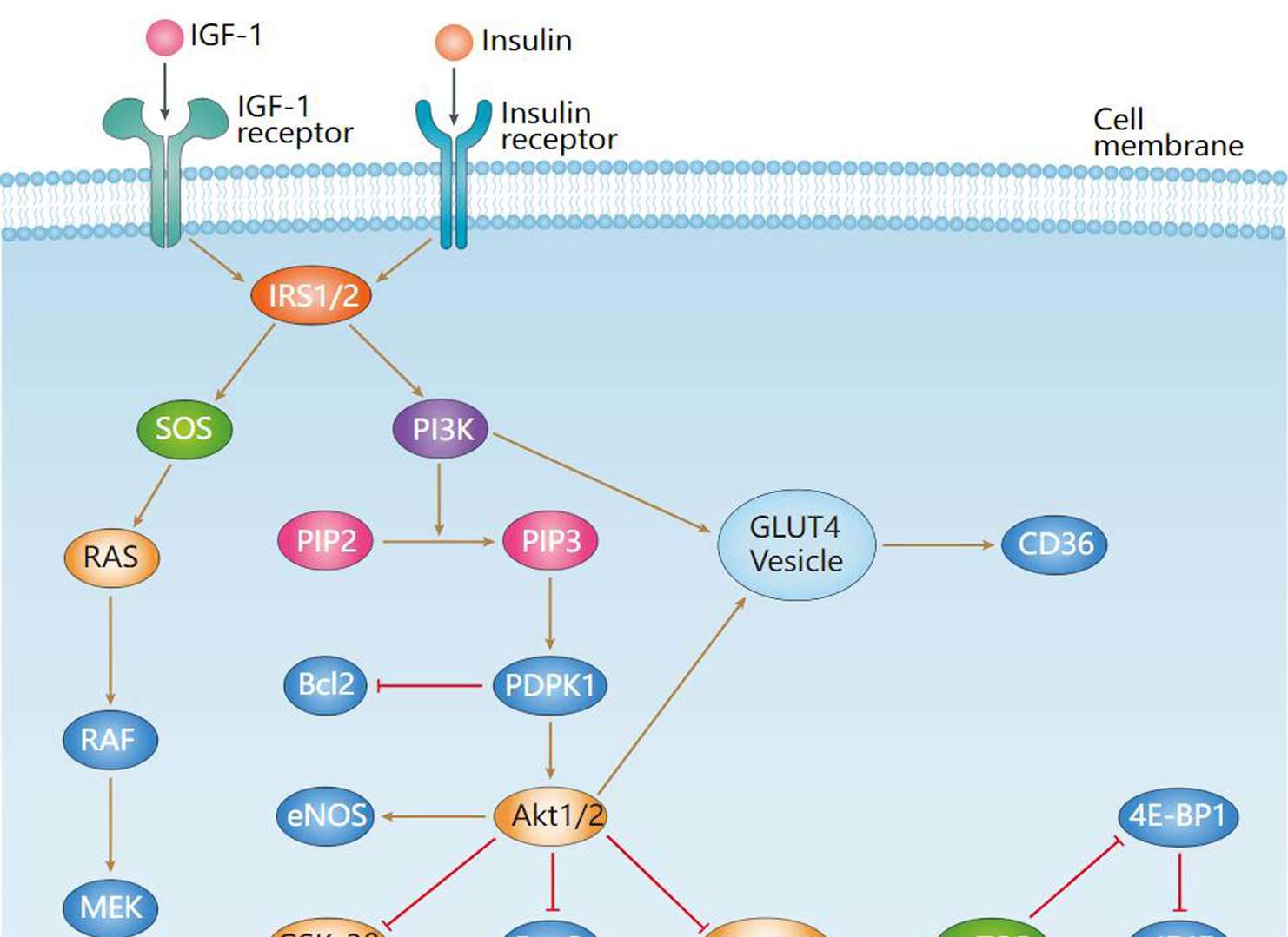 Insulin Signaling Pathway
Related Diseases
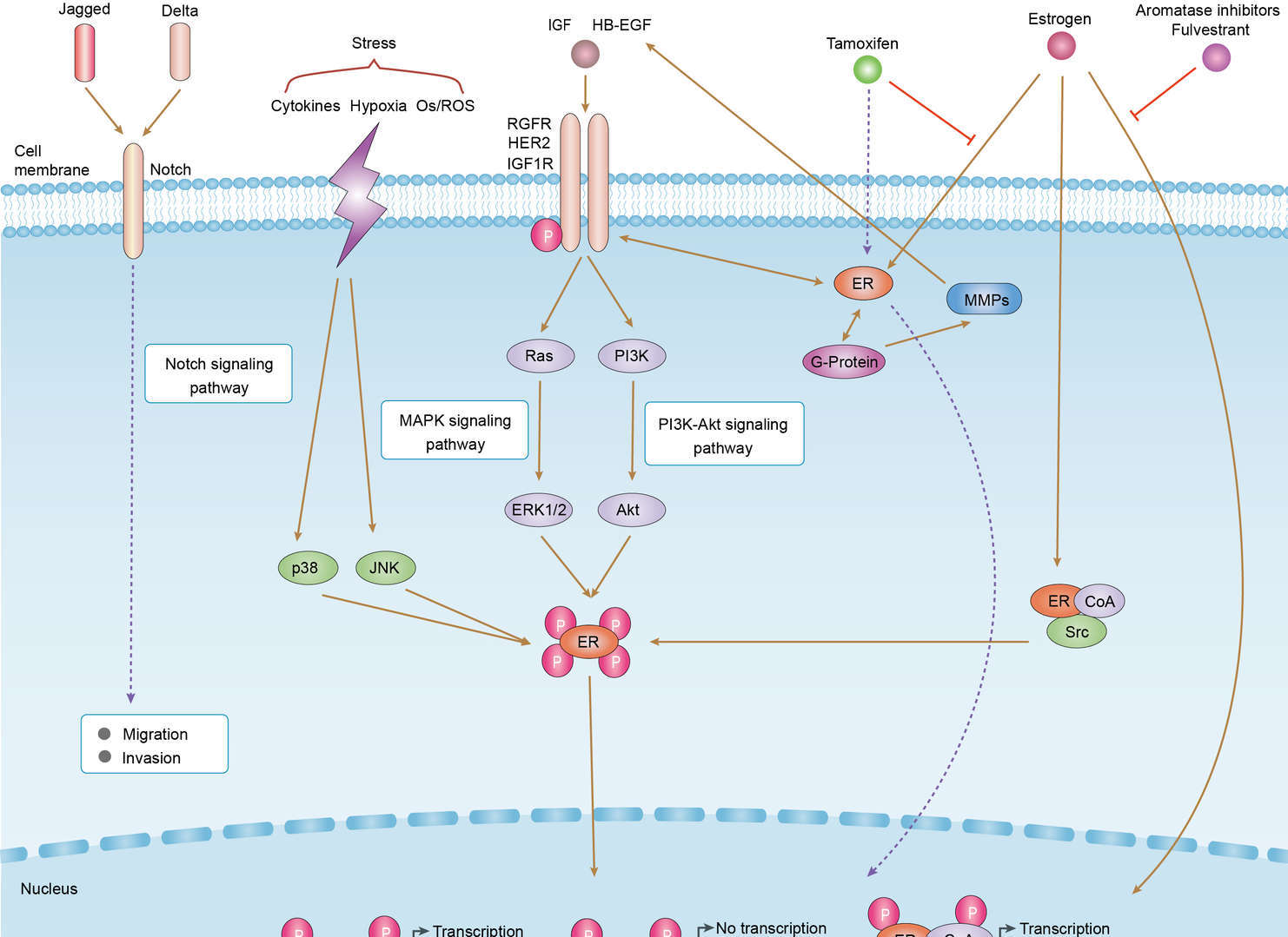 Endocrine Resistance
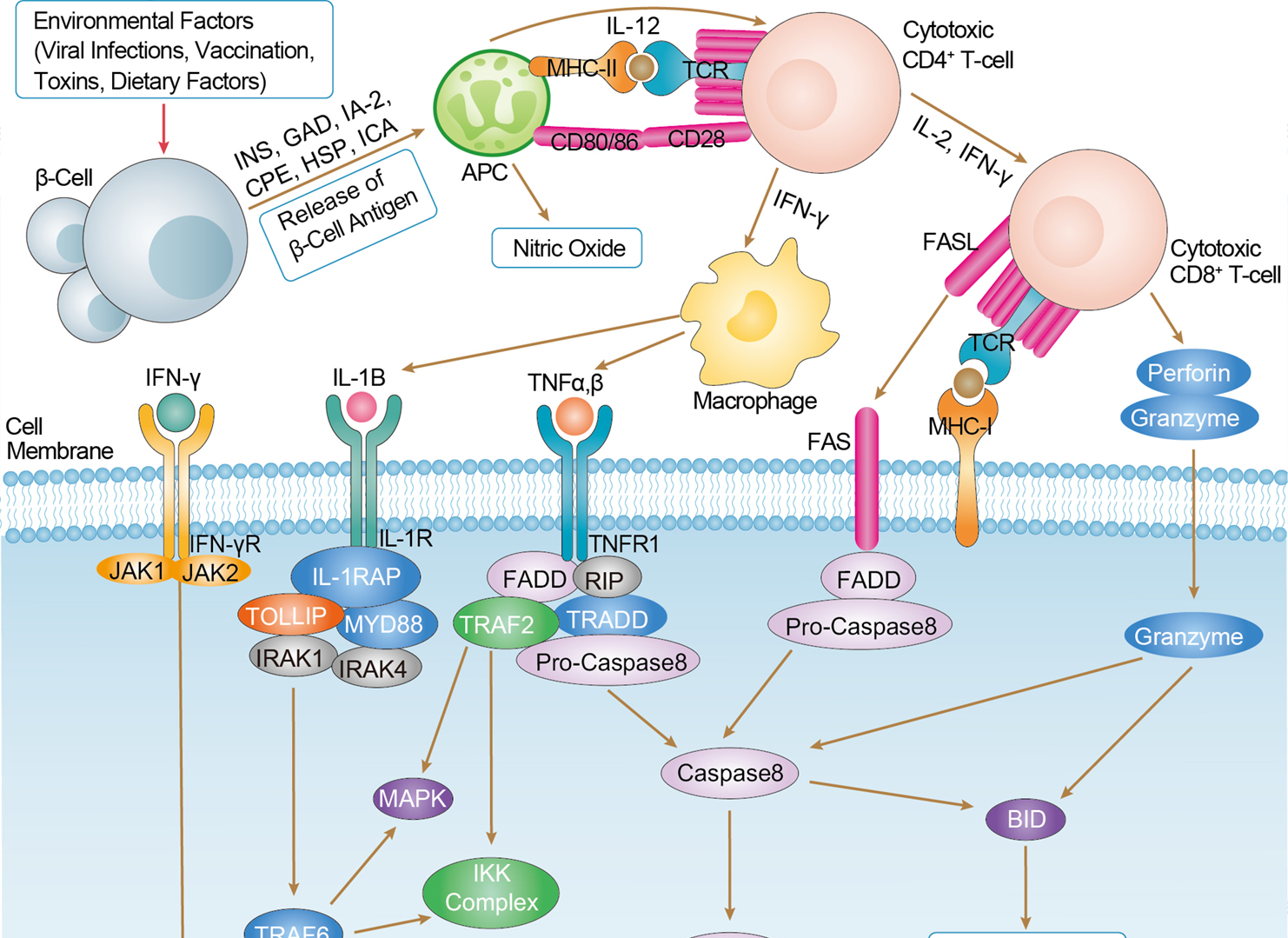 Type I Diabetes Mellitus
Downloadable Resources
Download resources about recombinant antibody development and antibody engineering to boost your research.
Product Notes
This is a product of Creative Biolabs' Hi-Affi™ recombinant antibody portfolio, which has several benefits including:
• Increased sensitivity
• Confirmed specificity
• High repeatability
• Excellent batch-to-batch consistency
• Sustainable supply
• Animal-free production
See more details about Hi-Affi™ recombinant antibody benefits.
Datasheet
MSDS
COA
Certificate of Analysis LookupTo download a Certificate of Analysis, please enter a lot number in the search box below. Note: Certificate of Analysis not available for kit components.
Lot Number:
See other products for "IGF1R"
Select a product category from the dropdown menu below to view related products.
| CAT | Product Name | Application | Type |
|---|---|---|---|
| NABL-091 | Recombinant Anti-human IGF1R VHH Single Domain Antibody | WB, ELISA, IHC, FC, FuncS | Llama VHH |
| TAB-096ZJ | Anti-Human IGF-1R Single Domain Antibody (TAB-096ZJ), Research Grade Biosimilar | ELISA, IHC, IF, WB | Single domain antibody |
| HPAB-1545-FY | Recombinant Llama Anti-IGF1R Single Domain Antibody (HPAB-1545-FY) | WB, ELISA | Llama VHH |
| HPAB-1546-FY | Recombinant Llama Anti-IGF1R Single Domain Antibody (HPAB-1546-FY) | WB, ELISA | Llama VHH |
| HPAB-1547-FY | Recombinant Llama Anti-IGF1R Single Domain Antibody (HPAB-1547-FY) | WB, ELISA | Llama VHH |
| CAT | Product Name | Application | Type |
|---|---|---|---|
| TAB-078 | Anti-Human IGF1 Receptor Recombinant Antibody (Cixutumumab) | IF, IP, Neut, FuncS, ELISA, FC, WB | IgG1 - lambda |
| TAB-209 | Anti-Human IGF1 Receptor Recombinant Antibody (TAB-209) | FuncS, IF, Neut, ELISA, FC, IP, ICC | IgG1 - kappa |
| TAB-052ZJ-F(E) | Anti-Human IGF-1R Recombinant Antibody Fab Fragment (TAB-052ZJ-F(E)) | ELISA, FC, IF, IP | Human antibody |
| TAB-053ZJ-F(E) | Anti-Human IGF-1R Recombinant Antibody Fab Fragment (IR3) | ELISA, FC, WB | Human antibody |
| TAB-054ZJ-F(E) | Anti-Human IGF-1R Recombinant Antibody Fab Fragment (IR10) | ELISA, FC, WB | Human antibody |
| CAT | Product Name | Application | Type |
|---|---|---|---|
| AGTO-L024E | IGF1-PE immunotoxin | Cytotoxicity assay, Functional assay | |
| AGTO-L024D | IGF1-DT immunotoxin | Cytotoxicity assay, Functional assay |
| CAT | Product Name | Application | Type |
|---|---|---|---|
| TAB-035ZJ-S(P) | Human Anti-IGF1R Recombinant Antibody; scFv Fragment (TAB-035ZJ-S(P)) | ELISA, FC | Humanized scFv |
| TAB-036ZJ-S(P) | Anti-Human IGF-1R Recombinant Antibody scFv Fragment (H0L0 IgGIm(AA)) | ELISA, Neut, FC, IHC | Humanized antibody |
| TAB-037ZJ-S(P) | Anti-Human IGF-1R Recombinant Antibody scFv Fragment (H1L0 IgGIm(AA)) | ELISA, Neut, FC, IHC | Humanized antibody |
| TAB-038ZJ-S(P) | Human Anti-IGF1R Recombinant Antibody; scFv Fragment (TAB-038ZJ-S(P)) | ELISA, FC, Inhib, IP | Humanized scFv |
| TAB-051ZJ-S(P) | Human Anti-IGF1R Recombinant Antibody; scFv Fragment (TAB-051ZJ-S(P)) | ELISA, IHC, IF, WB | Human scFv |
| CAT | Product Name | Application | Type |
|---|---|---|---|
| TAB-084ZJ-S(P) | Mouse Anti-IGF1R Recombinant Antibody; scFv Fragment (TAB-084ZJ-S(P)) | ELISA, FC | Mouse scFv |
| TAB-085ZJ-S(P) | Anti-Human IGF-1R Recombinant Antibody scFv Fragment (6E11c) | ELISA, Neut, FC, IHC | Chimeric antibody (mouse/human) |
| TAB-086ZJ-S(P) | Mouse Anti-IGF1R Recombinant Antibody; scFv Fragment (TAB-086ZJ-S(P)) | ELISA, FC, Inhib, IP | Mouse scFv |
| TAB-087ZJ-S(P) | Anti-Human IGF-1R Recombinant Antibody scFv Fragment (ch7C2) | ELISA | Chimeric antibody (mouse/human) |
| TAB-093ZJ-S(P) | Anti-Human IGF-1R Recombinant Antibody scFv Fragment (ch9E11) | ELISA | Chimeric antibody (mouse/human) |
| CAT | Product Name | Application | Type |
|---|---|---|---|
| TAB-009ZJ-F(E) | Mouse Anti-IGF1R Recombinant Antibody; Fab Fragment (TAB-009ZJ-F(E)) | ELISA, FC | Mouse Fab |
| TAB-010ZJ-F(E) | Anti-Human IGF-1R Recombinant Antibody Fab Fragment (6E11) | ELISA, Neut, FC, IHC | |
| TAB-011ZJ-F(E) | Mouse Anti-IGF1R Recombinant Antibody; Fab Fragment (TAB-011ZJ-F(E)) | ELISA, FC, WB, Inhib | Mouse Fab |
| TAB-012ZJ-F(E) | Mouse Anti-IGF1R Recombinant Antibody; Fab Fragment (TAB-012ZJ-F(E)) | ELISA, FC, WB, Inhib | Mouse Fab |
| TAB-014ZJ-F(E) | Mouse Anti-IGF1R Recombinant Antibody; Fab Fragment (TAB-014ZJ-F(E)) | ELISA, FC, Inhib, IP | Mouse Fab |
| CAT | Product Name | Application | Type |
|---|---|---|---|
| BRD-0103MZ | Chicken Anti-CD221 Polyclonal IgY | WB | Chicken antibody |
| CAT | Product Name | Application | Type |
|---|---|---|---|
| NEUT-1075CQ | Mouse Anti-IGF1R Recombinant Antibody (clone alphaIR3) | FC, IP, Neut, ICC, IF | Mouse IgG1 |
| NEUT-1076CQ | Mouse Anti-IGF1R Recombinant Antibody (clone 33255.111) | ELISA, Neut, WB | Mouse IgG1 |
| NEUT-1081CQ | Mouse Anti-IGF1R Recombinant Antibody (clone CBL453) | WB, ELISA, Neut | Mouse IgG1 |
| NEUT-1082CQ | Mouse Anti-IGF1R Recombinant Antibody (clone CBL080) | Neut, WB | Mouse IgG1 |
| CAT | Product Name | Application | Type |
|---|---|---|---|
| NEUT-1077CQ | Mouse Anti-IGF1R Recombinant Antibody (clone 1H7) | FC, Block, IHC, IP, WB | Mouse IgG1, κ |
| NEUT-1078CQ | Mouse Anti-IGF1R Recombinant Antibody (clone 24-60) | Inhib, ICC, IF, IP, WB | Mouse IgG2a, κ |
| NEUT-1079CQ | Mouse Anti-IGF1R Recombinant Antibody (clone 17-69) | Inhib, IP | Mouse IgG1 |
| NEUT-1080CQ | Mouse Anti-IGF1R Recombinant Antibody (clone 24-57) | Inhib, IP | Mouse IgG1, κ |
| CAT | Product Name | Application | Type |
|---|---|---|---|
| MOR-1755 | Rabbit Anti-IGF1R Recombinant Antibody (clone DS1755AB) | IP, ELISA | Rabbit IgG |
| MOR-4684 | Rabbit Anti-IGF1R Recombinant Antibody (clone TH198DS) | WB, ELISA | Rabbit IgG |
| MOR-4685 | Rabbit Anti-IGF1R Recombinant Antibody (clone TH199DS) | WB | Rabbit IgG |
| CAT | Product Name | Application | Type |
|---|---|---|---|
| HPAB-0084-YC-F(E) | Human Anti-IGF1R Recombinant Antibody; Fab Fragment (HPAB-0084-YC-F(E)) | ELISA, FC | Humanized Fab |
| HPAB-0085-YC-F(E) | Human Anti-IGF1R Recombinant Antibody; Fab Fragment (HPAB-0085-YC-F(E)) | Block, FuncS, FC, ELISA | Human Fab |
| HPAB-1441LY-F(E) | Human Anti-IGF1R Recombinant Antibody; Fab Fragment (HPAB-1441LY-F(E)) | ELISA, IHC | Humanized Fab |
| HPAB-1442LY-F(E) | Human Anti-IGF1R Recombinant Antibody; Fab Fragment (HPAB-1442LY-F(E)) | ELISA, IHC | Humanized Fab |
| HPAB-1277WJ-F(E) | Mouse Anti-IGF1R Recombinant Antibody (clone 1H7); Fab Fragment | ELISA | Mouse Fab |
| CAT | Product Name | Application | Type |
|---|---|---|---|
| NS-041CN | Mouse Anti-IGF1R Recombinant Antibody (NS-041CN) | ELISA, WB, FC | Mouse IgG |
| NS-041CN-F(E) | Mouse Anti-IGF1R Recombinant Antibody; Fab Fragment (NS-041CN-F(E)) | ELISA, WB, FC | Mouse Fab |
| NS-041CN-S(P) | Mouse Anti-IGF1R Recombinant Antibody; scFv Fragment (NS-041CN-S(P)) | ELISA, WB, FC | Mouse scFv |
| CAT | Product Name | Application | Type |
|---|---|---|---|
| HPAB-0210CQ | Human Anti-IGF1R Recombinant Antibody (clone 15H12) | ELISA, FC, Neut | Human IgG3, κ |
| HPAB-0211CQ | Human Anti-IGF1R Recombinant Antibody (clone 1H3) | ELISA, FC, Neut | Human IgG |
| HPAB-0212CQ | Human Anti-IGF1R Recombinant Antibody (clone 11A4) | ELISA, FC, Block | Human IgG1 |
| HPAB-0213CQ | Human Anti-IGF1R Recombinant Antibody (clone 8A1) | ELISA, FC, Block | Human IgG1 |
| HPAB-0214CQ | Human Anti-IGF1R Recombinant Antibody (clone PINT-9A2) | ELISA, FC, Block | Human IgG1 |
| CAT | Product Name | Application | Type |
|---|---|---|---|
| AFC-TAB-199 | Afuco™ Anti-IGF1R Recombinant Antibody (AFC-TAB-199), ADCC Enhanced | FC, IP, ELISA, Neut, FuncS, IF | Human IgG1, κ |
| AFC-TAB-209 | Afuco™ Anti-IGF1R ADCC Recombinant Antibody, ADCC Enhanced (AFC-TAB-209) | FuncS, IF, Neut, ELISA, FC, IP | ADCC enhanced antibody |
| AFC-TAB-736 | Afuco™ Anti-IGF1R ADCC Recombinant Antibody, ADCC Enhanced (AFC-TAB-736) | FC, IP, ELISA, Neut, FuncS, IF | ADCC enhanced antibody |
| AFC-TAB-078 | Afuco™ Anti-IGF1R ADCC Recombinant Antibody, ADCC Enhanced (AFC-TAB-078) | IF, IP, Neut, FuncS, ELISA, FC | ADCC enhanced antibody |
| AFC-TAB-232 | Afuco™ Anti-IGF1R ADCC Recombinant Antibody, ADCC Enhanced (AFC-TAB-232) | IF, IP, Neut, FuncS, ELISA | ADCC enhanced antibody |
| CAT | Product Name | Application | Type |
|---|---|---|---|
| HPAB-0069-YJ-S(P) | Human Anti-IGF1R Recombinant Antibody; scFv Fragment (HPAB-0069-YJ-S(P)) | ELISA, Block, FC, FuncS | Human scFv |
| HPAB-0712-FY-F(E) | Mouse Anti-IGF1R Recombinant Antibody (clone AB011); scFv Fragment | FC | Mouse scFv |
| HPAB-0839-FY-F(E) | Mouse Anti-IGF1R Recombinant Antibody; Fab Fragment (HPAB-0839-FY-F(E)) | IHC | Mouse Fab |
| HPAB-0840-FY-F(E) | Mouse Anti-IGF1R Recombinant Antibody; Fab Fragment (HPAB-0840-FY-F(E)) | IHC | Mouse Fab |
| HPAB-0841-FY-F(E) | Mouse Anti-IGF1R Recombinant Antibody; Fab Fragment (HPAB-0841-FY-F(E)) | IHC | Mouse Fab |
| CAT | Product Name | Application | Type |
|---|---|---|---|
| VS-0424-XY145 | AbPlus™ Anti-IGF1R Magnetic Beads (9E11) | IP, Protein Purification |
| CAT | Product Name | Application | Type |
|---|---|---|---|
| VS-0125-FY20 | Human Anti-IGF1R (clone 1H3) scFv-Fc Chimera | ELISA, FC, Neut, Inhib | Human IgG1, scFv-Fc |
| VS-0325-FY85 | Human Anti-IGF1R (clone 7C10) scFv-Fc Chimera | ELISA, FC, Inhib | Human IgG1, scFv-Fc |
| CAT | Product Name | Application | Type |
|---|---|---|---|
| VS-0325-XY1101 | Anti-IGF1R Immunohistochemistry Kit | IHC | |
| VS-0525-XY3431 | Anti-Mouse IGF1R Immunohistochemistry Kit | IHC | |
| VS-0525-XY3430 | Anti-Human IGF1R Immunohistochemistry Kit | IHC | |
| VS-0525-XY3432 | Anti-Rat IGF1R Immunohistochemistry Kit | IHC |
| CAT | Product Name | Application | Type |
|---|---|---|---|
| VS-0425-YC366 | Recombinant Anti-IGF1R Vesicular Antibody, EV Displayed (VS-0425-YC366) | ELISA, FC, Neut, Cell-uptake |
| CAT | Product Name | Application | Type |
|---|---|---|---|
| VS-0525-YC108 | Recombinant Anti-IGF1R (AA 62-184 x AA 283-440) Biparatopic Antibody, Tandem scFv (Clone 4-52 x Clone 45742) | ELISA | Tandem scFv |
| VS-0525-YC107 | Recombinant Anti-IGF1R (AA 283-440 x AA 440-514) Biparatopic Antibody, Tandem scFv (Clone 45742 x Clone 24-55) | ELISA | Tandem scFv |
Specific Inquiry

See Our Custom Production in Action


Popular Products


Application: Neut, ELISA, IF, IP, FuncS, FC, IHC

Application: ELISA, FC, IP, FuncS, IF, Neut, ICC

Application: WB, IF, IP, Neut, FuncS, ELISA, FC


![Figure 2 Anti-Human CD19 Recombinant Antibody Fab Fragment [TAB-1611CL-F(E)] in HPLC](https://img.creativebiolabs.net/productimages/COA-TAB-1611CL-F(E)-2.png)
Application: Depletion, FuncS
-CB2006C17L-4.jpg)
Application: WB, ELISA

Application: ELISA, FC, WB, Block
-3.jpg)
Application: Neut

Application: ELISA, WB, FC, IHC, IP

Application: ELISA, FC, Inhib


Application: WB, FC, IF, Inhib, ELISA, IHC


For research use only. Not intended for any clinical use. No products from Creative Biolabs may be resold, modified for resale or used to manufacture commercial products without prior written approval from Creative Biolabs.
Send Inquiry
This site is protected by reCAPTCHA and the Google Privacy Policy and Terms of Service apply.