

Afuco™ Anti-Human ICAM1 ADCC Recombinant Antibody (BI-505), ADCC Enhanced
CAT#: AFC-361CL
Anti-ICAM1 ADCC Enhanced Antibody (BI-505) is an ADCC enhanced antibody produced by our Afuco™ platform. BI-505 is a fully human high-affinity IgG1 antibody, which was isolated from a highly diverse human phage-antibody library based on its functional ability to induce programmed cell death of tumor cells. BI-505 binds to intercellular adhesion molecule-1 (ICAM-1), suggesting a previously unrecognized role for this receptor as a therapeutic target in cancer.
Gene Expression

Subcellular Location and Protein Expression
Figure 1 IF staining of human cell line U-2 OS
Immunofluorescent staining of human cell line U-2 OS shows localization to plasma membrane & cytosol.
* Image credit: Image credit: Human Protein Atlas https://v21.proteinatlas.org/images/2126/29_G3_2_selected.jpg

Subcellular Location and Protein Expression
Figure 2 IF staining of human cell line U-251 MG
Immunofluorescent staining of human cell line U-251 MG shows localization to plasma membrane.
* Image credit: Image credit: Human Protein Atlas https://v21.proteinatlas.org/images/2142/203_G3_1_selected.jpg

Subcellular Location and Protein Expression
Figure 3 IF staining of human cell line EFO-21
Immunofluorescent staining of human cell line EFO-21 shows localization to plasma membrane & cytosol.
* Image credit: Image credit: Human Protein Atlas https://v21.proteinatlas.org/images/2126/1937_A10_1_blue_red_green.jpg

Subcellular Location and Protein Expression
Figure 4 IF staining of human cell line GAMG
Immunofluorescent staining of human cell line GAMG shows localization to plasma membrane, vesicles, nucleoplasm & centrosome.
* Image credit: Image credit: Human Protein Atlas https://v21.proteinatlas.org/images/2126/2058_A10_4_blue_red_green.jpg

Normal Tissue
Figure 5 Cerebral cortex
Endothelial cells
Staining:Medium
Intensity: Moderate
Quantity:>75%
Location: Cytoplasmic/membranous
* Image credit: Image credit: Human Protein Atlas https://v21.proteinatlas.org/images/2126/7230_B_9_5.jpg

Normal Tissue
Figure 6 Lung
Alveolar cells
Staining:High
Intensity: Strong
Quantity:>75%
Location: Cytoplasmic/membranous
* Image credit: Image credit: Human Protein Atlas https://v21.proteinatlas.org/images/2126/7230_A_3_4.jpg

Normal Tissue
Figure 7 Colon
Endothelial cells
Staining:High
Intensity: Strong
Quantity:>75%
Location: Cytoplasmic/membranous
* Image credit: Image credit: Human Protein Atlas https://v21.proteinatlas.org/images/4877/13410_A_8_3.jpg

Normal Tissue
Figure 8 Kidney
Cells in glomeruli
Staining:High
Intensity: Strong
Quantity:>75%
Location: Cytoplasmic/membranous
* Image credit: Image credit: Human Protein Atlas https://v21.proteinatlas.org/images/4877/13410_A_8_5.jpg

Normal Tissue
Figure 9 Lymph node
Non-germinal center cells
Staining:Medium
Intensity: Strong
Quantity: <25%
Location: Cytoplasmic/membranous
* Image credit: Image credit: Human Protein Atlas https://v21.proteinatlas.org/images/4877/13410_A_9_8.jpg

RNA Expression
Figure 10 RNA cell line category: Cell line enhanced (HHSteC, Karpas-707, RPMI-8226)
Cell lines ordered by descending RNA expression order.
* Image credit: Image credit: Human Protein Atlas https://v21.proteinatlas.org/ENSG00000090339-ICAM1
❮


 ❯
❯
Specifications
- Host Species
- Human
- Derivation
- Human
- Type
- ADCC enhanced antibody
- Antibody Isotype
- IgG1
- Species Reactivity
- Human
- Related Disease
- Multiple Myeloma
Product Property
- Purity
- >95% as judged by SDS-polyacrylamide gel electrophoresis
- Storage
- ≥1 year at -20°C. If the reconstituted antibody cannot be used within two weeks, it should be aliquoted into smaller vials and stored at -20°C
Target
REVIEWS AND Q&AS
CITATIONS
RESOURCES
DOWNLOADS
RELATED PRODUCTS
Inquiry
Navs
Customer Review

There are currently no Customer reviews or questions for AFC-361CL. Click the button above to contact us or submit your feedback about this product.

Enhanced ADCC Activity
We were interested in the ADCC capabilities of this antibody, and the Afuco™ platform really delivers. Clone BI-505 showed enhanced ADCC activity in our assays compared to standard antibodies. It binds high-affinity to ICAM1 and induces cell death effectively in our tumor models.
High Affinity Human Antibody
This fully human IgG1 antibody is of excellent quality. We isolated it for its functional ability to bind ICAM-1, and it has proven to be a robust reagent. The purity is>95% by SDS-PAGE, and it has been stable when stored at -20°C.
Q&As
-
What makes this antibody "ADCC Enhanced"?
A: This Anti-ICAM1 Antibody (BI-505) is produced using our Afuco™ platform, which utilizes a fucosyltransferase-engineered cell line. This results in an afucosylated antibody that exhibits enhanced Antibody-Dependent Cellular Cytotoxicity (ADCC) compared to standard IgG1 antibodies.
-
Is this a fully human antibody?
A: Yes, BI-505 is a fully human high-affinity IgG1 antibody. It was isolated from a highly diverse human phage-antibody library based on its functional ability to induce programmed cell death of tumor cells by binding to ICAM-1.
View the frequently asked questions answered by Creative Biolabs Support.
Submit Your Publication
Published with our product? Submit your paper and receive a 10% discount on your next order! Share your research to earn exclusive rewards.
Related Diseases
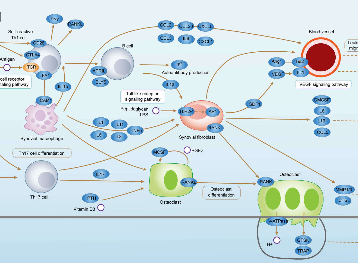 Rheumatoid Arthritis
 AGE-RAGE Signaling Pathway in Diabetic Complications
Related Signaling Pathways
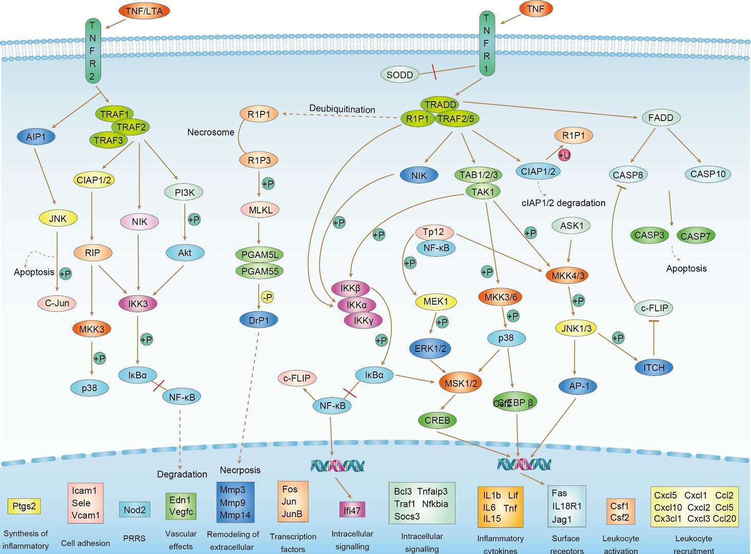 TNF Signaling Pathway
Downloadable Resources
Download resources about recombinant antibody development and antibody engineering to boost your research.
Product Notes
This is a product of Creative Biolabs' Hi-Affi™ recombinant antibody portfolio, which has several benefits including:
• Increased sensitivity
• Confirmed specificity
• High repeatability
• Excellent batch-to-batch consistency
• Sustainable supply
• Animal-free production
See more details about Hi-Affi™ recombinant antibody benefits.
Datasheet
MSDS
COA
Certificate of Analysis LookupTo download a Certificate of Analysis, please enter a lot number in the search box below. Note: Certificate of Analysis not available for kit components.
Lot Number:
See other products for "ICAM1"
Select a product category from the dropdown menu below to view related products.
| CAT | Product Name | Application | Type |
|---|---|---|---|
| NAB-1937-VHH | Recombinant Anti-human ICAM1 VHH Single Domain Antibody | FA, FC, ICC, ELISA | Llama VHH |
| CAT | Product Name | Application | Type |
|---|---|---|---|
| DrMAB-1136 | Human Anti-ICAM1 Recombinant Antibody (clone YC170); scFv Fragment | ELISA, WB, IHC, IP | Human scFv |
| CAT | Product Name | Application | Type |
|---|---|---|---|
| DrMAB-1139 | Human Anti-ICAM1 Recombinant Antibody (clone YC171); scFv Fragment, HRP | ELISA, WB, IHC, IP | Human scFv |
| CAT | Product Name | Application | Type |
|---|---|---|---|
| TAB-241 | Anti-Human ICAM-1 Recombinant Antibody Fab Fragment (TAB-241) | WB, IF, IP, Neut, FuncS, ELISA, FC | Fab' - G2a |
| CAT | Product Name | Application | Type |
|---|---|---|---|
| TAB-H26 | Anti-Human ICAM1 Recombinant Antibody (TAB-H26) | Neut, ELISA, IF, IP, FuncS, FC, WB | IgG2a |
| CAT | Product Name | Application | Type |
|---|---|---|---|
| AGTO-L023R | anti-ICAM1 immunotoxin UV3 (IgG)-RTA | Cytotoxicity assay, Functional assay |
| CAT | Product Name | Application | Type |
|---|---|---|---|
| TAB-376CL | Human Anti-ICAM1 Recombinant Antibody (TAB-376CL) | IHC, FuncS | Human IgG1 |
| CAT | Product Name | Application | Type |
|---|---|---|---|
| PABZ-079 | Mouse Anti-ICAM1 Recombinant Antibody (clone CA7) | FC | Mouse IgG |
| CAT | Product Name | Application | Type |
|---|---|---|---|
| PFBZ-079 | Mouse Anti-ICAM1 Recombinant Antibody (clone CA7); Fab Fragment | FC | Mouse Fab |
| CAT | Product Name | Application | Type |
|---|---|---|---|
| PSBZ-079 | Mouse Anti-ICAM1 Recombinant Antibody (clone CA7); scFv Fragment | FC | Mouse scFv |
| CAT | Product Name | Application | Type |
|---|---|---|---|
| TAB-121CT | Anti-Human ICAM-1 Recombinant Antibody (MD-2) | ELISA, FC | Human antibody |
| CAT | Product Name | Application | Type |
|---|---|---|---|
| TAB-122CT | Anti-Human ICAM-1 Recombinant Antibody (R6-5-D6) | ELISA, FC | Human antibody |
| CAT | Product Name | Application | Type |
|---|---|---|---|
| TAB-123CT | Anti-Human ICAM-1 Recombinant Antibody (h1A6) | ELISA | Humanized antibody |
| CAT | Product Name | Application | Type |
|---|---|---|---|
| TAB-124CT | Anti-Human ICAM-1 Recombinant Antibody (m1A6) | ELISA |
| CAT | Product Name | Application | Type |
|---|---|---|---|
| TAB-125CT | Anti-Human ICAM-1 Recombinant Antibody (ICM 10064) | Inhibit, ELISA, WB | Human antibody |
| CAT | Product Name | Application | Type |
|---|---|---|---|
| TAB-121CT-S(P) | Anti-Human ICAM-1 Recombinant Antibody scFv Fragment (MD-2) | ELISA, FC | Human antibody |
| CAT | Product Name | Application | Type |
|---|---|---|---|
| TAB-123CT-S(P) | Anti-Human ICAM-1 Recombinant Antibody scFv Fragment (h1A6) | ELISA | Humanized antibody |
| CAT | Product Name | Application | Type |
|---|---|---|---|
| TAB-124CT-S(P) | Anti-Human ICAM-1 Recombinant Antibody scFv Fragment (m1A6) | ELISA |
| CAT | Product Name | Application | Type |
|---|---|---|---|
| TAB-123CT-F(E) | Anti-Human ICAM-1 Recombinant Antibody Fab Fragment (h1A6) | ELISA | Humanized antibody |
| CAT | Product Name | Application | Type |
|---|---|---|---|
| TAB-124CT-F(E) | Anti-Human ICAM-1 Recombinant Antibody Fab Fragment (m1A6) | ELISA |
| CAT | Product Name | Application | Type |
|---|---|---|---|
| TAB-126CT | Llama Anti-ICAM1 Recombinant Antibody (TAB-126CT) | ELISA, IF | Llama VHH |
| CAT | Product Name | Application | Type |
|---|---|---|---|
| Gly-104LC | Recombinant Anti-Human ICAM1 Antibody (Fc glycosylation) | ELISA | Mouse antibody |
| CAT | Product Name | Application | Type |
|---|---|---|---|
| MOB-1243MZ | Recombinant Mouse Anti-Human ICAM1 Antibody (clone 26.3), PE-Conjugated | ICC | Mouse antibody |
| CAT | Product Name | Application | Type |
|---|---|---|---|
| NEUT-923CQ | Mouse Anti-ICAM1 Recombinant Antibody (clone CBL236) | Neut, IF, FC, WB, IHC | Mouse IgG1 |
| CAT | Product Name | Application | Type |
|---|---|---|---|
| NEUT-924CQ | Mouse Anti-ICAM1 Recombinant Antibody (clone HCD54) | FC, IF, Block | Mouse IgG1, κ |
| CAT | Product Name | Application | Type |
|---|---|---|---|
| NEUT-925CQ | Mouse Anti-ICAM1 Recombinant Antibody (NEUT-925CQ) | FC, FuncS, IHC, Neut, WB | Mouse IgG1, κ |
| CAT | Product Name | Application | Type |
|---|---|---|---|
| NEUT-926CQ | Mouse Anti-ICAM1 Recombinant Antibody (VMC182) | FC, IF, IHC, ICC, Neut | Mouse IgG2b |
| CAT | Product Name | Application | Type |
|---|---|---|---|
| NEUT-927CQ | Mouse Anti-ICAM1 Recombinant Antibody (NEUT-927CQ) | FC, IF, IHC, Neut, WB, ELISA | Mouse IgG1 |
| CAT | Product Name | Application | Type |
|---|---|---|---|
| NEUT-928CQ | Mouse Anti-ICAM1 Recombinant Antibody (VMC183) | Block, FC, IHC, IP | Mouse IgG1 |
| CAT | Product Name | Application | Type |
|---|---|---|---|
| NEUT-929CQ | Mouse Anti-ICAM1 Recombinant Antibody (NEUT-929CQ) | Block, WB, IP, ICC | Mouse IgG1 |
| CAT | Product Name | Application | Type |
|---|---|---|---|
| NEUT-930CQ | Rat Anti-Icam1 Recombinant Antibody (NEUT-930CQ) | FC, FuncS, IHC-Fr, Neut, IP | Rat IgG2a, κ |
| CAT | Product Name | Application | Type |
|---|---|---|---|
| MOR-1723 | Rabbit Anti-ICAM1 Recombinant Antibody (clone DS1723AB), Biotin | IHC-Fr, IP, WB, ELISA | Rabbit IgG |
| CAT | Product Name | Application | Type |
|---|---|---|---|
| MOR-1724 | Rabbit Anti-Icam1 Recombinant Antibody (clone DS1724AB) | ELISA | Rabbit IgG |
| CAT | Product Name | Application | Type |
|---|---|---|---|
| MOR-4598 | Rabbit Anti-ICAM1 Recombinant Antibody (clone TH111DS) | WB, FC | Rabbit IgG |
| CAT | Product Name | Application | Type |
|---|---|---|---|
| HPAB-0053-YC | Human Anti-ICAM1 Recombinant Antibody (HPAB-0053-YC) | Inhib, FC, IHC | Human IgG1 |
| CAT | Product Name | Application | Type |
|---|---|---|---|
| HPAB-0349-YC-VHH | Recombinant Llama Anti-ICAM1 Single Domain Antibody (HPAB-0349-YC-VHH) | ELISA | Llama VHH |
| CAT | Product Name | Application | Type |
|---|---|---|---|
| HPAB-0053-YC-S(P) | Human Anti-ICAM1 Recombinant Antibody scFv Fragment (HPAB-0053-YC-S(P)) | FC, IP | Human scFv |
| CAT | Product Name | Application | Type |
|---|---|---|---|
| HPAB-0053-YC-F(E) | Human Anti-ICAM1 Recombinant Antibody Fab Fragment (HPAB-0053-YC-F(E)) | FC, IP | Human Fab |
| CAT | Product Name | Application | Type |
|---|---|---|---|
| EPAF-1635LC | Mouse Anti-ICAM1 Recombinant Antibody (clone c78.4A) | FC | Mouse IgG |
| CAT | Product Name | Application | Type |
|---|---|---|---|
| HPAB-0043-FY | Human Anti-ICAM1 Recombinant Antibody (HPAB-0043-FY) | ELISA | Human IgG1, λ |
| CAT | Product Name | Application | Type |
|---|---|---|---|
| HPAB-0043-FY-S(P) | Human Anti-ICAM1 Recombinant Antibody scFv Fragment (HPAB-0043-FY-S(P)) | ELISA | Human scFv |
| CAT | Product Name | Application | Type |
|---|---|---|---|
| HPAB-0043-FY-F(E) | Human Anti-ICAM1 Recombinant Antibody Fab Fragment (HPAB-0043-FY-F(E)) | ELISA | Human Fab, λ |
| CAT | Product Name | Application | Type |
|---|---|---|---|
| HPAB-0859-CN | Human Anti-ICAM1 Recombinant Antibody (clone BI-AB) | ELISA, FC, Neut | Human IgG1 |
| CAT | Product Name | Application | Type |
|---|---|---|---|
| HPAB-0859-CN-F(E) | Human Anti-ICAM1 Recombinant Antibody (clone BI-AB); Fab Fragment | ELISA, FC, Neut | Human Fab |
| CAT | Product Name | Application | Type |
|---|---|---|---|
| HPAB-662-FY-F(E) | Human Anti-ICAM1 Recombinant Antibody Fab Fragment (HPAB-662-FY-F(E)) | Inhib | Human Fab |
| CAT | Product Name | Application | Type |
|---|---|---|---|
| VS-0424-XY141 | AbPlus™ Anti-ICAM1 Magnetic Beads (15.2) | IP, Protein Purification |
| CAT | Product Name | Application | Type |
|---|---|---|---|
| VS-0724-YC1353 | AbPlus™ Anti-Icam1 Magnetic Beads (VS-0724-YC1353) | IP, Protein Purification |
| CAT | Product Name | Application | Type |
|---|---|---|---|
| VS-0125-FY45 | Human Anti-ICAM1 (clone 1A6) scFv-Fc Chimera | ADCC | Human IgG1, scFv-Fc |
| CAT | Product Name | Application | Type |
|---|---|---|---|
| VS-0225-XY121 | CytoStream™ Mouse Anti-ICAM1 Recombinant Antibody (clone HA58) | FC | Mouse IgG1, kappa |
| CAT | Product Name | Application | Type |
|---|---|---|---|
| VS-0425-YC540 | Recombinant Anti-ICAM1 Vesicular Antibody, EV Displayed (VS-0425-YC540) | ELISA, FC, Neut, Cell-uptake |
| CAT | Product Name | Application | Type |
|---|---|---|---|
| VS-0525-XY3367 | Anti-Mouse ICAM1 Immunohistochemistry Kit | IHC |
| CAT | Product Name | Application | Type |
|---|---|---|---|
| VS-0525-XY3370 | Anti-Rat ICAM1 Immunohistochemistry Kit | IHC |
| CAT | Product Name | Application | Type |
|---|---|---|---|
| VS-0825-YC159 | SmartAb™ Recombinant Anti-ICAM1 pH-dependent Antibody (VS-0825-YC159) | Neut, ELISA, IF, IP, FC, WB | Mouse IgG2a |
Specific Inquiry

See Our Custom Production in Action


Popular Products


Application: WB, FuncS, IF, Neut, ELISA, FC, IP

Application: ELISA, FC, IP, FuncS, IF, Neut, ICC

Application: ELISA, Neut, IF, IP, FC, FuncS


-2.png)

Application: ELISA, Neut, IF, IP, FC, FuncS


Application: ELISA, FC, IP, FuncS, IF, Neut, ICC

Application: FC, IP, ELISA, Neut, FuncS, IF, IHC

Application: ELISA, WB, BLI, SPR


For research use only. Not intended for any clinical use. No products from Creative Biolabs may be resold, modified for resale or used to manufacture commercial products without prior written approval from Creative Biolabs.
Send Inquiry
This site is protected by reCAPTCHA and the Google Privacy Policy and Terms of Service apply.