

Ubiquitin-Proteasome Pathway
Product List
+ Filter
 Loading...
- AbPlus™ Anti-Uchl1 Magnetic Beads (VS-0724-YC1237) (VS-0724-YC1237)
-
- Target: Uchl1
- Target Species: Rat
- Application: IP, Protein Purification
 Compare
- AbPlus™ Anti-CDC34 Magnetic Beads (VS-0724-YC1215) (VS-0724-YC1215)
-
- Target: CDC34
- Target Species: Human
- Application: IP, Protein Purification
Compare
- AbPlus™ Anti-UCHL1 Magnetic Beads (VS-0724-YC1164) (VS-0724-YC1164)
-
- Target: UCHL1
- Target Species: Human
- Application: IP, Protein Purification
Compare
- AbPlus™ Anti-Uchl1 Magnetic Beads (VS-0724-YC1162) (VS-0724-YC1162)
-
- Target: Uchl1
- Target Species: Mouse
- Application: IP, Protein Purification
Compare
- AbPlus™ Anti-USP7 Magnetic Beads (D545) (VS-0424-XY271)
-
- Target: USP7
- Target Species: Human
- Application: IP-MS, Protein Purification
Compare
- AbPlus™ Anti-UCHL1 Magnetic Beads (JF93-08) (VS-0424-XY270)
-
- Target: UCHL1
- Target Species: Human, Mouse, Rat
- Application: IP, Protein Purification
Compare
- Mouse Anti-UCHL1 Recombinant Antibody (VS3-FY2912) (VS3-FY2912)
-
- Species Reactivity: Human, Monkey
- Type: Mouse IgG
- Application: WB, ICC
Compare
-
- Species Reactivity: Human, Mouse, Rat
- Type: Rabbit IgG
- Application: WB, IHC-P, ICC, IF, IP, FC
Compare
- Recombinant Mouse Anti-UCHL1 Antibody (VS-0923-FY154) (VS-0923-FY154)
-
- Species Reactivity: Human
- Type: Mouse IgG2a
- Application: ELISA, WB
Compare
- Rabbit Anti-UCHL1 Recombinant Antibody (VS-0723-WK91) (VS-0723-WK91)
-
- Species Reactivity: Human
- Type: Rabbit IgG
- Application: ELISA
Compare
-
- Species Reactivity: Human, Mouse, Rat
- Type: Rabbit IgG
- Application: WB, IHC-Fr, IHC-P, ICC, IF, IP
Compare
- Recombinant Mouse Anti-UCHL1 Antibody (clone 1D1B12) (VS3-FY1518)
-
- Type: Mouse IgG2b
- Application: ELISA, WB, IHC, FC
Compare
- Recombinant Mouse Anti-UCHL1 Antibody (clone 1B4H3) (VS3-FY1517)
-
- Type: Mouse IgG2b
- Application: ELISA, WB, IHC, FC
Compare
- Mouse Anti-MDM2 Recombinant Antibody (VS3-WK838) (VS3-WK838)
-
- Derivation: Mouse
- Species Reactivity: Human
- Type: Mouse IgG
- Application: WB
Compare
- Mouse Anti-USP7 Recombinant Antibody (clone 5D5B8) (VS3-XY1581)
-
- Species Reactivity: Human
- Type: Mouse IgG1
- Application: ELISA, WB
Compare
- Mouse Anti-MDM2 Recombinant Antibody (clone 3G2) (VS3-XY1056)
-
- Species Reactivity: Human
- Type: Mouse IgG1
- Application: WB, IP, IF, IHC
Compare
- Mouse Anti-USP14 Recombinant Antibody (VS3-CJ1037) (VS3-CJ1037)
-
- Species Reactivity: Human, Mouse, Rat
- Type: Mouse IgG1
- Application: WB, FC
Compare
- Mouse Anti-USP5 Recombinant Antibody (VS3-CJ968) (VS3-CJ968)
-
- Species Reactivity: Human, Mouse, Rat
- Type: Mouse IgG1, κ
- Application: WB
Compare
- Recombinant Anti-human MDM2 Antibody (MOB-648)
-
- Derivation: Mouse
- Species Reactivity: Human
- Type: IgG
- Application: ELISA, WB, IF, FuncS
Compare
-
- Derivation: Mouse
- Species Reactivity: Human
- Type: Mouse IgG1
- Application: WB, ICC, IF, IHC, ELISA
Compare
- Mouse Anti-CRBN Recombinant Antibody (clone 8C1) (MOB-3265z)
-
- Derivation: Mouse
- Species Reactivity: Human
- Type: Mouse IgG1
- Application: WB, IHC, ELISA
Compare
-
- Derivation: Mouse
- Species Reactivity: Human
- Type: Mouse IgG2a, κ
- Application: WB, ELISA, IHC
Compare
-
- Derivation: Mouse
- Species Reactivity: Human
- Type: Mouse IgG1, κ
- Application: WB, IP, IF, IHC, ELISA
Compare
- Recombinant Mouse Anti-Human NAE1 Antibody (MOB-0691MZ)
-
- Species Reactivity: Human
- Type: Mouse antibody
- Application: WB
Compare
- Recombinant Human Anti-MDM2 Soluble TCR (clone 1bd2) (scTCR-205CQ-S(P))
-
- Species Reactivity: Human
- Epitope: aa 81-88
- MHC: HLA-A2.1
Compare
- Recombinant Mouse Anti-MDM2 Soluble TCR (clone MDM2) (scTCR-206CQ-S(P))
-
- Species Reactivity: Human
- Epitope: aa 81-88
- MHC: H-2Kb
Compare
- Recombinant Mouse Anti-MDM2 Soluble TCR (clone TCR 1) (scTCR-207CQ-S(P))
-
- Species Reactivity: Human
- Epitope: aa 81-88
- MHC: H-2Kb
Compare
- Rabbit Anti-CDC34 Recombinant Antibody (clone CBACN-128) (MRO-0327-CN)
-
- Species Reactivity: Human, Mouse, Rat
- Type: Rabbit IgG
- Application: WB, IF, IP, FC
Compare
- Mouse Anti-UCHL1 Recombinant Antibody (clone B1-5-6) (MRO-1216-CN)
-
- Species Reactivity: Human, Mouse, Rat
- Type: Mouse IgG2a
- Application: WB, IHC, IF, FC
Compare
- Rabbit Anti-UCHL1 Recombinant Antibody (clone JF93-08) (MRO-1217-CN)
-
- Species Reactivity: Human, Mouse, Rat
- Type: Rabbit IgG
- Application: WB, IF, IHC, IP
Compare
- Rabbit Anti-UCHL1 Recombinant Antibody (clone JM10-59) (MRO-1218-CN)
-
- Species Reactivity: Human, Mouse, Rat
- Type: Rabbit IgG
- Application: WB, IF, IHC, IP
Compare
- Rabbit Anti-USP14 Recombinant Antibody (clone JU30-49) (MRO-1562-CN)
-
- Species Reactivity: Human, Zebrafish
- Type: Rabbit IgG
- Application: WB, IF, IHC, FC
Compare
- Rabbit Anti-USP7 Recombinant Antibody (clone JB80-36) (MRO-1564-CN)
-
- Species Reactivity: Human, Mouse, Rat
- Type: Rabbit IgG
- Application: WB, IHC, FC
Compare
- Rabbit Anti-FBXW7 Polyclonal Antibody (MRO-1858-CN) (MRO-1858-CN)
-
- Species Reactivity: Human, Mouse, Rat
- Type: Rabbit IgG
- Application: WB, IF, FC, IHC
Compare
- Rabbit Anti-UCHL1 Polyclonal Antibody (MRO-2116-CN) (MRO-2116-CN)
-
- Species Reactivity: Human, Mouse, Rat
- Type: Rabbit IgG
- Application: WB, IF, IHC, FC
Compare
- Rabbit Anti-USP14 Polyclonal Antibody (MRO-2258-CN) (MRO-2258-CN)
-
- Species Reactivity: Human
- Type: Rabbit IgG
- Application: WB, FC
Compare
- PE-A*02:01/Human MDM2 (LLGDLFGV) MHC Tetramer (MHC-CN1420)
-
- Class: Class I
- Antigen: MDM2
- Antigen Species: Human
- Peptide: LLGDLFGV
- Conjugate: PE
Compare
- A*02:01/Human MDM2 (LLGDLFGV) MHC Monomer (MHC-YF287)
-
- Class: Class I
- Antigen: MDM2
- Antigen Species: Human
- Peptide: LLGDLFGV
- Conjugate: Biotin
Compare
- Mouse Anti-VHL Recombinant Antibody (MOB-0233F) (MOB-0233F)
-
- Species Reactivity: Human, Mouse
- Type: Mouse IgG
- Application: IHC-P, IF
Compare
- Mouse Anti-VHL Recombinant Antibody (MOB-0234F) (MOB-0234F)
-
- Species Reactivity: Human, Mouse
- Type: Mouse IgG
- Application: IHC-P, IF
Compare
- Recombinant Human Anti-MDM2 Soluble TCR (1bd2) (ZIP-Fos/ZIP-Jun) (VS-0622-YF205)
-
- Species Reactivity: Human
- Epitope: aa 81-88
- MHC: HLA-A2.1
Compare
- Recombinant Mouse Anti-MDM2 Soluble TCR (MDM2) (ZIP-Fos/ZIP-Jun) (VS-0622-YF206)
-
- Species Reactivity: Human
- Epitope: aa 81-88
- MHC: H-2Kb
Compare
- Recombinant Human Anti-MDM2 Soluble TCR (1bd2) (C-Cys) (VS-0622-YF2913)
-
- Species Reactivity: Human
- Epitope: aa 81-88
- MHC: HLA-A2.1
Compare
- Recombinant Mouse Anti-MDM2 Soluble TCR (MDM2) (C-Cys) (VS-0622-YF2914)
-
- Species Reactivity: Human
- Epitope: aa 81-88
- MHC: H-2Kb
Compare
- Recombinant Mouse Anti-MDM2 Soluble TCR (TCR 1) (C-Cys) (VS-0622-YF2915)
-
- Species Reactivity: Human
- Epitope: aa 81-88
- MHC: H-2Kb
Compare
- Recombinant Human Anti-MDM2 Soluble TCR (1bd2) (KIH) (VS-0622-YF5621)
-
- Species Reactivity: Human
- Epitope: aa 81-88
- MHC: HLA-A2.1
Compare
- Recombinant Mouse Anti-MDM2 Soluble TCR (MDM2) (KIH) (VS-0622-YF5622)
-
- Species Reactivity: Human
- Epitope: aa 81-88
- MHC: H-2Kb
Compare
- Recombinant Mouse Anti-MDM2 Soluble TCR (TCR 1) (KIH) (VS-0622-YF5623)
-
- Species Reactivity: Human
- Epitope: aa 81-88
- MHC: H-2Kb
Compare
-
- Derivation: Mouse
- Species Reactivity: Human
- Type: Mouse IgG1
- Application: WB, IHC, ICC, FC, ELISA
Compare
View More Products
Our customer service representatives are available 24 hours a day, from Monday to Sunday. Contact Us
Can't find the products you're looking for? Try to filter in the left sidebar.Filter By Tag
Representative Targets in Ubiquitin-Proteasome Pathway Full List of Targets in Ubiquitin-Proteasome Pathway Tested Data-Supported Products for Targeting Ubiquitin-Proteasome Pathway
The ubiquitin-proteasome pathway (UPP) is a pivotal cellular mechanism, fundamental for maintaining cellular homeostasis and regulating a multitude of cellular processes including the regulation of cell cycle progression, DNA repair mechanisms, apoptosis, and immune responses. This highly conserved and intricate system is central to the degradation of intracellular proteins, a process crucial for the removal of misfolded, damaged, or unneeded proteins, thereby preventing the accumulation of toxic protein aggregates that can lead to cellular dysfunction. At its core, the UPP involves the tagging of target proteins with ubiquitin molecules, a small regulatory protein found in all eukaryotic cells. This tagging process is highly regulated and involves a cascade of enzymatic activities: E1 ubiquitin-activating enzymes, E2 ubiquitin-conjugating enzymes, and E3 ubiquitin ligases, which confer substrate specificity to the pathway. Once polyubiquitinated, the tagged proteins are recognized and degraded by the 26S proteasome, a large proteolytic complex, releasing ubiquitin for reuse and peptides that can be further degraded or presented for immune surveillance. By controlling the abundance and activity of various cellular proteins, the UPP directly influences signal transduction pathways and gene expression. It also plays a vital role in the quality control of proteins, ensuring that only properly folded and functional proteins persist within the cell.
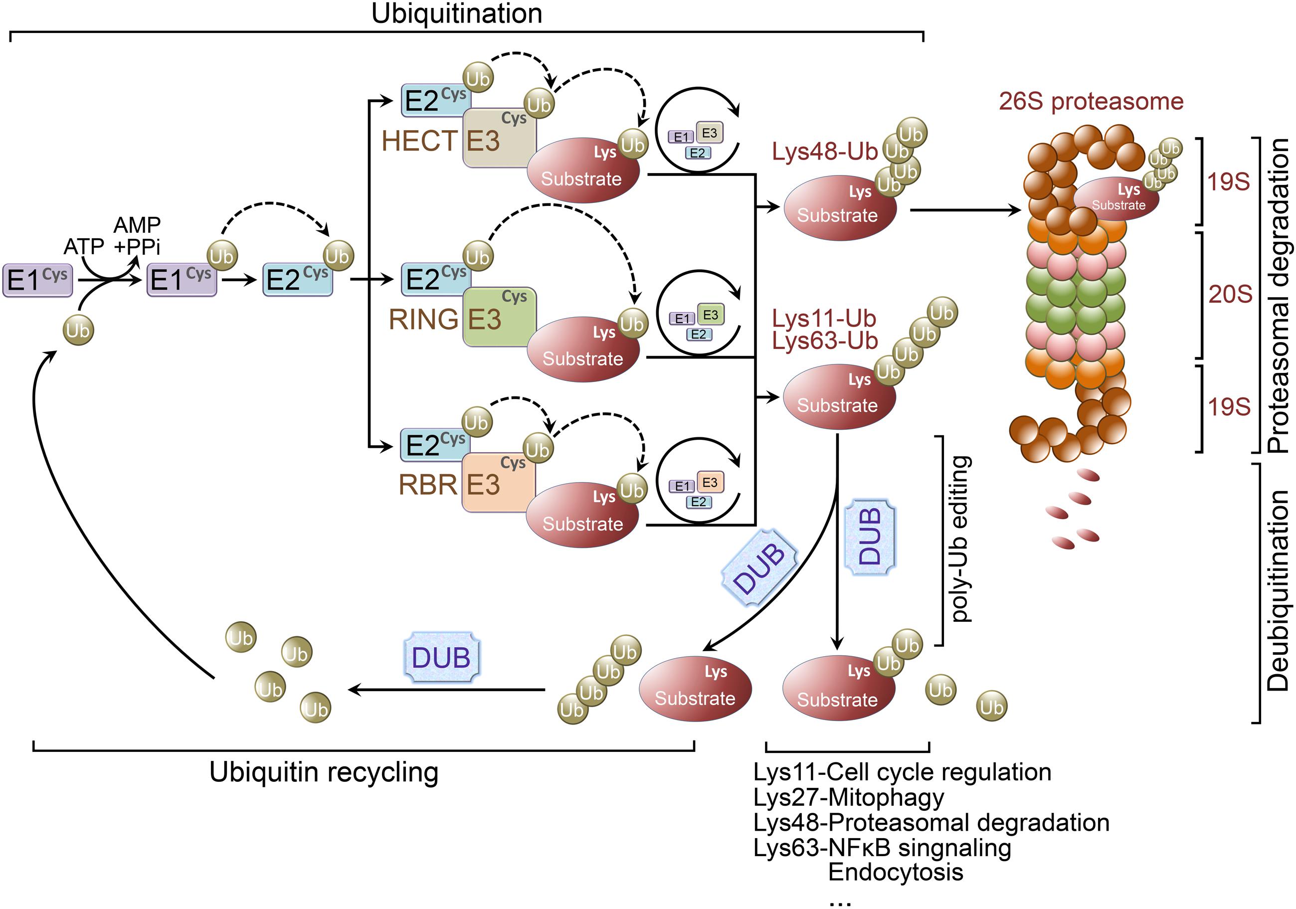 Figure 1 Ubiquitination and ubiquitin-proteasome system. (Zheng, 2016)
Representative Targets in Ubiquitin-Proteasome Pathway
UCHL1
UCHL1 (Ubiquitin C-Terminal Hydrolase L1), also known as PGP9.5, is a protein that plays a critical role in the ubiquitin-proteasome system (UPS). This enzyme is highly expressed in neuronal tissues, where it is involved in the processing of ubiquitin precursors as well as the recycling of ubiquitin from degraded proteins, thereby maintaining cellular protein homeostasis. UCHL1's activity is essential for the removal of misfolded or damaged proteins, preventing protein aggregation that can lead to cellular dysfunction and disease. In addition to its well-established function in the UPS, UCHL1 has been implicated in various cellular processes including apoptosis, cell cycle regulation, and synaptic function. The dysregulation of UCHL1 activity has been associated with a number of neurodegenerative diseases, such as Parkinson's disease (PD) and Alzheimer's disease (AD), where its altered expression and mutations are linked to the accumulation of toxic protein aggregates and neuronal cell death. Furthermore, UCHL1 has been found to be involved in cancer progression, where it can function either as an oncogene or a tumor suppressor, depending on the cellular context and type of cancer.
Recommended Mouse Anti-UCHL1 mAb (CAT#: MOB-0186F)
 Figure 2 Mouse Anti-UCHL1 Recombinant Antibody (MOB-0186F) in IHC. Immunohistochemical analysis of paraffin-embedded Kindey. 1. The antibody is diluted 1:200 (overnight at 4°C). 2. Use pH8.0 TRIS-EDTA for antigen retrieval. 3. Dilute the secondary antibody at 1:200 (room temperature, 30min).
Recommended Mouse Anti-UCHL1 mAb (CAT#: MOB-0187F)
 Figure 3 Mouse Anti-UCHL1 Recombinant Antibody (MOB-0187F) in IHC. Immunohistochemical analysis of paraffin-embedded pancreas. 1. The antibody is diluted 1:200 (overnight at 4°C). 2. Tris-EDTA, pH8.0 is used for antigen retrieval. 3. Dilute the secondary antibody at 1:200 (room temperature, 30min).
Recommended Mouse Anti-UCHL1 mAb (CAT#: ZG-0493F)
 Figure 4 Mouse Anti-UCHL1 Recombinant Antibody (ZG-0493F) in ICC. UCHL1/PGP9.5 mouse mAb (1:300) was used for immunocytochemical staining of COS7.
USP7
USP7 (Ubiquitin-Specific Protease 7), also known as HAUSP (Herpesvirus-Associated Ubiquitin-Specific Protease), plays a critical role in cellular processes through its function in the ubiquitin-proteasome system. By specifically cleaving ubiquitin moieties from ubiquitinated proteins, USP7 regulates the stability and function of its target substrates, which include key regulatory proteins involved in cell cycle control, apoptosis, DNA repair, and immune responses. This modulation of protein stability by USP7 is crucial for maintaining cellular homeostasis and responding to stress signals. The significance of USP7 extends to its roles in disease progression, particularly in cancer, where its activity influences tumor growth and survival through the regulation of tumor suppressor proteins like p53 and PTEN, as well as oncoproteins such as MDM2. Aberrant expression or mutation of USP7 has been linked to the development and progression of various cancers, underscoring its potential as a therapeutic target. Moreover, USP7's involvement in the regulation of immune responses highlights its importance in autoimmune diseases and inflammation. The enzyme's role in DNA repair mechanisms also implicates it in the response to genomic stress and the prevention of mutations that could lead to malignancies.
Recommended Mouse Anti-USP7 mAb (CAT#: ZG-0408J)
 Figure 5 Mouse Anti-USP7 Recombinant Antibody (ZG-0408J) in IP. Immunoprecipitation analysis of Hela cell lysate using Ikaros (C-terminus) mouse mAb.
Recommended Rabbit Anti-USP7 mAb (CAT#: ZG-0618U)
 Figure 6 Rabbit Anti-USP7 Antibody (ZG-0618U) in IHC. IHC image of ZG-0618U diluted at 1:100 and staining in paraffin-embedded human prostate cancer performed on a Leica Bond™ system. After dewaxing and hydration, antigen retrieval was mediated by high pressure in a citrate buffer (pH 6.0). Section was blocked with 10% normal goat serum 30min at RT. Then primary antibody (1% BSA) was incubated at 4°C overnight. The primary is detected by a Goat anti-rabbit IgG polymer labeled by HRP and visualized using 0.05% DAB.
VHL
The Von Hippel-Lindau (VHL) protein is a pivotal tumor suppressor involved in the regulation of gene expression, maintenance of cellular oxygen homeostasis, and the ubiquitination and subsequent proteasomal degradation of hypoxia-inducible factors (HIFs). Through its central role in the oxygen-sensing pathway, VHL mediates the degradation of HIFs under normoxic conditions, thereby preventing the activation of oxygen-regulated genes involved in angiogenesis, erythropoiesis, and glucose metabolism. The loss of VHL function, due to mutations or inactivation, leads to the stabilization of HIFs under normoxic conditions, triggering the aberrant activation of genes that promote tumor growth, vascularization, and metabolic reprogramming, hallmarks of cancer progression. VHL's significance extends beyond its role in oxygen sensing; it is also implicated in the extracellular matrix formation, microtubule dynamics, and primary cilium maintenance, which are essential for cellular polarity and signaling. Dysregulation of these processes due to VHL mutations contributes to the pathogenesis of Von Hippel-Lindau disease, a hereditary cancer syndrome characterized by the development of tumors and cysts in multiple organs, including the brain, spine, retina, and kidneys, particularly clear cell renal cell carcinoma (ccRCC).
Recommended Mouse Anti-VHL mAb (CAT#: MOB-0233F)
 Figure 7 Mouse Anti-VHL Recombinant Antibody (MOB-0233F) in IHC. Immunohistochemical analysis of paraffin-embedded Kindey. 1. The antibody is diluted 1:200 (overnight at 4°C). 2. Tris-EDTA, pH8.0 is used for antigen retrieval. 3. Dilute the secondary antibody at 1:200 (room temperature, 30min).
Recommended Mouse Anti-VHL mAb (CAT#: MOB-0234F)
 Figure 8 Mouse Anti-VHL Recombinant Antibody (MOB-0234F) in IHC. Immunohistochemical analysis of paraffin-embedded Kindey. 1. The antibody is diluted 1:200 (overnight at 4°C). 2. Tris-EDTA, pH8.0 is used for antigen retrieval. 3. Dilute the secondary antibody at 1:200 (room temperature, 30min).
Full List of Targets in Ubiquitin-Proteasome Pathway
| Biomarker | Alternative Names | Gene ID | UniProt ID | Roles |
| Cdc34 | UBC3; UBCH3; UBE2R1; E2-CDC34 | 997 | P49427 | The protein encoded by this gene is a member of the ubiquitin-conjugating enzyme family. Ubiquitin-conjugating enzyme catalyzes the covalent attachment of ubiquitin to other proteins. This protein is a part of the large multiprotein complex, which is required for ubiquitin-mediated degradation of cell cycle G1 regulators, and for the initiation of DNA replication. |
| CRBN | CRBN; Protein X 0001; MRT2A; Mental Retardation, Non-Syndromic, Autosomal Recessive, 2A; Cereblon | 51185 | Q96SW2 | This gene encodes a protein related to the Lon protease protein family. In rodents and other mammals this gene product is found in the cytoplasm localized with a calcium channel membrane protein, and is thought to play a role in brain development. Mutations in this gene are associated with autosomal recessive nonsyndromic mental retardation. Multiple transcript variants encoding different isoforms have been found for this gene. |
| FBXW7 | F-Box And WD Repeat Domain Containing 7; F-Box And WD Repeat Domain Containing 7, E3 Ubiquitin Protein Ligase; F-Box And WD-40 Domain Protein 7 (Archipelago Homolog, Drosophila); F-Box Protein FBX30; SEL-10; Fbx30; SEL10; HCdc4; FBW7; HAgo; F-Box And WD-40 Domain-Containing Protein 7; F-Box/WD Repeat-Containing Protein 7; | 55294 | Q969H0 | This gene encodes a member of the F-box protein family which is characterized by an approximately 40 amino acid motif, the F-box. The F-box proteins constitute one of the four subunits of ubiquitin protein ligase complex called SCFs (SKP1-cullin-F-box), which function in phosphorylation-dependent ubiquitination. The F-box proteins are divided into 3 classes: Fbws containing WD-40 domains, Fbls containing leucine-rich repeats, and Fbxs containing either different protein-protein interaction modules or no recognizable motifs. The protein encoded by this gene was previously referred to as FBX30, and belongs to the Fbws class; in addition to an F-box, this protein contains 7 tandem WD40 repeats. This protein binds directly to cyclin E and probably targets cyclin E for ubiquitin-mediated degradation. Mutations in this gene are detected in ovarian and breast cancer cell lines, implicating the gene's potential role in the pathogenesis of human cancers. Multiple transcript variants encoding different isoforms have been found for this gene. [provided by RefSeq, Mar 2012] |
| MDM2 | MDM2 Proto-Oncogene; MDM2 Proto-Oncogene, E3 Ubiquitin Protein Ligase; Oncoprotein Mdm2; Hdm2; Mdm2, Transformed 3T3 Cell Double Minute 2, P53 Binding Protein (Mouse); Mdm2, Transformed 3T3 Cell Double Minute 2, P53 Binding Protein; Mouse Double Minute 2, Human Homolog Of; P53-Binding Protein; Double Minute 2, Human Homolog Of; P53-Binding Protein; Mdm2, P53 E3 Ubiquitin Protein Ligase Homolog; MDM2 Oncogene, E3 Ubiquitin Protein Ligase | 4193 | A7UKX8 | This gene encodes a nuclear-localized E3 ubiquitin ligase. The encoded protein can promote tumor formation by targeting tumor suppressor proteins, such as p53, for proteasomal degradation. This gene is itself transcriptionally-regulated by p53. Overexpression or amplification of this locus is detected in a variety of different cancers. There is a pseudogene for this gene on chromosome 2. Alternative splicing results in a multitude of transcript variants, many of which may be expressed only in tumor cells. [provided by RefSeq, Jun 2013] |
| MDM4 | HDMX; MDMX; MRP1 | 4194 | O15151 | This gene encodes a nuclear protein that contains a p53 binding domain at the N-terminus and a RING finger domain at the C-terminus, and shows structural similarity to p53-binding protein MDM2. Both proteins bind the p53 tumor suppressor protein and inhibit its activity, and have been shown to be overexpressed in a variety of human cancers. However, unlike MDM2 which degrades p53, this protein inhibits p53 by binding its transcriptional activation domain. This protein also interacts with MDM2 protein via the RING finger domain, and inhibits the latter's degradation. So this protein can reverse MDM2-targeted degradation of p53, while maintaining suppression of p53 transactivation and apoptotic functions. Alternatively spliced transcript variants encoding different isoforms have been noted for this gene. [provided by RefSeq, Feb 2011] |
| NAE1 | NAE1; Human NAE1 | 8883 | Q13564 | |
| UCHL1 | Ubiquitin C-Terminal Hydrolase L1; Ubiquitin Carboxyl-Terminal Esterase L1 (Ubiquitin Thiolesterase); Neuron Cytoplasmic Protein 9.5; Ubiquitin Thioesterase L1; Ubiquitin Thiolesterase; PGP 9.5; Uch-L1; PGP9.5; Ubiquitin Carboxyl-Terminal Hydrolase Isozyme L1; Epididymis Secretory Protein Li 53 | 7345 | P09936 | The protein encoded by this gene belongs to the peptidase C12 family. This enzyme is a thiol protease that hydrolyzes a peptide bond at the C-terminal glycine of ubiquitin. This gene is specifically expressed in the neurons and in cells of the diffuse neuroendocrine system. Mutations in this gene may be associated with Parkinson disease.[provided by RefSeq, Sep 2009] |
| UCHL5 | Ubiquitin C-Terminal Hydrolase L5; Ubiquitin Carboxyl-Terminal Hydrolase L5; Ubiquitin C-Terminal Hydrolase UCH37; Ubiquitin Thioesterase L5; INO80 Complex Subunit R; EC 3.4.19.12; UCH-L5 | 51377 | Q9Y5K5 | Protease that specifically cleaves Lys-48-linked polyubiquitin chains. Deubiquitinating enzyme associated with the 19S regulatory subunit of the 26S proteasome. Putative regulatory component of the INO80 complex; however is inactive in the INO80 complex and is activated by a transient interaction of the INO80 complex with the proteasome via ADRM1. |
| USP14 | TGT; Ubp6 | 9097 | P54578 | This gene encodes a member of the ubiquitin-specific processing (UBP) family of proteases that is a deubiquitinating enzyme (DUB) with His and Cys domains. This protein is located in the cytoplasm and cleaves the ubiquitin moiety from ubiquitin-fused precursors and ubiquitinylated proteins. Mice with a mutation that results in reduced expression of the ortholog of this protein are retarded for growth, develop severe tremors by 2 to 3 weeks of age followed by hindlimb paralysis and death by 6 to 10 weeks of age. Alternate transcriptional splice variants, encoding different isoforms, have been characterized. |
| USP5 | ISOT | 8078 | P45974 | Ubiquitin (see MIM 191339)-dependent proteolysis is a complex pathway of protein metabolism implicated in such diverse cellular functions as maintenance of chromatin structure, receptor function, and degradation of abnormal proteins. A late step of the process involves disassembly of the polyubiquitin chains on degraded proteins into ubiquitin monomers. USP5 disassembles branched polyubiquitin chains by a sequential exo mechanism, starting at the proximal end of the chain (Wilkinson et al., 1995 [PubMed 7578059]). |
| USP7 | Ubiquitin Specific Peptidase 7; Ubiquitin Specific Peptidase 7 (Herpes Virus-Associated); Ubiquitin-Specific-Processing Protease 7; Deubiquitinating Enzyme 7; Ubiquitin Thioesterase 7; HAUSP; Ubiquitin Specific Protease 7 (Herpes Virus-Associated) | 7874 | B7ZAX6 | The protein encoded by this gene belongs to the peptidase C19 family, which includes ubiquitinyl hydrolases. This protein deubiquitinates target proteins such as p53 (a tumor suppressor protein) and WASH (essential for endosomal protein recycling), and regulates their activities by counteracting the opposing ubiquitin ligase activity of proteins such as HDM2 and TRIM27, involved in the respective process. Mutations in this gene have been implicated in a neurodevelopmental disorder. [provided by RefSeq, Mar 2016] |
| VHL | Von Hippel-Lindau Tumor Suppressor; Von Hippel-Lindau Tumor Suppressor, E3 Ubiquitin Protein Ligase; Protein G7; PVHL; Von Hippel-Lindau Disease Tumor Suppressor; Von Hippel-Lindau Syndrome | 7428 | P40337 | Von Hippel-Lindau syndrome (VHL) is a dominantly inherited familial cancer syndrome predisposing to a variety of malignant and benign tumors. A germline mutation of this gene is the basis of familial inheritance of VHL syndrome. The protein encoded by this gene is a component of the protein complex that includes elongin B, elongin C, and cullin-2, and possesses ubiquitin ligase E3 activity. This protein is involved in the ubiquitination and degradation of hypoxia-inducible-factor (HIF), which is a transcription factor that plays a central role in the regulation of gene expression by oxygen. RNA polymerase II subunit POLR2G/RPB7 is also reported to be a target of this protein. Alternatively spliced transcript variants encoding distinct isoforms have been observed. [provided by RefSeq, Jul 2008] |
Tested Data-Supported Products for Targeting Ubiquitin-Proteasome Pathway


Reference
- Zheng, Qiuyang, et al. "Dysregulation of ubiquitin-proteasome system in neurodegenerative diseases." Frontiers in aging neuroscience 8 (2016): 303. Distributed under Open Access license CC BY 4.0, without modification.
For Research Use Only. Not For Clinical Use.