
Human Anti-IGF2 Recombinant Antibody (clone DX-2647) (CAT#: PABL-231)
Recombinant Human Antibody (DX-2647) is capable of binding to IGF-II, expressed in HEK 293 cells. Expressed as the combination of a heavy chain (HC) containing VH from anti-IGF-II mAb and CH1-3 region of human IgG1 and a light chain (LC) encoding VL from anti-IGF-II mAb and CL of human kappa light chain. Exists as a disulfide linked dimer of the HC and LC hetero-dimer under non-reducing condition.
Specific Inquiry

Figure 1 HepG2 cells are resistant to DX-2647 therapy in vivo.
Hep3B (left panel) and HepG2 (right panel) xenografts were established to 100 mm3 and treated with vehicle or DX-2647 for 32 days. Data are presented as the mean tumor volume (mm3) at each time point postinoculation ± S.E.M.
Greenall, S. A., Donoghue, J., Johns, T. G., & Adams, T. E. (2018). Differential Sensitivity of Human Hepatocellular Carcinoma Xenografts to an IGF-II Neutralizing Antibody May Involve Activated STAT3. Translational oncology, 11(4), 971-978.

Figure 2 Characterization of the IGF-II axis in Hep3B and HepG2 cells.
(A) Cells were treated with vehicle or 20 μg/ml DX-2647 for 48 hours and then tested on activated RTK arrays. Black dots indicate a positive activation signal. (1) p-IR, (2) p-IGF-IR, and (3) p-HER3. Signals in each array corner are positive controls. (B) Western analyses of IGF-II species contained within concentrated and conditioned cell supernatants, treated with vehicle or an O-glycosyltransferase inhibitor (OGTi). IGF-II isoforms are indicated by black arrows. (C) Western analyses of IGFBP expression in cell lysates isolated from Hep3B and HepG2 cells. IGFBP2, IGFBP5, and IGFBP6 were not detected. (D) Immunoprecipitation analysis of bioavailable IGF-II bound from concentrated and conditioned cell supernatants followed by IGF-II blotting. IGF-II species are indicated by black arrows.
Greenall, S. A., Donoghue, J., Johns, T. G., & Adams, T. E. (2018). Differential Sensitivity of Human Hepatocellular Carcinoma Xenografts to an IGF-II Neutralizing Antibody May Involve Activated STAT3. Translational oncology, 11(4), 971-978.

Figure 3 Western blotting of IR and IGF-IR receptor status and downstream signaling status in cell lysates isolated from Hep3B and HepG2 cells treated with vehicle or 20 μg/ml DX-2647 for 48 hours.
Greenall, S. A., Donoghue, J., Johns, T. G., & Adams, T. E. (2018). Differential Sensitivity of Human Hepatocellular Carcinoma Xenografts to an IGF-II Neutralizing Antibody May Involve Activated STAT3. Translational oncology, 11(4), 971-978.

Figure 4 DX-2647 blocks IGF-II-stimulated proliferation.
A, IGF-II/IGF-IIE synergizes with IL-4 to stimulate Ba/F3 cell proliferation. Ba/F3 cells were stimulated with the indicated cytokines, growth factors, or combinations thereof in the presence or absence of lipopolysaccharide (LPS). Proliferation was assessed after 2 d. B, representative plot of DX-2647-induced inhibition of IGF-II-stimulated and IGF-IIE (100 ng/mL)-stimulated Ba/F3 cell proliferation.
Dransfield, D. T., Cohen, E. H., Chang, Q., Sparrow, L. G., Bentley, J. D., Dolezal, O.,... & Phan, T. (2010). A human monoclonal antibody against insulin-like growth factor-II blocks the growth of human hepatocellular carcinoma cell lines in vitro and in vivo. Molecular cancer therapeutics, 9(6), 1809-1819.

Figure 5 DX-2647 blocks IGF-driven cell signaling events.
Dose-dependent inhibition of IGF-II stimulated phosphorylation of IGF-IR (A) or IR-A (B) in transfected NIH-3T3 cells. C, DX-2647 blocks exogenous and endogenous IGF-II/IGF-I-mediated phosphorylation of IGF-IR in select cancer cell lines.
Dransfield, D. T., Cohen, E. H., Chang, Q., Sparrow, L. G., Bentley, J. D., Dolezal, O.,... & Phan, T. (2010). A human monoclonal antibody against insulin-like growth factor-II blocks the growth of human hepatocellular carcinoma cell lines in vitro and in vivo. Molecular cancer therapeutics, 9(6), 1809-1819.

Figure 6 DX-2647 inhibits anchorage-independent and anchorage-dependent colony formation in HCC cell lines.
A, HepG2 cell anchorage-independent colony formation. Representative images of colonies are shown (inset). B, Hep3B-cell anchorage-dependent colony formation. Representative images for each respective dose of DX-2647 tested are shown above each bar.
Dransfield, D. T., Cohen, E. H., Chang, Q., Sparrow, L. G., Bentley, J. D., Dolezal, O.,... & Phan, T. (2010). A human monoclonal antibody against insulin-like growth factor-II blocks the growth of human hepatocellular carcinoma cell lines in vitro and in vivo. Molecular cancer therapeutics, 9(6), 1809-1819.

Figure 7 DX-2647 slows tumor progression in the Hep3B hepatocellular xenograft model.
A, tumor burden graph depicting slowing of tumor progression by DX-2647. B, tumor wet weight after tumor excision (day 39). C, animal body weight over time indicates no significant changes due to treatment regimen.
Dransfield, D. T., Cohen, E. H., Chang, Q., Sparrow, L. G., Bentley, J. D., Dolezal, O.,... & Phan, T. (2010). A human monoclonal antibody against insulin-like growth factor-II blocks the growth of human hepatocellular carcinoma cell lines in vitro and in vivo. Molecular cancer therapeutics, 9(6), 1809-1819.

Figure 8 Immunohistochemical analysis of tumors from DX-2647-treated animals reveals decreased CD31 staining and reduced levels of IGF-IIE and IGF-IR phosphorylation levels.
A, H&E and immunohistochemical evaluation of human xenograft Hep3B tumors. Tumor sections were stained with H&E and immunohistochemistry for human IgG1 (hIgG1), TUNEL assay (brown), and CD31 (red). Pictures are representative of four animals from each group. Apo, apoptosis. Dotted line delineates apoptotic areas. Magnifications, ×400 (a, b, c1-c3, and g-o) and ×100 (d-f). B, immunohistochemical evaluation IGF pathway molecules. Tumor sections were stained for IGF-IIE, IGF-II, IGF-I, IGF-IR, and IGF-IR-phospho.
Dransfield, D. T., Cohen, E. H., Chang, Q., Sparrow, L. G., Bentley, J. D., Dolezal, O.,... & Phan, T. (2010). A human monoclonal antibody against insulin-like growth factor-II blocks the growth of human hepatocellular carcinoma cell lines in vitro and in vivo. Molecular cancer therapeutics, 9(6), 1809-1819.


Specifications
- Immunogen
- Human insulin-like growth factor 2
- Host Species
- Human
- Type
- Human IgG
- Specificity
- Human IGF2
- Species Reactivity
- Human
- Clone
- DX-2647
- Applications
- WB, ELISA, FuncS
Product Property
- Purity
- >95% as determined by SDS-PAGE and HPLC analysis
- Concentration
- Please refer to the vial label for the specific concentration.
- Storage
- Centrifuge briefly prior to opening vial. Store at +4°C short term (1-2 weeks). Aliquot and store at -20°C long term. Avoid repeated freeze/thaw cycles.
Applications
- Application Notes
- The antibody was validated for Block, Inhibition and Function Assay. For details, refer to Published Data.
Target
- Alternative Names
- IGF2; insulin-like growth factor 2; IGF-II; PP9974; C11orf43; insulin-like growth factor II; insulin-like growth factor type 2; insulin-like growth factor 2 (somatomedin A)
- Gene ID
- 3481
- UniProt ID
- P01344
Related Resources
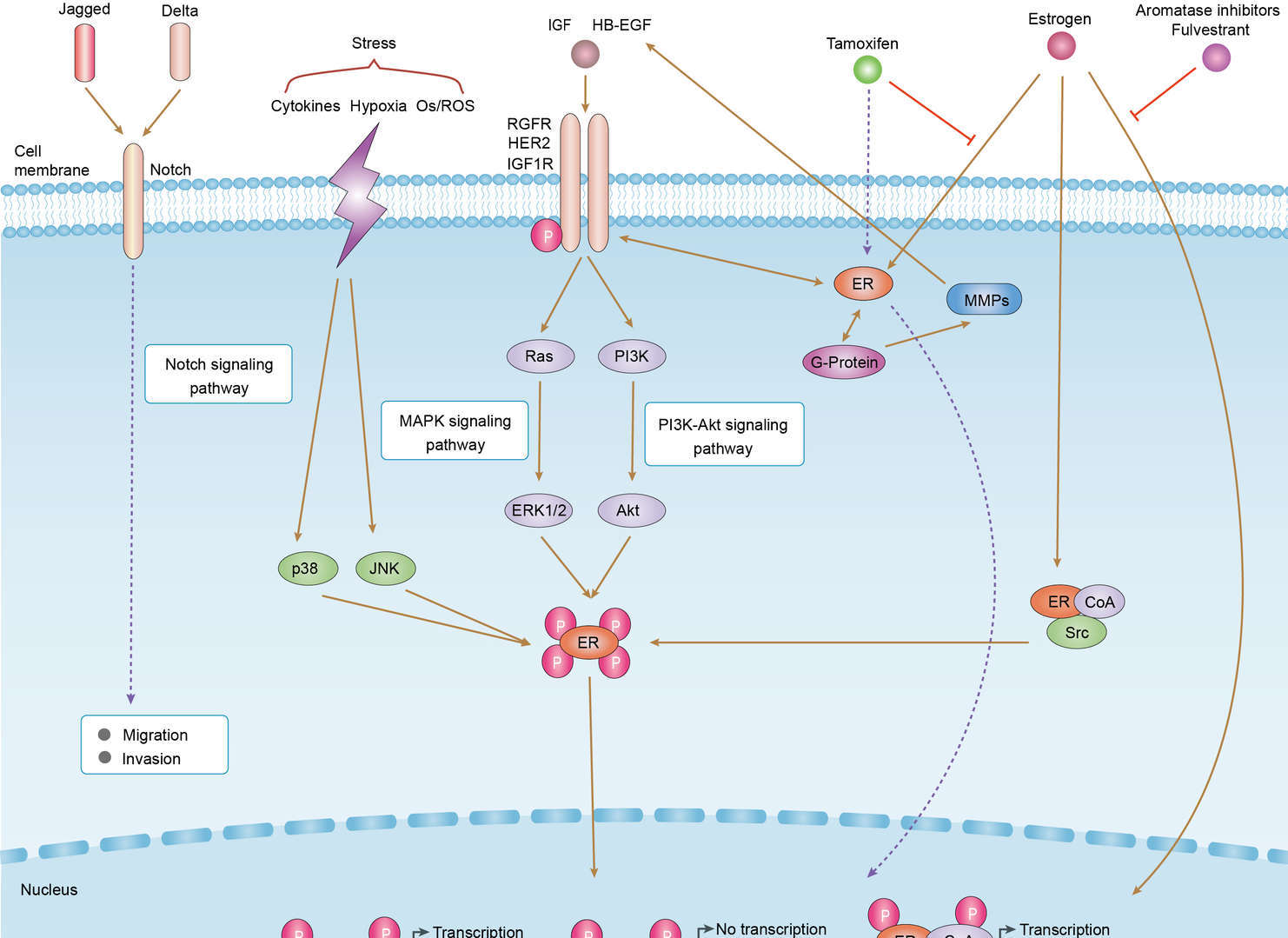 Endocrine Resistance
Product Notes
This is a product of Creative Biolabs' Hi-Affi™ recombinant antibody portfolio, which has several benefits including:
• Increased sensitivity
• Confirmed specificity
• High repeatability
• Excellent batch-to-batch consistency
• Sustainable supply
• Animal-free production
See more details about Hi-Affi™ recombinant antibody benefits.
Downloads
Download resources about recombinant antibody development and antibody engineering to boost your research.
See other products for "Clone DX-2647"
See other products for "IGF2"
Human Antibody
| CAT | Product Name | Application | Type |
|---|---|---|---|
| TAB-740 | Anti-IGF2 Recombinant Antibody (Dusigitumab) | Neut, ELISA, IF, IP, FuncS, FC, ICC | IgG2 |
| TAB-387CL | Anti-Human IGF2 Recombinant Antibody (DX-2647) | BL, Inhib | Antibody |
Neutralizing Antibody
| CAT | Product Name | Application | Type |
|---|---|---|---|
| NEUT-1083CQ | Mouse Anti-IGF2 Recombinant Antibody (clone CBL493) | Neut | Mouse IgG1 |
Rabbit Monoclonal Antibody
| CAT | Product Name | Application | Type |
|---|---|---|---|
| MOR-1756 | Rabbit Anti-IGF2 Recombinant Antibody (clone DS1756AB) | ELISA | Rabbit IgG |
scFv Fragment Antibody
| CAT | Product Name | Application | Type |
|---|---|---|---|
| HPAB-0037LY-S(P) | Human Anti-IGF2 Recombinant Antibody; scFv Fragment (HPAB-0037LY-S(P)) | WB | Human scfv |
| HPAB-0613-WJ-S(P) | Human Anti-IGF2 Recombinant Antibody (clone m610.27); scFv Fragment | ELISA, WB | Humanized scFv |
| HPAB-1733-FY-F(E) | Human Anti-IGF2 Recombinant Antibody (clone M606); scFv Fragment | Inhib, WB, ELISA | Human scFv |
| HPAB-1734-FY-F(E) | Human Anti-IGF2 Recombinant Antibody (clone m610); scFv Fragment | Inhib, WB, ELISA | Human scFv |
| HPAB-1735-FY-F(E) | Human Anti-IGF2 Recombinant Antibody (clone m616); scFv Fragment | Inhib, WB, ELISA | Human scFv |
Fab Fragment Antibody
| CAT | Product Name | Application | Type |
|---|---|---|---|
| HPAB-0037LY-F(E) | Human Anti-IGF2 Recombinant Antibody; Fab Fragment (HPAB-0037LY-F(E)) | WB | Human Fab |
| HPAB-0613-WJ-F(E) | Human Anti-IGF2 Recombinant Antibody (clone m610.27); Fab Fragment | ELISA, WB | Humanized Fab |
| HPAB-1733-FY-S(P) | Human Anti-IGF2 Recombinant Antibody (clone M606); Fab Fragment | Inhib, WB, ELISA | Human Fab |
| HPAB-1734-FY-S(P) | Human Anti-IGF2 Recombinant Antibody (clone m610); Fab Fragment | Inhib, WB, ELISA | Human Fab |
| HPAB-1735-FY-S(P) | Human Anti-IGF2 Recombinant Antibody (clone m616); Fab Fragment | Inhib, WB, ELISA | Human Fab |
Recombinant Antibody
| CAT | Product Name | Application | Type |
|---|---|---|---|
| MRO-0818-CN | Rabbit Anti-IGF2 Recombinant Antibody (clone CBACN-300) | WB, IF, IHC | Rabbit IgG |
| HPAB-1733-FY | Human Anti-IGF2 Recombinant Antibody (clone M606) | Inhib, WB, ELISA | Human IgG |
| HPAB-1734-FY | Human Anti-IGF2 Recombinant Antibody (clone m610) | Inhib, WB, ELISA | Human IgG |
| HPAB-1735-FY | Human Anti-IGF2 Recombinant Antibody (clone m616) | ELISA | Human IgG |
| MOB-251YJL | Rabbit Anti-IGF2 Recombinant Antibody (clone CBI236) | ELISA | Rabbit IgG |
ADCC Enhanced Antibody
| CAT | Product Name | Application | Type |
|---|---|---|---|
| AFC-TAB-740 | Afuco™ Anti-IGF2 ADCC Recombinant Antibody (Dusigitumab), ADCC Enhanced | Neut, ELISA, IF, IP, FuncS, FC | ADCC enhanced antibody |
Customer Reviews and Q&As

There are currently no Customer reviews or questions for PABL-231. Click the button above to contact us or submit your feedback about this product.
View the frequently asked questions answered by Creative Biolabs Support.
For Research Use Only. Not For Clinical Use.
For research use only. Not intended for any clinical use. No products from Creative Biolabs may be resold, modified for resale or used to manufacture commercial products without prior written approval from Creative Biolabs.
Send Inquiry
This site is protected by reCAPTCHA and the Google Privacy Policy and Terms of Service apply.