

AbPlus™ Anti-RXRB Magnetic Beads (VS-0724-YC664)
CAT#: VS-0724-YC664
The AbPlus Anti-RXRB Magnetic Beads (VS-0724-YC664) is an innovative affinity resin which is bound with anti-RXRB specific antibody. The beads were designed for small-scale affinity purification and immunoprecipitation (IP) of RXRB protein under native and denaturing conditions.
Gene Expression

Subcellular Location
Figure 1 IF staining of human cell line SK-MEL-30
Immunofluorescent staining of human cell line SK-MEL-30 shows localization to nucleoplasm.
* Image credit: Image credit: Human Protein Atlas v21.proteinatlas.org/images/63653/1262_G9_2_selected.jpg

Normal Tissue
Figure 2 Cerebral cortex
Endothelial cells Staining: Medium Intensity: Moderate Quantity:>75% Location: Nuclear Glial cells Staining: High Intensity: Strong Quantity:>75% Location: nuclear Neuronal cells Staining: High Intensity: Strong Quantity:>75% Location: Nuclear
* Image credit: Image credit: Human Protein Atlas v21.proteinatlas.org/images/2003/5372_B_8_5.jpg

Normal Tissue
Figure 3 Colon
Endothelial cells Staining: Medium Intensity: Moderate Quantity:>75% Location: Nuclear Glandular cells Staining: High Intensity: Strong Quantity:>75% Location: Nuclear Peripheral nerve/ganglion Staining: Medium Intensity: Moderate Quantity:>75% Location: Nuclear
* Image credit: Image credit: Human Protein Atlas v21.proteinatlas.org/images/2003/5372_A_9_3.jpg

Normal Tissue
Figure 4 Liver
Cholangiocytes Staining: Medium Intensity: Moderate Quantity:>75% Location: Nuclear Hepatocytes Staining: Low Intensity: Weak Quantity:>75% Location: Cytoplasmic/ membranous
* Image credit: Image credit: Human Protein Atlas v21.proteinatlas.org/images/2003/5372_A_8_4.jpg

Normal Tissue
Figure 5 Kidney
Cells in glomeruli Staining: High Intensity: Strong Quantity:>75% Location: nuclear Cells in tubules Staining: Medium Intensity: Moderate Quantity:>75% Location: Nuclear
* Image credit: Image credit: Human Protein Atlas v21.proteinatlas.org/images/2003/5372_A_9_5.jpg

Normal Tissue
Figure 6 Testis
Cells in seminiferous ducts Staining: Low Intensity: Weak Quantity: 75%-25% Location: nuclear Leydig cells Staining: High Intensity: Strong Quantity: 75%-25% Location: Cytoplasmic/ membranous nuclear
* Image credit: Image credit: Human Protein Atlas v21.proteinatlas.org/images/2003/5372_A_4_6.jpg

Normal Tissue
Figure 7 Lymph node
Germinal center cells Staining: High Intensity: Strong Quantity:>75% Location: Nuclear Non-germinal center cells Staining: High Intensity: Strong Quantity:>75% Location: Nuclear
* Image credit: Image credit: Human Protein Atlas v21.proteinatlas.org/images/2003/5372_A_8_8.jpg

RNA Expression
Figure 8 RNA cell line category: Cell line enhanced (GAMG, RH-30, RT4, T-47d, U-266/70, U-266/84)
Cell lines ordered by descending RNA expression order
* Image credit: Image credit: Human Protein Atlas v21.proteinatlas.org/ENSG00000204231-RXRB
❮


 ❯
❯
Specifications
- Applications
- Immunoprecipitation, Protein Purification
- Matrix
- Magnetic bead
- Bead Ligand
- Anti-RXRB specific antibody
- Target
- RXRB
- Immunogen
- Recombinant human Rxrb fragment derived from E. coli.
- Target Species
- Human
- Bead Capacity
- 40 mg/mL
- Bead size
- 25 μm
- Format
- Suspension
- Concentration
- 2 mg/mL
- Buffer
- PBS, pH 7.4
- Preservative
- 0.1% Sodium azide
- Storage
- Stored at 4°C, and is stable for up to 2 years. Do not centrifuge, dry or freeze the magnetic beads.
Applications
- Application Notes
- The beads are in suspension and will settle upon storage. Prior to use, mix the vial gently (do not vortex) to ensure delivery of proper bead volume.
Target
- Introduction
- This gene encodes a member of the retinoid X receptor (RXR) family of nuclear receptors which are involved in mediating the effects of retinoic acid (RA). The encoded protein forms homodimers with the retinoic acid, thyroid hormone, and vitamin D receptors, increasing both DNA binding and transcriptional function on their respective response elements. This gene lies within the major histocompatibility complex (MHC) class II region on chromosome 6. Alternatively spliced transcript variants encoding multiple isoforms have been observed for this gene. [provided by RefSeq, Jul 2012]
- Alternative Names
- Retinoid X Receptor Beta; Nuclear Receptor Subfamily 2 Group B Member 2; NR2B2; MHC Class I Promoter Binding Protein; Retinoic Acid Receptor RXR-Beta; Retinoid X Receptor, Beta; H-2RIIBP; DAUDI6; RCoR-1;
- Gene ID
- 6257
- UniProt ID
- P28702
REVIEWS AND Q&AS
CITATIONS
RESOURCES
DOWNLOADS
RELATED PRODUCTS
Inquiry
Navs
Customer Review

There are currently no Customer reviews or questions for VS-0724-YC664. Click the button above to contact us or submit your feedback about this product.
Submit Your Publication
Published with our product? Submit your paper and receive a 10% discount on your next order! Share your research to earn exclusive rewards.
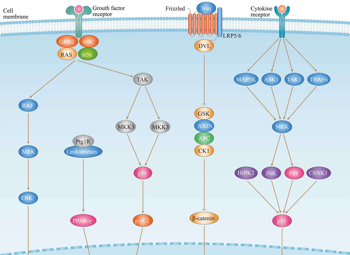
Related Signaling Pathways
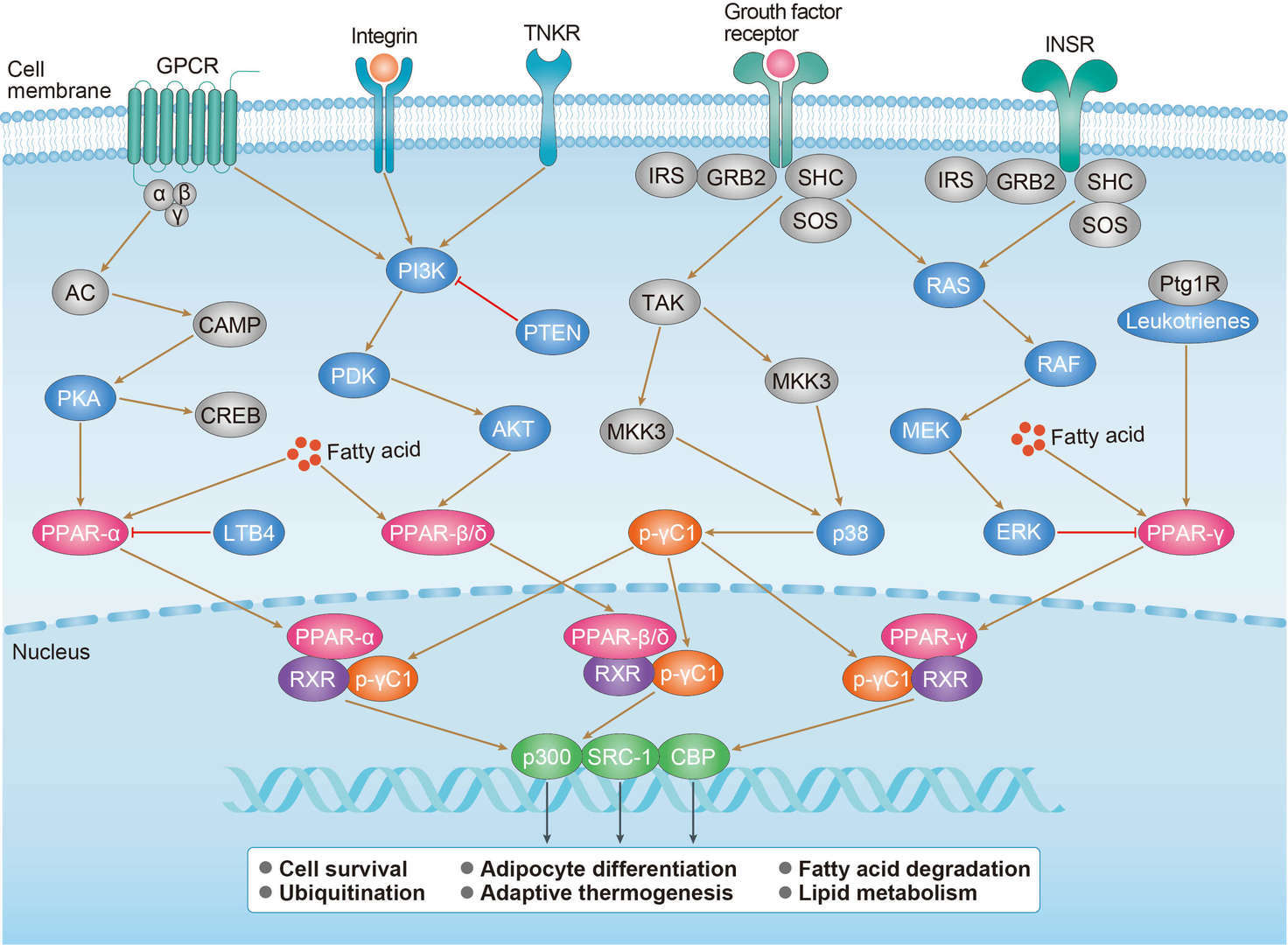 PPAR Signaling Pathway
Related Diseases
 Throid Cancer
Downloadable Resources
Download resources about recombinant antibody development and antibody engineering to boost your research.
Datasheet
MSDS
COA
Certificate of Analysis LookupTo download a Certificate of Analysis, please enter a lot number in the search box below. Note: Certificate of Analysis not available for kit components.
Lot Number:
See other products for "RXRB"
Select a product category from the dropdown menu below to view related products.
| CAT | Product Name | Application | Type |
|---|---|---|---|
| MOB-3864z | Mouse Anti-RXRB Recombinant Antibody (clone 11C1) | WB, IP, IF, ELISA | Mouse IgG1, κ |
| CAT | Product Name | Application | Type |
|---|---|---|---|
| MOB-1812CT | Recombinant Mouse anti-Human RXRB Monoclonal antibody (NPL24-28) | GSA, ICC, IF, IP, WB |
| CAT | Product Name | Application | Type |
|---|---|---|---|
| VS-0325-XY1984 | Anti-RXRB Immunohistochemistry Kit | IHC |
| CAT | Product Name | Application | Type |
|---|---|---|---|
| VS-0525-XY6285 | Anti-Human RXRB Immunohistochemistry Kit | IHC |
Specific Inquiry
See Our Custom Production in Action


Popular Products

Application: IP, IF, FuncS, FC, Neut, ELISA, ICC

Application: ELISA, FC, IP, FuncS, IF, Neut, ICC


Application: IP, IF, FuncS, FC, Neut, ELISA, ICC


Application: ELISA, IP, FC, FuncS, Neut, IF, ICC

Application: FuncS, IF, Neut, ELISA, FC, IP, ICC

Application: ELISA, FC, IP, FuncS, IF, Neut, ICC


Application: WB, Neut, FuncS


For research use only. Not intended for any clinical use. No products from Creative Biolabs may be resold, modified for resale or used to manufacture commercial products without prior written approval from Creative Biolabs.
Send Inquiry
This site is protected by reCAPTCHA and the Google Privacy Policy and Terms of Service apply.