

Recombinant Mouse anti-Human PLD1 Monoclonal antibody (EML673)
CAT#: MOB-0172CT
Recombinant Mouse monoclonal antibody to Human PLD1, expressed in Chinese Hamster Ovary cells(CHO).
Gene Expression

Normal Tissue
Figure 7 Kidney
Cells in glomeruli
Staining: Medium
Intensity: Moderate
Quantity:>75%
Location: Cytoplasmic/membranous
Cells in tubules
Staining: Low
Intensity: Weak
Quantity:>75%
Location: Cytoplasmic/membranous
* Image credit: Image credit: Human Protein Atlas v21.proteinatlas.org/images/42396/116392_A_7_5.jpg

Normal Tissue
Figure 8 Testis
Cells in seminiferous ducts
Staining: Medium
Intensity: Strong
Quantity: <25%
Location: Cytoplasmic/membranous
Leydig cells
Staining: Medium
Intensity: Moderate
Quantity:>75%
Location: Cytoplasmic/membranous
* Image credit: Image credit: Human Protein Atlas v21.proteinatlas.org/images/42396/116392_A_6_6.jpg

Normal Tissue
Figure 9 Lymph node
Germinal center cells
Staining: Low
Intensity: Moderate
Quantity: <25%
Location: Cytoplasmic/membranous
Non-germinal center cells
Staining: Medium
Intensity: Moderate
Quantity: 75%-25%
Location: Cytoplasmic/membranous
* Image credit: Image credit: Human Protein Atlas v21.proteinatlas.org/images/42396/116392_A_9_8.jpg

RNA Expression
Figure 10 RNA cell line category: Cell line enhanced (HUVEC TERT2, SK-MEL-30, TIME)
Cell lines ordered by descending RNA expression order.
* Image credit: Image credit: Human Protein Atlas v21.proteinatlas.org/ENSG00000075651-PLD1/subcellular

Subcellular Location and Protein Expression
Figure 1 IF staining of human cell line GAMG
Immunofluorescent staining of human cell line GAMG shows localization to vesicles.
* Image credit: Image credit: Human Protein Atlas v21.proteinatlas.org/images/66938/2058_H7_4_selected.jpg

Subcellular Location and Protein Expression
Figure 2 IHC staining of human gallbladder
Immunohistochemical staining of human gallbladder shows strong membranous and cytoplasmic positivity in glandular cells.
* Image credit: Image credit: Human Protein Atlas v21.proteinatlas.org/images/4527/12161_A_5_4_selected.jpg

Normal Tissue
Figure 3 Cerebral cortex
Endothelial cells
Staining: Medium
Intensity: Moderate
Quantity:>75%
Location: Cytoplasmic/membranous
Glial cells
Staining: Low
Intensity: Weak
Quantity: 75%-25%
Location: Cytoplasmic/membranous
Neuronal cells
Staining: Not detected
Intensity: Weak
Quantity: <25%
Location: Nuclear
Neuropil
Staining: Low
Intensity: Weak
Quantity:>75%
Location: Cytoplasmic/membranous
* Image credit: Image credit: Human Protein Atlas v21.proteinatlas.org/images/42396/116392_B_8_5.jpg

Normal Tissue
Figure 4 Colon
Endothelial cells
Staining: Medium
Intensity: Moderate
Quantity:>75%
Location: Cytoplasmic/membranous
Glandular cells
Staining: Medium
Intensity: Moderate
Quantity:>75%
Location: Cytoplasmic/membranous
Peripheral nerve/ganglion
Staining: Medium
Intensity: Moderate
Quantity:>75%
Location: Cytoplasmic/membranous
* Image credit: Image credit: Human Protein Atlas v21.proteinatlas.org/images/42396/116392_A_8_3.jpg

Normal Tissue
Figure 5 Liver
Cholangiocytes
Staining: High
Intensity: Strong
Quantity:>75%
Location: Cytoplasmic/membranous
* Image credit: Image credit: Human Protein Atlas v21.proteinatlas.org/images/42396/116392_A_8_4.jpg

Normal Tissue
Figure 6 Gallbladder
Glandular cells
Staining: High
Intensity: Strong
Quantity:>75%
Location: Cytoplasmic/membranous
* Image credit: Image credit: Human Protein Atlas v21.proteinatlas.org/images/42396/116392_A_6_4.jpg
❮


 ❯
❯
Specifications
- Immunogen
- Recombinant fragment, corresponding to amino acids 965-1075 of Human Phospholipase D1
- Host Species
- Mouse
- Antibody Isotype
- IgG1
- Species Reactivity
- Human
- Clone
- EML673
- Applications
- Used for immunoassay techniques such as: Western Blot
- Conjugate
- Unconjugated
Product Property
- Purity
- Protein G purified
- Format
- Liquid
- Concentration
- Batch dependent within range:0.37 -0.37 mg/ml
- Buffer
- Preservative: None PBS, pH 7.2
- Storage
- Shipped at 4°C. Upon delivery aliquot and store at -20°C or -80°C. Avoid repeated freeze / thaw cycles.
Target
- Alternative Names
- PLD1; phospholipase D1, phosphatidylcholine-specific; phospholipase D1; choline phosphatase 1; phosphatidylcholine-hydrolyzing phospholipase D1;
- Gene ID
- 5337
- UniProt ID
- Q13393
- Protein Refseq
- NP_001123553
- Function
- NAPE-specific phospholipase D activity; hydrolase activity; phosphatidylinositol binding; phospholipase D activity;
REVIEWS AND Q&AS
CITATIONS
RESOURCES
DOWNLOADS
RELATED PRODUCTS
Inquiry
Navs
Customer Review

There are currently no Customer reviews or questions for MOB-0172CT. Click the button above to contact us or submit your feedback about this product.
Submit Your Publication
Published with our product? Submit your paper and receive a 10% discount on your next order! Share your research to earn exclusive rewards.
Related Signaling Pathways
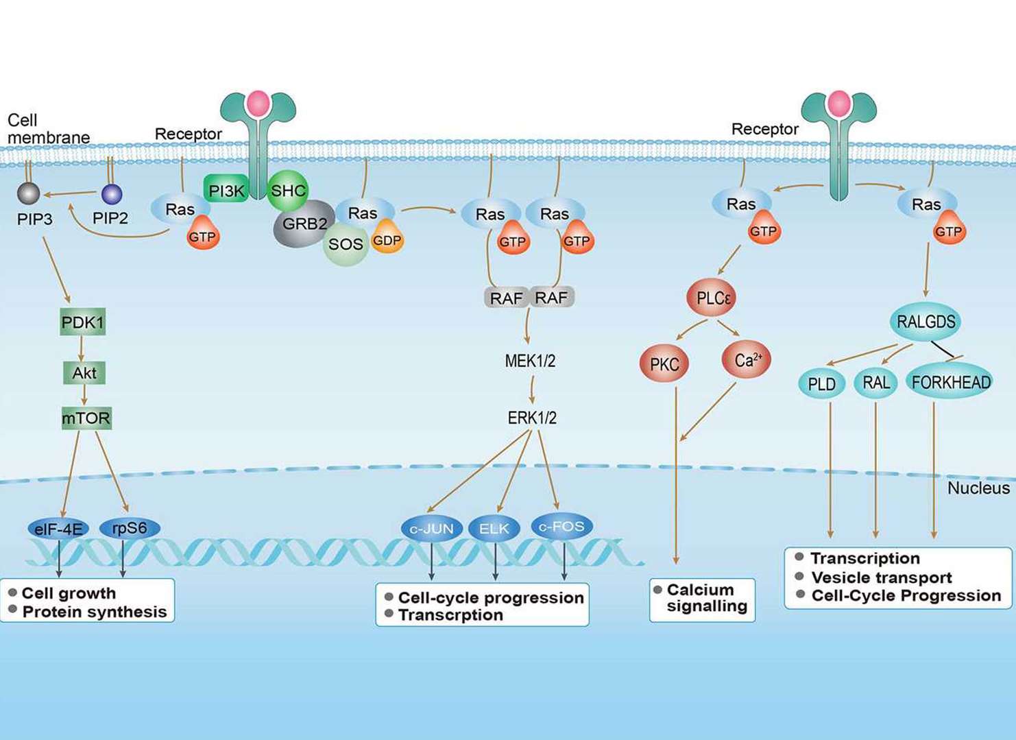 Ras Signaling Pathway
Downloadable Resources
Download resources about recombinant antibody development and antibody engineering to boost your research.
Product Notes
This is a product of Creative Biolabs' Hi-Affi™ recombinant antibody portfolio, which has several benefits including:
• Increased sensitivity
• Confirmed specificity
• High repeatability
• Excellent batch-to-batch consistency
• Sustainable supply
• Animal-free production
See more details about Hi-Affi™ recombinant antibody benefits.
Datasheet
MSDS
COA
Certificate of Analysis LookupTo download a Certificate of Analysis, please enter a lot number in the search box below. Note: Certificate of Analysis not available for kit components.
Lot Number:
See other products for "PLD1"
Select a product category from the dropdown menu below to view related products.
| CAT | Product Name | Application | Type |
|---|---|---|---|
| MOR-2744 | Hi-Affi™ Recombinant Rabbit Anti-PLD1 Monoclonal Antibody (DS2744AB) | WB, FC, IP | IgG |
| CAT | Product Name | Application | Type |
|---|---|---|---|
| VS-0325-XY1670 | Anti-PLD1 Immunohistochemistry Kit | IHC |
Specific Inquiry

See Our Custom Production in Action


Popular Products

Application: IP, IF, FuncS, FC, Neut, ELISA, IHC


Application: FC, IP, ELISA, Neut, FuncS, IF, IHC

Application: ELISA, FC, IP, FuncS, IF, Neut, ICC


Application: ELISA, IP, FC, FuncS, Neut, IF, ICC

Application: FC, IHC, FuncS, Inhib, Cyt

Application: Inhib, Cyt

Application: FuncS, IF, Neut, ELISA, FC, IP, ICC

Application: Neut, ELISA, IF, IP, FuncS, FC, ICC

Application: IF, IP, Neut, FuncS, ELISA, FC, ICC

Application: IF, IP, Neut, FuncS, ELISA, FC, ICC

Application: ELISA, FC, IP, FuncS, IF, Neut, ICC


Application: WB, ELISA, FuncS


For research use only. Not intended for any clinical use. No products from Creative Biolabs may be resold, modified for resale or used to manufacture commercial products without prior written approval from Creative Biolabs.
Send Inquiry
This site is protected by reCAPTCHA and the Google Privacy Policy and Terms of Service apply.