

AbPlus™ Anti-ATM Magnetic Beads (4E11)
CAT#: VS-0424-XY24
The AbPlus Anti-ATM Magnetic Beads (4E11) is an innovative affinity resin which is bound with anti-ATM specific antibody (4E11). The beads were designed for small-scale affinity purification and immunoprecipitation (IP) of ATM protein under native and denaturing conditions.
Gene Expression

Normal Tissue
Figure 1 IHC staining of human kidney
Immunohistochemical staining of human kidney shows strong nuclear positivity in tubules and glomeruli.
* Image credit: Image credit: Human Protein Atlas v21.proteinatlas.org/images/102/2822_A_9_5_selected.jpg

Normal Tissue
Figure 2 Cerebral cortex
Endothelial cells Staining: Medium Intensity: Moderate Quantity:>75% Location: Nuclear Glial cells Staining: Medium Intensity: Moderate Quantity:>75% Location: Cytoplasmic/ membranous nuclear Neuronal cells Staining: Medium Intensity: Moderate Quantity: 75%-25% Location: Cytoplasmic/ membranous nuclear Neuropil Staining: Low Intensity: Weak Quantity:>75% Location: Cytoplasmic/ membranous
* Image credit: Image credit: Human Protein Atlas v21.proteinatlas.org/images/102/2822_B_8_5.jpg

Normal Tissue
Figure 3 Colon
Endocrine cells Staining: Medium Intensity: Moderate Endothelial cells Staining: Medium Intensity: Moderate Quantity: 75%-25% Enterocytes Staining: Medium Intensity: Moderate Quantity: 75%-25% Enterocytes - Microvilli Staining: Medium Intensity: Moderate Goblet cells Staining: High Intensity: Strong Quantity:>75% Mucosal lymphoid cells Staining: Low Intensity: Moderate Quantity: <25% Peripheral nerve/ganglion Staining: High Intensity: Strong
* Image credit: Image credit: Human Protein Atlas v21.proteinatlas.org/images/102/2822_A_8_3.jpg

Normal Tissue
Figure 4 Liver
Cholangiocytes Staining: Medium Intensity: Moderate Quantity:>75% Location: Cytoplasmic/ membranous nuclear Hepatocytes Staining: Low Intensity: Weak Quantity:>75% Location: Cytoplasmic/ membranous
* Image credit: Image credit: Human Protein Atlas v21.proteinatlas.org/images/102/2822_A_9_4.jpg

Normal Tissue
Figure 5 Pancreas
Exocrine glandular cells Staining: High Intensity: Strong Quantity:>75% Location: Cytoplasmic/ membranous nuclear Pancreatic endocrine cells Staining: Medium Intensity: Moderate Quantity:>75% Location: Nuclear
* Image credit: Image credit: Human Protein Atlas v21.proteinatlas.org/images/102/2822_A_2_3.jpg

Normal Tissue
Figure 6 Kidney
Cells in glomeruli Staining: High Intensity: Strong Quantity:>75% Location: Nuclear Cells in tubules Staining: High Intensity: Strong Quantity:>75% Location: Cytoplasmic/ membranous nuclear
* Image credit: Image credit: Human Protein Atlas v21.proteinatlas.org/images/102/2822_A_9_5.jpg

Normal Tissue
Figure 7 Testis
Leydig cells Staining: Medium Intensity: Moderate Quantity:>75% Pachytene spermatocytes Staining: Medium Intensity: Moderate Quantity: 75%-25% Preleptotene spermatocytes Staining: Medium Intensity: Moderate Quantity: 75%-25% Round or early spermatids Staining: Medium Intensity: Moderate Quantity: 75%-25% Sertoli cells Staining: Low Intensity: Weak Quantity: 75%-25% Spermatogonia cells Staining: Low Intensity: Moderate
* Image credit: Image credit: Human Protein Atlas v21.proteinatlas.org/images/102/2822_A_6_6.jpg

Normal Tissue
Figure 8 Lymph node
Germinal center cells Staining: High Intensity: Strong Quantity:>75% Location: Cytoplasmic/ membranous nuclear Non-germinal center cells Staining: High Intensity: Strong Quantity:>75% Location: Cytoplasmic/ membranous nuclear
* Image credit: Image credit: Human Protein Atlas v21.proteinatlas.org/images/102/2822_A_8_8.jpg

RNA Expression
Figure 9 RNA cell line category: Cell line enhanced (U-698)
Cell lines ordered by descending RNA expression order.
* Image credit: Image credit: Human Protein Atlas v21.proteinatlas.org/ENSG00000149311-ATM
❮


 ❯
❯
Specifications
- Applications
- Immunoprecipitation, Protein Purification
- Matrix
- Magrose bead (> 50 μmol/mL gel)
- Bead Ligand
- Anti-ATM specific antibody (4E11)
- Target
- ATM
- Immunogen
- Synthesized peptide derived from human ATM
- Target Species
- Human
- Antibody Clone
- 4E11
- Bead Capacity
- 20-30 mg/mL binding antibody
- Bead size
- 10-37 μm
- Stability
- pH 2-14
- Format
- 20% Suspension
- Buffer
- PBS, pH 7.4, with 1% BSA
- Preservative
- 0.03% Proclin 300
- Storage
- Stored at 4°C, and is stable for up to 2 years. Do not centrifuge, dry or freeze the magnetic beads.
Applications
- Application Notes
- The beads are in suspension and will settle upon storage. Prior to use, mix the vial gently (do not vortex) to ensure delivery of proper bead volume.
Target
- Introduction
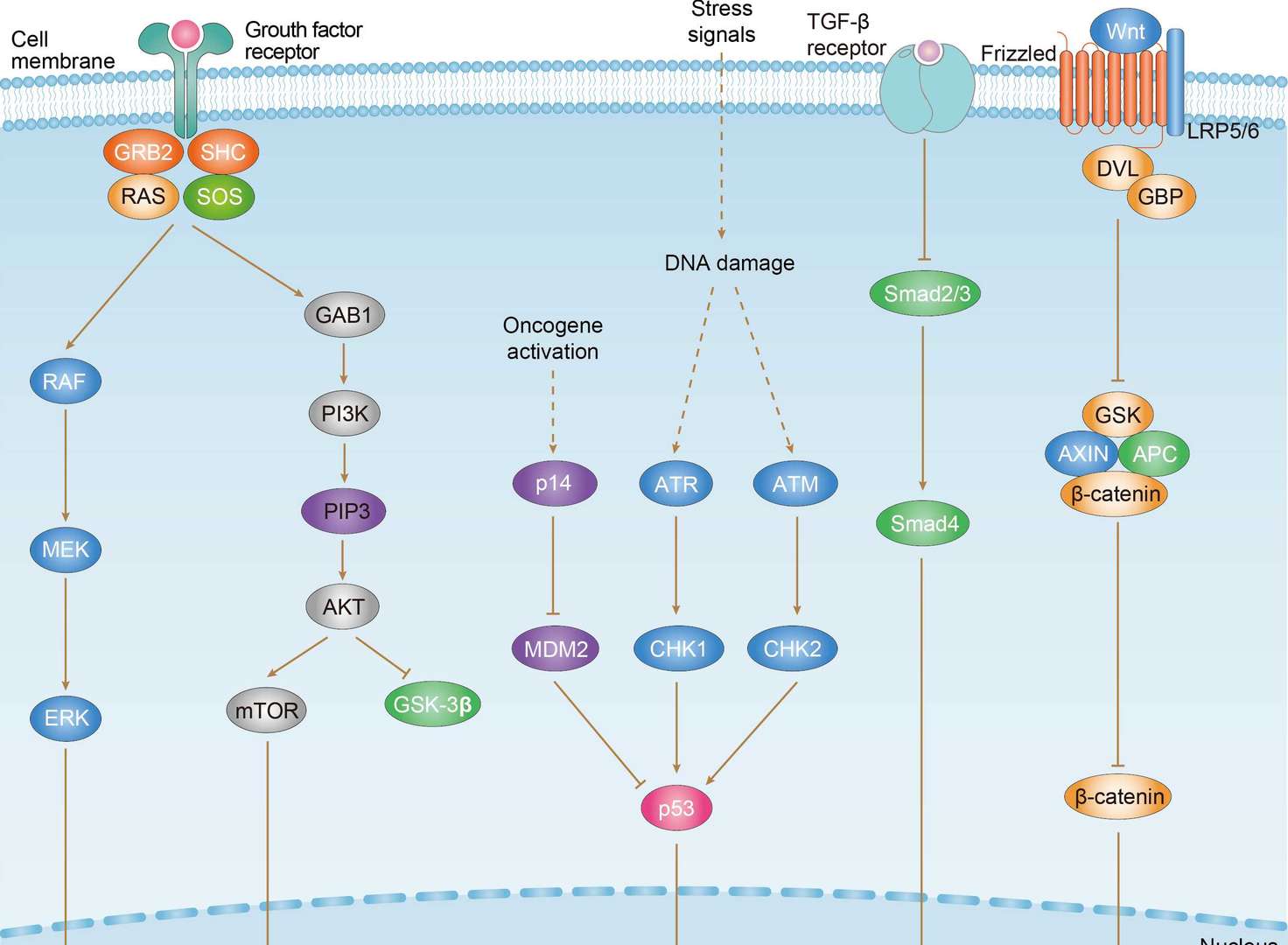
- The protein encoded by this gene belongs to the PI3/PI4-kinase family. This protein is an important cell cycle checkpoint kinase that phosphorylates; thus, it functions as a regulator of a wide variety of downstream proteins, including tumor suppressor proteins p53 and BRCA1, checkpoint kinase CHK2, checkpoint proteins RAD17 and RAD9, and DNA repair protein NBS1. This protein and the closely related kinase ATR are thought to be master controllers of cell cycle checkpoint signaling pathways that are required for cell response to DNA damage and for genome stability. Mutations in this gene are associated with ataxia telangiectasia, an autosomal recessive disorder.
- Alternative Names
- AT1; ATA; ATC; ATD; ATE; ATDC; TEL1; TELO1; serine-protein kinase ATM; A-T mutated; AT mutated; TEL1, telomere maintenance 1, homolog; ataxia telangiectasia mutated
- Gene ID
- 472
- UniProt ID
- Q13315
REVIEWS AND Q&AS
CITATIONS
RESOURCES
DOWNLOADS
RELATED PRODUCTS
Inquiry
Navs
Customer Review

There are currently no Customer reviews or questions for VS-0424-XY24. Click the button above to contact us or submit your feedback about this product.
Submit Your Publication
Published with our product? Submit your paper and receive a 10% discount on your next order! Share your research to earn exclusive rewards.
Related Diseases
 Gastric Cancer
Downloadable Resources
Download resources about recombinant antibody development and antibody engineering to boost your research.
Datasheet
MSDS
COA
Certificate of Analysis LookupTo download a Certificate of Analysis, please enter a lot number in the search box below. Note: Certificate of Analysis not available for kit components.
Lot Number:
See other products for "ATM"
Select a product category from the dropdown menu below to view related products.
| CAT | Product Name | Application | Type |
|---|---|---|---|
| MOB-0634MZ | Mouse Anti-ATM Recombinant Antibody (clone NBU4-5H20/9) | ELISA, WB | Mouse IgG1 |
| CAT | Product Name | Application | Type |
|---|---|---|---|
| BRD-0059MZ | Chicken Anti-ATM Polyclonal IgY | WB | Chicken antibody |
| CAT | Product Name | Application | Type |
|---|---|---|---|
| MOR-0286 | Hi-Affi™ Rabbit Anti-ATM Recombinant Antibody (clone DS286AB) | IF, IHC-P, IP, WB | Rabbit IgG |
| CAT | Product Name | Application | Type |
|---|---|---|---|
| MRO-0139-CN | Rabbit Anti-ATM Recombinant Antibody (clone CBACN-053) | WB, IF, IHC | Rabbit IgG |
| CAT | Product Name | Application | Type |
|---|---|---|---|
| MRO-2302-CN | Rabbit Anti-ATM Recombinant Antibody (clone JM93-25) | WB, IF, IHC, IP | Rabbit IgG |
| CAT | Product Name | Application | Type |
|---|---|---|---|
| ZG-0317F | Mouse Anti-ATM Recombinant Antibody (ZG-0317F) | WB, ELISA | Mouse IgG |
| CAT | Product Name | Application | Type |
|---|---|---|---|
| ZG-0041J | Mouse Anti-ATM Recombinant Antibody (clone 1D1) | IHC-P, IF | Mouse IgG |
| CAT | Product Name | Application | Type |
|---|---|---|---|
| VS-1024-XY36 | Mouse Anti-NHP ATM Recombinant Antibody (clone 2C1) | WB, IHC, IP | Mouse IgG1 |
| CAT | Product Name | Application | Type |
|---|---|---|---|
| VS-0325-XY197 | Anti-ATM Immunohistochemistry Kit | IHC |
| CAT | Product Name | Application | Type |
|---|---|---|---|
| VS-0525-XY599 | Anti-Mouse ATM Immunohistochemistry Kit | IHC |
| CAT | Product Name | Application | Type |
|---|---|---|---|
| VS-0525-XY598 | Anti-Human ATM Immunohistochemistry Kit | IHC |
Specific Inquiry
See Our Custom Production in Action


Popular Products

Application: ELISA, IHC

Application: FC, IP, ELISA, Neut, FuncS, IF, ICC

Application: Neut, ELISA, IF, IP, FuncS, FC, ICC

Application: WB, FC, IP, ELISA, Neut, FuncS, IF

Application: FuncS, IF, Neut, ELISA, FC, IP, ICC

Application: IF, IP, Neut, FuncS, ELISA, FC, ICC


Application: ELISA, WB, BLI, SPR


Application: ELISA, Neut, FuncS

Application: Neut, ELISA, FuncS


For research use only. Not intended for any clinical use. No products from Creative Biolabs may be resold, modified for resale or used to manufacture commercial products without prior written approval from Creative Biolabs.
Send Inquiry
This site is protected by reCAPTCHA and the Google Privacy Policy and Terms of Service apply.