

Anti-CXCL5 Immunohistochemistry Kit
CAT#: VS-0325-XY592
Our CXCL5 IHC Kit is a remarkable choice for immunohistochemical analysis. It is equipped with all the required reagents, so you can focus on your experiment without worrying about reagent shortages. The user - friendly design simplifies the operation, reducing the likelihood of errors. The kit provides high - resolution staining, allowing for clear visualization of CXCL5 in tissues and facilitating in - depth research. This kit can be applied to paraffin sections and frozen sections.
Gene Expression

Subcellular Location
Figure 1 IF staining of human cell line A549
Immunofluorescent staining of human cell line A549 shows localization to vesicles.
* Image credit: Image credit: Human Protein Atlas v21.proteinatlas.org/images/65474/1873_B10_6_selected.jpg

Normal Tissue
Figure 2 IHC staining of human lymph node
Immunohistochemical staining of human lymph node shows strong cytoplasmic positivity in non-germinal center cells.
* Image credit: Image credit: Human Protein Atlas v21.proteinatlas.org/images/65474/ihc_selected.jpg

Normal Tissue
Figure 3 Cerebral cortex
Glial cells Staining: Medium Intensity: Strong Quantity: <25% Location: Cytoplasmic/ membranous Neuronal cells Staining: Medium Intensity: Strong Quantity: <25% Location: Cytoplasmic/ membranous
* Image credit: Image credit: Human Protein Atlas v21.proteinatlas.org/images/65474/146785_B_7_5.jpg

Normal Tissue
Figure 4 Pancreas
Pancreatic endocrine cells Staining: Medium Intensity: Strong Quantity: <25% Location: Cytoplasmic/ membranous
* Image credit: Image credit: Human Protein Atlas v21.proteinatlas.org/images/65474/146785_A_2_3.jpg

Normal Tissue
Figure 5 Testis
Cells in seminiferous ducts Staining: Low Intensity: Weak Quantity: 75%-25% Location: Cytoplasmic/ membranous Leydig cells Staining: Low Intensity: Weak Quantity: 75%-25% Location: Cytoplasmic/ membranous
* Image credit: Image credit: Human Protein Atlas v21.proteinatlas.org/images/65474/146785_A_5_6.jpg

Normal Tissue
Figure 6 Lymph node
Non-germinal center cells Staining: Low Intensity: Moderate Quantity: <25% Location: Cytoplasmic/ membranous
* Image credit: Image credit: Human Protein Atlas v21.proteinatlas.org/images/65474/146785_A_7_8.jpg

RNA Expression
Figure 7 RNA cell line category: Cell line enhanced.
Cell lines ordered by descending RNA expression order.
* Image credit: Image credit: Human Protein Atlas v21.proteinatlas.org/ENSG00000163735-CXCL5
❮


 ❯
❯
Specifications
- Application
- IHC
- Size
- 50 Tests
- Species Reactivity
- Human
- Target
- CXCL5
- Primary Antibody
- Rabbit Anti-CXCL5 Antibody
- Secondary Antibody
- Goat anti-Rabbit Antibody, HRP
- Sample Type
- FFPE tissue; Frozen section tissue
- Kit Storage
- All reagents should be kept at 2-8°C. The kit remains stable for up to 6 months after arrival.
REVIEWS AND Q&AS
CITATIONS
RESOURCES
DOWNLOADS
RELATED PRODUCTS
Inquiry
Navs
Customer Review

There are currently no Customer reviews or questions for VS-0325-XY592. Click the button above to contact us or submit your feedback about this product.
Submit Your Publication
Published with our product? Submit your paper and receive a 10% discount on your next order! Share your research to earn exclusive rewards.
Related Diseases
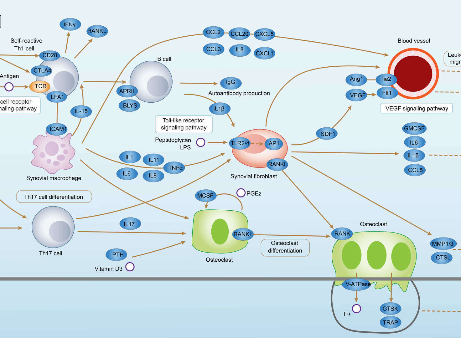 Rheumatoid Arthritis
Related Signaling Pathways
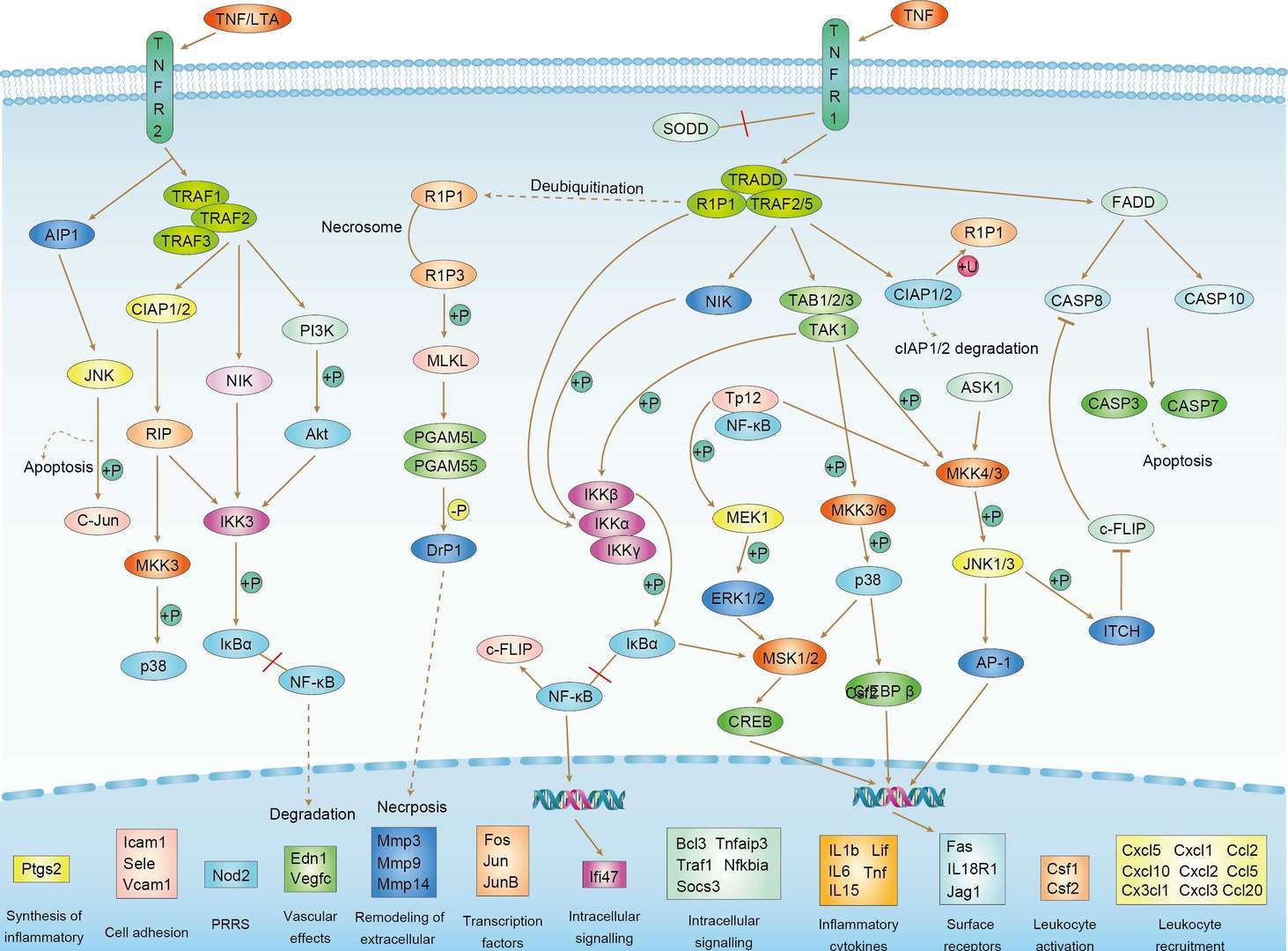 TNF Signaling Pathway
Downloadable Resources
Download resources about recombinant antibody development and antibody engineering to boost your research.
Datasheet
MSDS
COA
Certificate of Analysis LookupTo download a Certificate of Analysis, please enter a lot number in the search box below. Note: Certificate of Analysis not available for kit components.
Lot Number:
Protocol & Troubleshooting
We have outlined the assay protocols, covering reagents, solutions, procedures, and troubleshooting tips for common issues in order to better assist clients in conducting experiments with our products. View the full list of Protocol & Troubleshooting.
See other products for "CXCL5"
Select a product category from the dropdown menu below to view related products.
| CAT | Product Name | Application | Type |
|---|---|---|---|
| MOB-3276z | Mouse Anti-CXCL5 Recombinant Antibody (clone 42D6) | WB, IF, IHC, ELISA | Mouse IgG1 |
| CAT | Product Name | Application | Type |
|---|---|---|---|
| MOB-2065MZ | Recombinant Mouse Anti-Human CXCL5 Antibody (clone NN0327-20M20) | IHC-P, Neut, WB | Mouse antibody |
| CAT | Product Name | Application | Type |
|---|---|---|---|
| NEUT-632CQ | Mouse Anti-CXCL5 Recombinant Antibody (clone CBL039) | WB, IHC, Neut | Mouse IgG1 |
| CAT | Product Name | Application | Type |
|---|---|---|---|
| NEUT-633CQ | Mouse Anti-CXCL5 Recombinant Antibody (clone MM0216-10L19) | WB, IHC-P, Neut | Mouse IgG1 |
| CAT | Product Name | Application | Type |
|---|---|---|---|
| NEUT-634CQ | Mouse Anti-CXCL5 Recombinant Antibody (clone CBL752) | WB, IHC, Neut | Mouse IgG1 |
| CAT | Product Name | Application | Type |
|---|---|---|---|
| NEUT-635CQ | Mouse Anti-CXCL5 Recombinant Antibody (clone CBL754) | CyTOF®, ELISA, ICFC, Neut | Mouse IgG1 |
| CAT | Product Name | Application | Type |
|---|---|---|---|
| NEUT-636CQ | Rat Anti-Cxcl5 Recombinant Antibody (clone 32B3) | WB, Neut, ELISA | Rat IgG2 |
| CAT | Product Name | Application | Type |
|---|---|---|---|
| MOR-0886 | Hi-Affi™ Rabbit Anti-CXCL5 Recombinant Antibody (clone DS886AB) | WB, ICC, IP | Rabbit IgG |
| CAT | Product Name | Application | Type |
|---|---|---|---|
| MOR-4563 | Hi-Affi™ Rabbit Anti-CXCL5 Recombinant Antibody (clone TH73DS) | WB, IHC-P, IP | Rabbit IgG |
| CAT | Product Name | Application | Type |
|---|---|---|---|
| VS-0525-XY1820 | Anti-Human CXCL5 Immunohistochemistry Kit | IHC |
Specific Inquiry
See Our Custom Production in Action


Popular Products


Application: WB, FuncS, IF, Neut, ELISA, FC, IP

Application: FC, IP, ELISA, Neut, FuncS, IF, IHC

Application: Neut, ELISA, IF, IP, FuncS, FC, ICC

Application: WB, IP, IF, FuncS, FC, Neut, ELISA


Application: ELISA, IP, FC, FuncS, Neut, IF, ICC

Application: IF, IP, Neut, FuncS, ELISA, FC, ICC

Application: Neut, ELISA, IF, IP, FuncS, FC, WB

Application: Neut, ELISA, IF, IP, FuncS, FC, ICC


Application: ELISA, WB, BLI, SPR

Application: ELISA, Neut, FuncS


Application: ELISA, SPR, Inhib, FuncS

For research use only. Not intended for any clinical use. No products from Creative Biolabs may be resold, modified for resale or used to manufacture commercial products without prior written approval from Creative Biolabs.
Send Inquiry
This site is protected by reCAPTCHA and the Google Privacy Policy and Terms of Service apply.