

PE-A*02:01/Human ABL1 (QQAHCLWCV) MHC Tetramer
CAT#: MHC-CN0381
Gene Expression

Subcellular Location and Protein Expression
Figure 1 IF staining of human cell line A-431
Immunofluorescent staining of human cell line A-431 shows localization to nucleoplasm & nuclear bodies.
* Image credit: Image credit: Human Protein Atlas https://v21.proteinatlas.org/images/27280/259_B8_2_selected.jpg

Subcellular Location and Protein Expression
Figure 2 IF staining of human cell line U-2 OS
Immunofluorescent staining of human cell line U-2 OS shows localization to nucleoplasm.
* Image credit: Image credit: Human Protein Atlas https://v21.proteinatlas.org/images/28409/497_A9_2_selected.jpg

Subcellular Location and Protein Expression
Figure 3 IHC staining of human heart muscle
Immunohistochemical staining of human heart muscle shows strong cytoplasmic positivity in myocytes.
* Image credit: Image credit: Human Protein Atlas https://v21.proteinatlas.org/images/27251/ihc_selected.jpg

Subcellular Location and Protein Expression
Figure 4 IHC staining of human prostate
Immunohistochemical staining of human prostate shows cytoplasmic and membranous positivity in glandular cells.
* Image credit: Image credit: Human Protein Atlas https://v21.proteinatlas.org/images/27280/ihc_selected.jpg

Subcellular Location and Protein Expression
Figure 5 IHC staining of human adrenal gland
Immunohistochemical staining of human adrenal gland shows strong nuclear positivity in cortical cells.
* Image credit: Image credit: Human Protein Atlas https://v21.proteinatlas.org/images/28409/ihc_selected.jpg

Subcellular Location and Protein Expression
Figure 6 IHC staining of human duodenum
Immunohistochemical staining of human duodenum shows moderate cytoplasmic positivity in glandular cells.
* Image credit: Image credit: Human Protein Atlas https://v21.proteinatlas.org/images/2686/ihc_selected.jpg

Subcellular Location and Protein Expression
Figure 7 IF staining of human cell line U-251 MG
Immunofluorescent staining of human cell line U-251 MG shows localization to nucleoplasm & nuclear bodies.
* Image credit: Image credit: Human Protein Atlas https://v21.proteinatlas.org/images/27280/258_B8_1_red_green.jpg

Normal Tissue
Figure 8 Cerebral cortex
Neuropil
Staining:Medium
Intensity: Moderate
Quantity:>75%
Location: Cytoplasmic/membranous
* Image credit: Image credit: Human Protein Atlas https://v21.proteinatlas.org/images/28409/61378_B_8_5.jpg

Normal Tissue
Figure 9 Adrenal gland
Glandular cells
Staining:High
Intensity: Strong
Quantity: 75%-25%
Location: Cytoplasmic/membranous nuclear
* Image credit: Image credit: Human Protein Atlas https://v21.proteinatlas.org/images/28409/61378_B_5_5.jpg

Normal Tissue
Figure 10 Colon
Endothelial cells
Staining:Medium
Intensity: Moderate
Quantity:>75%
Location: Cytoplasmic/membranous
Glandular cells
Staining:Medium
Intensity: Moderate
Quantity:>75%
Location: Cytoplasmic/membranous nuclear
Peripheral nerve/ganglion
Staining:Medium
Intensity: Moderate
Quantity:>75%
Location: Cytoplasmic/membranous
* Image credit: Image credit: Human Protein Atlas https://v21.proteinatlas.org/images/28409/61494_A_9_3.jpg

Normal Tissue
Figure 11 Liver
Hepatocytes
Staining:Medium
Intensity: Moderate
Quantity:>75%
Location: Cytoplasmic/membranous nuclear
* Image credit: Image credit: Human Protein Atlas https://v21.proteinatlas.org/images/28409/61494_A_9_4.jpg

Normal Tissue
Figure 12 Kidney
Cells in glomeruli
Staining:Medium
Intensity: Moderate
Quantity: 75%-25%
Location: Cytoplasmic/membranous nuclear
Cells in tubules
Staining:Medium
Intensity: Moderate
Quantity:>75%
Location: Cytoplasmic/membranous nuclear
* Image credit: Image credit: Human Protein Atlas https://v21.proteinatlas.org/images/28409/61494_A_9_5.jpg

Normal Tissue
Figure 13 Testis
Spermatogonia cells
Staining:Medium
Intensity: Moderate
Quantity: 75%-25%
* Image credit: Image credit: Human Protein Atlas https://v21.proteinatlas.org/images/28409/61494_A_6_6.jpg

RNA Expression
Figure 14 RNA cell line category: Low cell line specificity
Cell lines ordered by descending RNA expression order.
* Image credit: Image credit: Human Protein Atlas https://v21.proteinatlas.org/ENSG00000097007-ABL1
❮


 ❯
❯
Specifications
- Allele
- A*02:01
- Class
- Class I
- MHC Species
- Human
- Antigen
- ABL1
- Antigen Species
- Human
- Peptide
- QQAHCLWCV
- Conjugate
- PE
- Application
- FCM, IHC
Target
- Antigen Introduction
- This gene is a protooncogene that encodes a protein tyrosine kinase involved in a variety of cellular processes, including cell division, adhesion, differentiation, and response to stress. The activity of the protein is negatively regulated by its SH3 domain, whereby deletion of the region encoding this domain results in an oncogene. The ubiquitously expressed protein has DNA-binding activity that is regulated by CDC2-mediated phosphorylation, suggesting a cell cycle function. This gene has been found fused to a variety of translocation partner genes in various leukemias, most notably the t(9;22) translocation that results in a fusion with the 5' end of the breakpoint cluster region gene (BCR; MIM:151410). Alternative splicing of this gene results in two transcript variants, which contain alternative first exons that are spliced to the remaining common exons.
- Alternative Names
- ABL1; ABL proto-oncogene 1, non-receptor tyrosine kinase; ABL; JTK7; p150; c-ABL; v-abl; c-ABL1
- Gene ID
- 25
- UniProt ID
- Q13848
REVIEWS AND Q&AS
CITATIONS
RESOURCES
DOWNLOADS
RELATED PRODUCTS
Inquiry
Navs
Customer Review

There are currently no Customer reviews or questions for MHC-CN0381. Click the button above to contact us or submit your feedback about this product.
Submit Your Publication
Published with our product? Submit your paper and receive a 10% discount on your next order! Share your research to earn exclusive rewards.
Related Signaling Pathways
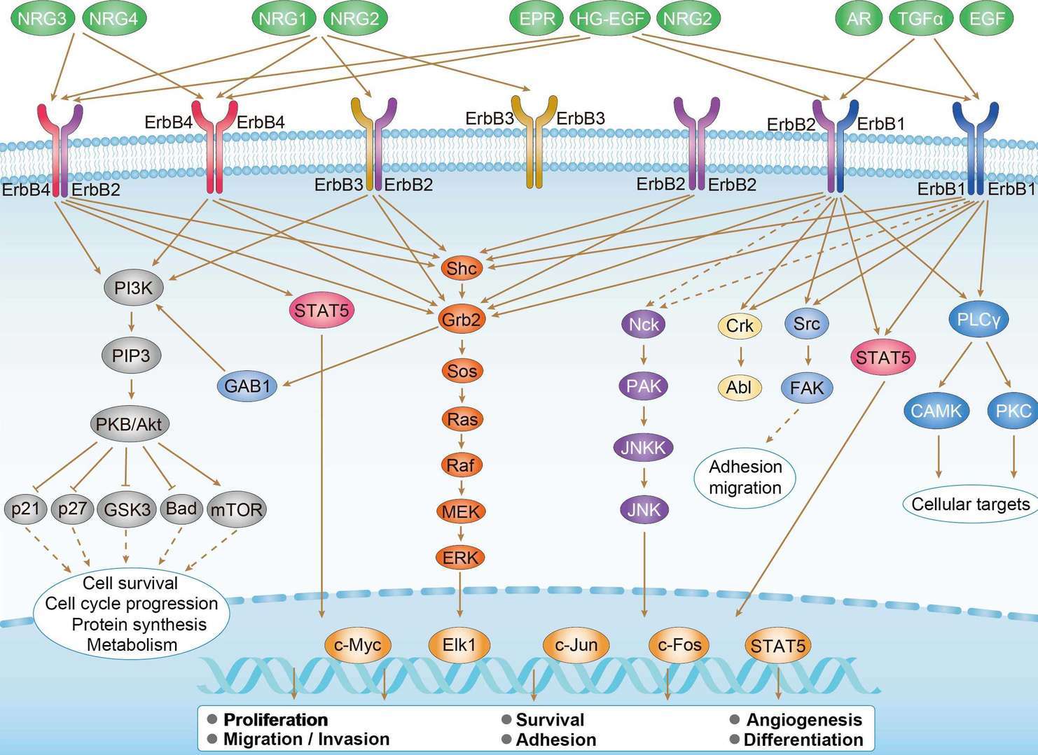 ErbB Signaling Pathway
Downloadable Resources
Download resources about recombinant antibody development and antibody engineering to boost your research.
Datasheet
MSDS
COA
Certificate of Analysis LookupTo download a Certificate of Analysis, please enter a lot number in the search box below. Note: Certificate of Analysis not available for kit components.
Lot Number:
Protocol & Troubleshooting
We have outlined the assay protocols, covering reagents, solutions, procedures, and troubleshooting tips for common issues in order to better assist clients in conducting experiments with our products. View the full list of Protocol & Troubleshooting.
See other products for "ABL1"
Select a product category from the dropdown menu below to view related products.
| CAT | Product Name | Application | Type |
|---|---|---|---|
| NAB-1-VHH | Recombinant Anti-human ABL1 VHH Single Domain Antibody | WB, ICC, ChiP, FA, ELISA | Llama VHH |
| CAT | Product Name | Application | Type |
|---|---|---|---|
| MOB-0964MZ | Mouse Anti-ABL1 Recombinant Antibody (clone TQN439) | IHC-P | Mouse IgG1 |
| CAT | Product Name | Application | Type |
|---|---|---|---|
| MHC-LC084 | PE-A*02:01/Human BCR-ABL (GVRGRVEEI) MHC Tetramer | FCM |
| CAT | Product Name | Application | Type |
|---|---|---|---|
| MHC-LC085 | APC-A*02:01/Human BCR-ABL (GVRGRVEEI) MHC Tetramer | FCM |
| CAT | Product Name | Application | Type |
|---|---|---|---|
| MHC-LC086 | BV421-A*02:01/Human BCR-ABL (GVRGRVEEI) MHC Tetramer | FCM |
| CAT | Product Name | Application | Type |
|---|---|---|---|
| MHC-LC2231 | PE-A*03:01/Human ABL1 (ATGFKQSSK) MHC Tetramer | FCM |
| CAT | Product Name | Application | Type |
|---|---|---|---|
| MHC-LC2232 | PE-A*11:01/Human ABL1 (ATGFKQSSK) MHC Tetramer | FCM |
| CAT | Product Name | Application | Type |
|---|---|---|---|
| ZG-0356F | Mouse Anti-ABL1 Recombinant Antibody (ZG-0356F) | WB, ELISA | Mouse IgG |
| CAT | Product Name | Application | Type |
|---|---|---|---|
| VS3-XY13 | Mouse Anti-ABL1 Recombinant Antibody (clone 7B11D6) | ELISA, WB | Mouse IgG1 |
| CAT | Product Name | Application | Type |
|---|---|---|---|
| VS7-HM11 | Mouse Anti-ABL1 Recombinant Antibody (clone CBL006HM) | WB, ELISA | Mouse IgG |
| CAT | Product Name | Application | Type |
|---|---|---|---|
| VS-0525-XY46 | Anti-ABL1 Immunohistochemistry Kit | IHC |
| CAT | Product Name | Application | Type |
|---|---|---|---|
| VS-0525-XY47 | Anti-Mouse ABL1 Immunohistochemistry Kit | IHC |
Specific Inquiry

See Our Custom Production in Action


Popular Products

Application: IP, IF, FuncS, FC, Neut, ELISA, IHC

Application: WB, FC, IP, ELISA, Neut, FuncS, IF

Application: ELISA, IP, FC, FuncS, Neut, IF, ICC

Application: FC, IP, ELISA, Neut, FuncS, IF, IHC

Application: IF, IP, Neut, FuncS, ELISA, FC, ICC

Application: WB, ELISA, IP, FC, FuncS, Neut, IF

Application: ELISA, IHC

Application: Neut, ELISA, IF, IP, FuncS, FC, ICC

Application: Inhib, Cyt

Application: FuncS, IF, Neut, ELISA, FC, IP, ICC

Application: ELISA, FC, IP, FuncS, IF, Neut, ICC

Application: FC, IP, ELISA, Neut, FuncS, IF, ICC

Application: ELISA, FC, IP, FuncS, IF, Neut, WB


Application: WB, Neut, FuncS


For research use only. Not intended for any clinical use. No products from Creative Biolabs may be resold, modified for resale or used to manufacture commercial products without prior written approval from Creative Biolabs.
Send Inquiry
This site is protected by reCAPTCHA and the Google Privacy Policy and Terms of Service apply.