

PE-A*02:01/Human Cyclin D1 (LLNDRVLRV) MHC Tetramer
CAT#: MHC-CN0166
Gene Expression

Subcellular Location
Figure 1 IF staining of human cell line BJ
Immunofluorescent staining of human cell line BJ shows localization to nucleoplasm.
* Image credit: Image credit: Human Protein Atlas https://v21.proteinatlas.org/images/27802/1667_B12_1_selected.jpg

Subcellular Location
Figure 2 IHC staining of human esophagus
Immunohistochemical staining of human esophagus shows strong nuclear positivity in a fraction of basal cells.
* Image credit: Image credit: Human Protein Atlas https://v21.proteinatlas.org/images/24/ihc_selected.jpg

Subcellular Location
Figure 3 IF staining of human cell line U-2 OS
Immunofluorescent staining of human cell line U-2 OS shows localization to nucleoplasm.
* Image credit: Image credit: Human Protein Atlas https://v21.proteinatlas.org/images/27802/1607_B12_2_red_green.jpg

Normal Tissue
Figure 4 Liver
Hepatocytes
Staining: High
Intensity: Strong
Quantity:>75%
Location: Cytoplasmic/membranous
* Image credit: Image credit: Human Protein Atlas https://v21.proteinatlas.org/images/24/70_A_9_4.jpg

Normal Tissue
Figure 5 Testis
Leydig cells
Staining: Medium
Intensity: Moderate
Quantity: 75%-25%
Location: Cytoplasmic/membranous
* Image credit: Image credit: Human Protein Atlas https://v21.proteinatlas.org/images/24/70_A_6_6.jpg

RNA Expression
Figure 6 RNA cell line category: Cell line enhanced (U-266/70, WM-115)
Cell lines ordered by descending RNA expression order.
* Image credit: Image credit: Human Protein Atlas https://v21.proteinatlas.org/ENSG00000110092-CCND1
❮


 ❯
❯
Specifications
- Allele
- A*02:01
- Class
- Class I
- MHC Species
- Human
- Antigen
- Cyclin D1
- Antigen Species
- Human
- Peptide
- LLNDRVLRV
- Conjugate
- PE
- Application
- FCM
Target
- Antigen Introduction
- The protein encoded by this gene belongs to the highly conserved cyclin family, whose members are characterized by a dramatic periodicity in protein abundance throughout the cell cycle. Cyclins function as regulators of CDK kinases. Different cyclins exhibit distinct expression and degradation patterns which contribute to the temporal coordination of each mitotic event. This cyclin forms a complex with and functions as a regulatory subunit of CDK4 or CDK6, whose activity is required for cell cycle G1/S transition. This protein has been shown to interact with tumor suppressor protein Rb and the expression of this gene is regulated positively by Rb. Mutations, amplification and overexpression of this gene, which alters cell cycle progression, are observed frequently in a variety of tumors and may contribute to tumorigenesis. [provided by RefSeq, Jul 2008]
- Alternative Names
- CCND1; Parathyroid Adenomatosis 1; G1/S-Specific Cyclin-D1; Cyclin D1 (PRAD1: Parathyroid Adenomatosis 1); PRAD1; B-Cell Lymphoma 1 Protein; D11S287E; U21B31
- Gene ID
- 595
- UniProt ID
- P24385
REVIEWS AND Q&AS
CITATIONS
RESOURCES
DOWNLOADS
RELATED PRODUCTS
Inquiry
Navs
Customer Review

There are currently no Customer reviews or questions for MHC-CN0166. Click the button above to contact us or submit your feedback about this product.
Submit Your Publication
Published with our product? Submit your paper and receive a 10% discount on your next order! Share your research to earn exclusive rewards.
Related Diseases
 Breast Cancer
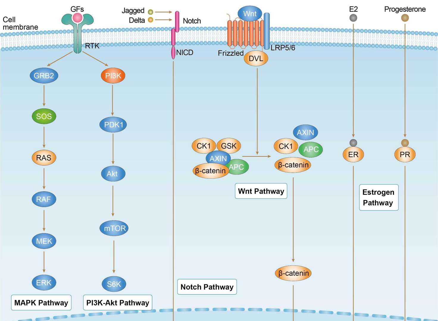
Related Signaling Pathways
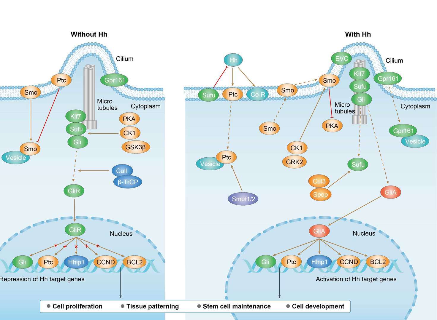 Hedgehog Signaling Pathway
Downloadable Resources
Download resources about recombinant antibody development and antibody engineering to boost your research.
Datasheet
MSDS
COA
Certificate of Analysis LookupTo download a Certificate of Analysis, please enter a lot number in the search box below. Note: Certificate of Analysis not available for kit components.
Lot Number:
See other products for "CCND1"
Select a product category from the dropdown menu below to view related products.
| CAT | Product Name | Application | Type |
|---|---|---|---|
| MOB-1231z | Mouse Anti-CCND1 Recombinant Antibody (clone 22F3) | WB, IP, IF, IHC, FuncS | Mouse IgG1, κ |
| CAT | Product Name | Application | Type |
|---|---|---|---|
| AGTO-G004R | Anti-Ccnd1 immunotoxin (IgG)-RTA | Cytotoxicity assay, Function study |
| CAT | Product Name | Application | Type |
|---|---|---|---|
| MOB-0110CT | Mouse Anti-CCND1 Recombinant Antibody (clone 4E9) | ELISA, WB | Mouse IgG1 |
| CAT | Product Name | Application | Type |
|---|---|---|---|
| NEUT-262CQ | Mouse Anti-CCND1 Recombinant Antibody (clone DCS-6) | IHC-Fr, WB, Block, IHC-P, IF, FC, IP | Mouse IgG2a, κ |
| CAT | Product Name | Application | Type |
|---|---|---|---|
| MOR-0515 | Hi-Affi™ Rabbit Anti-CCND1 Recombinant Antibody (clone DS515AB) | IHC | Rabbit IgG |
| CAT | Product Name | Application | Type |
|---|---|---|---|
| MOR-4566 | Hi-Affi™ Rabbit Anti-CCND1 Recombinant Antibody (clone TH76DS) | WB, IF, ICC, FC | Rabbit IgG |
| CAT | Product Name | Application | Type |
|---|---|---|---|
| MRO-0413-CN | Rabbit Anti-CCND1 Recombinant Antibody (clone CBACN-164) | WB, IF, IHC, IP | Rabbit IgG |
| CAT | Product Name | Application | Type |
|---|---|---|---|
| MRO-1799-CN | Rabbit Anti-CCND1 Polyclonal Antibody (MRO-1799-CN) | WB, IF, IHC, FC | Rabbit IgG |
| CAT | Product Name | Application | Type |
|---|---|---|---|
| MRO-1800-CN | Rabbit Anti-CCND1 Polyclonal Antibody (MRO-1800-CN) | WB, IHC | Rabbit IgG |
| CAT | Product Name | Application | Type |
|---|---|---|---|
| MHC-CN0163 | APC-A*02:01/Human Cyclin (LLGATCMFV) MHC Tetramer | FCM |
| CAT | Product Name | Application | Type |
|---|---|---|---|
| MHC-CN0164 | PE-A*02:01/Human Cyclin (LLGATCMFV) MHC Tetramer | FCM |
| CAT | Product Name | Application | Type |
|---|---|---|---|
| MHC-CN0165 | APC-A*02:01/Human Cyclin D1 (LLNDRVLRV) MHC Tetramer | FCM |
| CAT | Product Name | Application | Type |
|---|---|---|---|
| MHC-CN0359 | APC-A*02:01/Human Cyclin D1 (YLGATCMFV) MHC Tetramer | FCM |
| CAT | Product Name | Application | Type |
|---|---|---|---|
| MHC-CN0360 | PE-A*02:01/Human Cyclin D1 (YLGATCMFV) MHC Tetramer | FCM |
| CAT | Product Name | Application | Type |
|---|---|---|---|
| VS-1024-XY148 | Mouse Anti-NHP CCND1 Recombinant Antibody (clone DCS-6) | WB, IF | Mouse IgG2a, kappa |
| CAT | Product Name | Application | Type |
|---|---|---|---|
| VS-0525-XY1085 | Anti-CCND1 Immunohistochemistry Kit | IHC |
| CAT | Product Name | Application | Type |
|---|---|---|---|
| VS-0525-XY1086 | Anti-Mouse CCND1 Immunohistochemistry Kit | IHC |
| CAT | Product Name | Application | Type |
|---|---|---|---|
| VS-0525-XY1087 | Anti-Monkey CCND1 Immunohistochemistry Kit | IHC |
Specific Inquiry

See Our Custom Production in Action


Popular Products

Application: ELISA, IP, FC, FuncS, Neut, IF, ICC


Application: ELISA, Neut, IF, IP, FC, FuncS

Application: ELISA, IP, FC, FuncS, Neut, IF, ICC

Application: WB, IF, IP, Neut, FuncS, ELISA, FC

Application: ELISA, Neut, IF, IP, FC, FuncS


Application: IP, IF, FuncS, FC, Neut, ELISA, ICC

Application: Neut, ELISA, IF, IP, FuncS, FC, ICC

Application: FC, IP, ELISA, Neut, FuncS, IF, ICC


Application: Neut, ELISA, IF, IP, FuncS, FC, WB


For research use only. Not intended for any clinical use. No products from Creative Biolabs may be resold, modified for resale or used to manufacture commercial products without prior written approval from Creative Biolabs.
Send Inquiry
This site is protected by reCAPTCHA and the Google Privacy Policy and Terms of Service apply.