

Chicken Anti-α-1-Catenin (ab1) Polyclonal IgY
CAT#: BRD-0084MZ
This antibody is a chicken polyclonal antibody which specifically reacts with α-1-Catenin (ab1).
Gene Expression

Subcellular Location
Figure 1 IF staining of human cell line CACO-2
Immunofluorescent staining of human cell line CACO-2 shows localization to plasma membrane & cell junctions.
* Image credit: Image credit: Human Protein Atlas v21.proteinatlas.org/images/46119/1028_H10_1_selected.jpg

Normal Tissue
Figure 2 IHC staining of human prostate
Immunohistochemical staining of human prostate shows strong cytoplasmic positivity in glandular cells.
* Image credit: Image credit: Human Protein Atlas v21.proteinatlas.org/images/21089/ihc_selected.jpg

Normal Tissue
Figure 3 Testis
Cells in seminiferous ducts Staining: High Intensity: Strong Quantity: 75%-25% Location: Cytoplasmic/ membranous Leydig cells Staining: Medium Intensity: Moderate Quantity:>75% Location: Cytoplasmic/ membranous
* Image credit: Image credit: Human Protein Atlas v21.proteinatlas.org/images/46119/124809_A_4_6.jpg

Normal Tissue
Figure 4 Lymph node
Non-germinal center cells Staining: Medium Intensity: Moderate Quantity: 75%-25% Location: Cytoplasmic/ membranous
* Image credit: Image credit: Human Protein Atlas v21.proteinatlas.org/images/46119/124809_A_9_8.jpg

Normal Tissue
Figure 5 Colon
Endothelial cells Staining: Medium Intensity: Moderate Quantity:>75% Location: Cytoplasmic/ membranous Glandular cells Staining: High Intensity: Strong Quantity:>75% Location: Cytoplasmic/ membranous Peripheral nerve/ganglion Staining: Medium Intensity: Moderate Quantity:>75% Location: Cytoplasmic/ membranous
* Image credit: Image credit: Human Protein Atlas v21.proteinatlas.org/images/46119/124809_A_7_3.jpg

Normal Tissue
Figure 6 Liver
Hepatocytes Staining: Medium Intensity: Moderate Quantity: 75%-25% Location: Cytoplasmic/ membranous
* Image credit: Image credit: Human Protein Atlas v21.proteinatlas.org/images/46119/124809_A_8_4.jpg

Normal Tissue
Figure 7 Gallbladder
Glandular cells Staining: High Intensity: Strong Quantity:>75% Location: Cytoplasmic/ membranous
* Image credit: Image credit: Human Protein Atlas v21.proteinatlas.org/images/46119/124809_A_6_4.jpg

Normal Tissue
Figure 8 Kidney
Cells in glomeruli Staining: Low Intensity: Weak Quantity: 75%-25% Location: Cytoplasmic/ membranous Cells in tubules Staining: High Intensity: Strong Quantity:>75% Location: Cytoplasmic/ membranous
* Image credit: Image credit: Human Protein Atlas v21.proteinatlas.org/images/46119/124809_A_8_5.jpg

RNA Expression
Figure 9 RNA cell line category: Low cell line specificity
Cell lines ordered by descending RNA expression order
* Image credit: Image credit: Human Protein Atlas v21.proteinatlas.org/ENSG00000044115-CTNNA1
❮


 ❯
❯
Specifications
- Immunogen
- Recombinant protein of human catenin, α 1 (CTNNA1)
- Host Species
- Chicken
- Type
- Chicken antibody
- Antibody Isotype
- IgY
- Species Reactivity
- Human, Mouse, Rat
- Applications
- WB
Target
REVIEWS AND Q&AS
CITATIONS
RESOURCES
DOWNLOADS
RELATED PRODUCTS
Inquiry
Navs
Customer Review

There are currently no Customer reviews or questions for BRD-0084MZ. Click the button above to contact us or submit your feedback about this product.
Submit Your Publication
Published with our product? Submit your paper and receive a 10% discount on your next order! Share your research to earn exclusive rewards.
Related Signaling Pathways
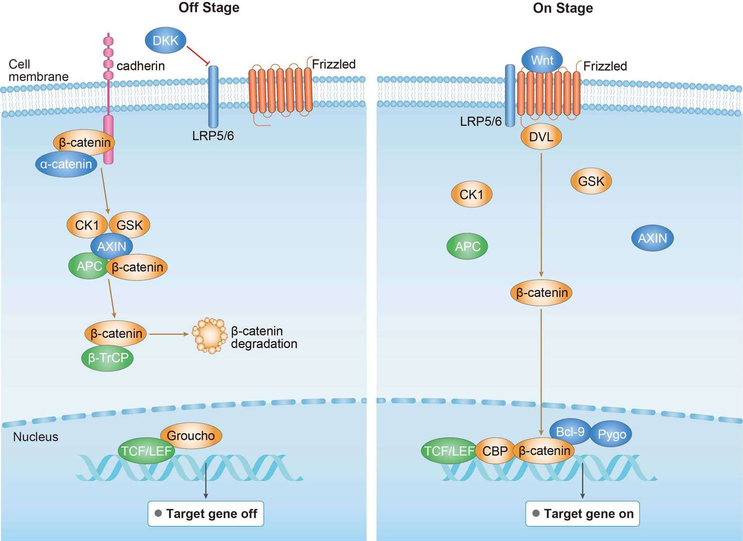 Canonical Wnt Signaling Pathway
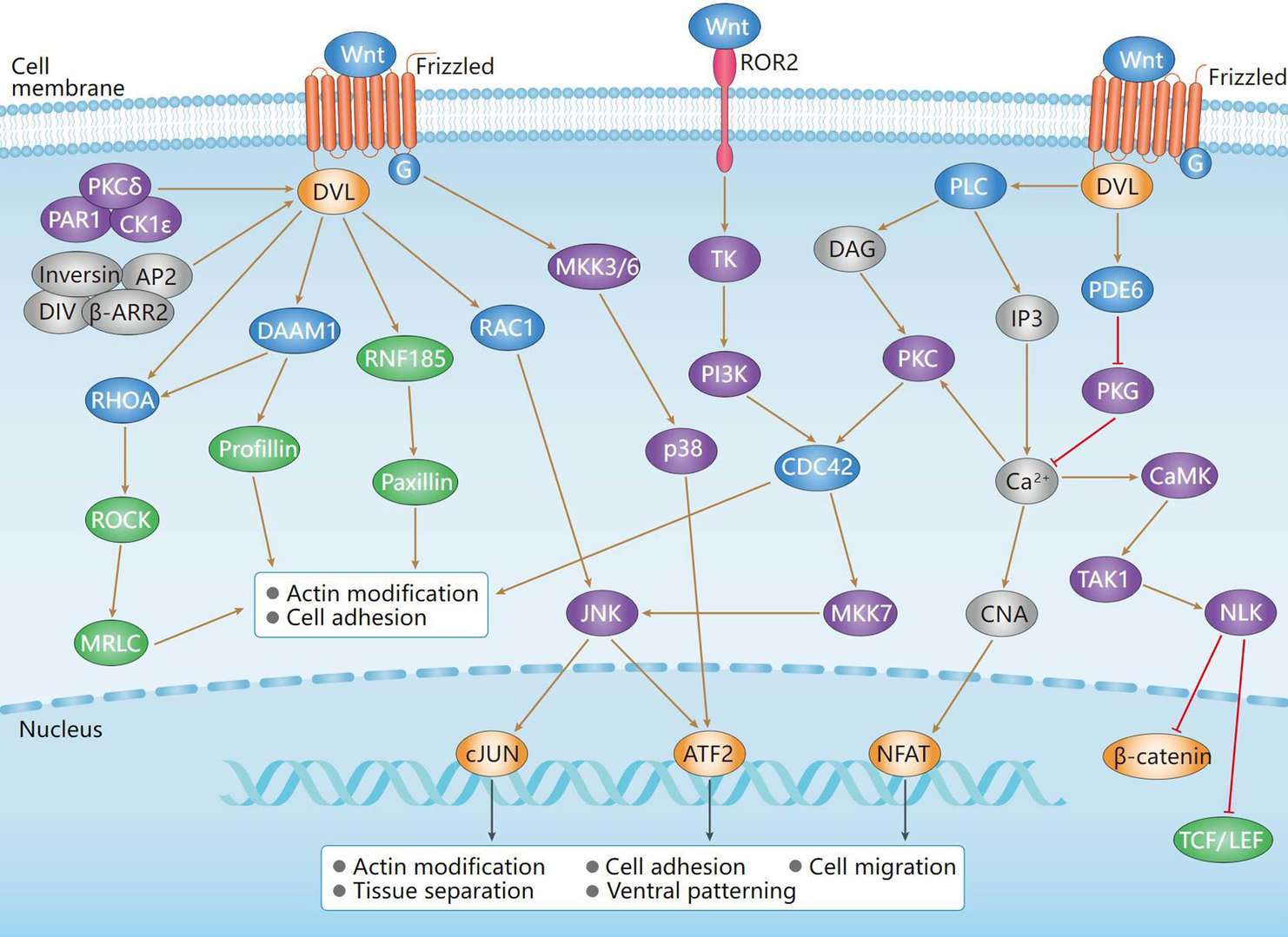 Non-Canonical Wnt Signaling Pathway
Downloadable Resources
Download resources about recombinant antibody development and antibody engineering to boost your research.
Product Notes
This is a product of Creative Biolabs' Hi-Affi™ recombinant antibody portfolio, which has several benefits including:
• Increased sensitivity
• Confirmed specificity
• High repeatability
• Excellent batch-to-batch consistency
• Sustainable supply
• Animal-free production
See more details about Hi-Affi™ recombinant antibody benefits.
Datasheet
MSDS
COA
Certificate of Analysis LookupTo download a Certificate of Analysis, please enter a lot number in the search box below. Note: Certificate of Analysis not available for kit components.
Lot Number:
See other products for "CTNNA1"
Select a product category from the dropdown menu below to view related products.
| CAT | Product Name | Application | Type |
|---|---|---|---|
| MOB-2020z | Mouse Anti-CTNNA1 Recombinant Antibody (clone 5G7) | WB, FC, ICC, IF | Mouse IgG1 |
| CAT | Product Name | Application | Type |
|---|---|---|---|
| MOB-0582MZ | Recombinant Mouse Anti-Human CTNNA1 Antibody (clone 36C2) | IHC-Fr, IHC-P, WB | Mouse antibody |
| CAT | Product Name | Application | Type |
|---|---|---|---|
| BRD-0085MZ | Chicken Anti-α-1-Catenin (ab2) Polyclonal IgY | WB | Chicken antibody |
| CAT | Product Name | Application | Type |
|---|---|---|---|
| MOR-0856 | Hi-Affi™ Rabbit Anti-CTNNA1 Recombinant Antibody (clone DS856AB) | WB, FC, ICC | Rabbit IgG |
| CAT | Product Name | Application | Type |
|---|---|---|---|
| MRO-0062-CN | Rabbit Anti-CTNNA1 Recombinant Antibody (clone CBACN-021) | WB, IF, IHC, IP, FC | Rabbit IgG |
| CAT | Product Name | Application | Type |
|---|---|---|---|
| VS3-CJ890 | Rabbit Anti-CTNNA1 Recombinant Antibody (VS3-CJ890) | WB, ICC, IF, IHC, IP, FC | Rabbit IgG |
| CAT | Product Name | Application | Type |
|---|---|---|---|
| VS3-FY422 | Recombinant Rabbit Anti-CTNNA1 Antibody (clone R07-7D5) | WB, IP | Rabbit IgG |
| CAT | Product Name | Application | Type |
|---|---|---|---|
| VS-1024-XY64 | Rabbit Anti-NHP CTNNA1 Recombinant Antibody (VS-1024-XY64) | WB, IP | Rabbit IgG |
| CAT | Product Name | Application | Type |
|---|---|---|---|
| VS-0325-XY574 | Anti-CTNNA1 Immunohistochemistry Kit | IHC |
| CAT | Product Name | Application | Type |
|---|---|---|---|
| VS-0525-XY1766 | Anti-Human CTNNA1 Immunohistochemistry Kit | IHC |
Specific Inquiry

See Our Custom Production in Action


Popular Products

Application: FuncS, IF, Neut, ELISA, FC, IP, ICC

Application: IF, IP, Neut, FuncS, ELISA, FC, ICC

Application: WB, ELISA, FC, IP, FuncS, IF, Neut

Application: ELISA, FC, IP, FuncS, IF, Neut, ICC

Application: IF, IP, Neut, FuncS, ELISA, FC, ICC

Application: ELISA, Neut, IF, IP, FC, FuncS

Application: FC, IP, ELISA, Neut, FuncS, IF, ICC

Application: ELISA, FC, IP, FuncS, IF, Neut, ICC

Application: FuncS, IF, Neut, ELISA, FC, IP, ICC

Application: ELISA, FC, IP, FuncS, IF, Neut, ICC

Application: FuncS, IF, Neut, ELISA, FC, IP, IHC


For research use only. Not intended for any clinical use. No products from Creative Biolabs may be resold, modified for resale or used to manufacture commercial products without prior written approval from Creative Biolabs.
Send Inquiry
This site is protected by reCAPTCHA and the Google Privacy Policy and Terms of Service apply.