

Rabbit Anti-CTNNA1 Recombinant Antibody (VS3-CJ890)
CAT#: VS3-CJ890
This product is a rabbit antibody that recognizes human, mouse, and rat CTNNA1.
Gene Expression

Subcellular Location
Figure 1 IF staining of human cell line CACO-2
Immunofluorescent staining of human cell line CACO-2 shows localization to plasma membrane & cell junctions.
* Image credit: Image credit: Human Protein Atlas v21.proteinatlas.org/images/46119/1028_H10_1_selected.jpg

Normal Tissue
Figure 2 IHC staining of human prostate
Immunohistochemical staining of human prostate shows strong cytoplasmic positivity in glandular cells.
* Image credit: Image credit: Human Protein Atlas v21.proteinatlas.org/images/21089/ihc_selected.jpg

Normal Tissue
Figure 3 Testis
Cells in seminiferous ducts Staining: High Intensity: Strong Quantity: 75%-25% Location: Cytoplasmic/ membranous Leydig cells Staining: Medium Intensity: Moderate Quantity:>75% Location: Cytoplasmic/ membranous
* Image credit: Image credit: Human Protein Atlas v21.proteinatlas.org/images/46119/124809_A_4_6.jpg

Normal Tissue
Figure 4 Lymph node
Non-germinal center cells Staining: Medium Intensity: Moderate Quantity: 75%-25% Location: Cytoplasmic/ membranous
* Image credit: Image credit: Human Protein Atlas v21.proteinatlas.org/images/46119/124809_A_9_8.jpg

Normal Tissue
Figure 5 Colon
Endothelial cells Staining: Medium Intensity: Moderate Quantity:>75% Location: Cytoplasmic/ membranous Glandular cells Staining: High Intensity: Strong Quantity:>75% Location: Cytoplasmic/ membranous Peripheral nerve/ganglion Staining: Medium Intensity: Moderate Quantity:>75% Location: Cytoplasmic/ membranous
* Image credit: Image credit: Human Protein Atlas v21.proteinatlas.org/images/46119/124809_A_7_3.jpg

Normal Tissue
Figure 6 Liver
Hepatocytes Staining: Medium Intensity: Moderate Quantity: 75%-25% Location: Cytoplasmic/ membranous
* Image credit: Image credit: Human Protein Atlas v21.proteinatlas.org/images/46119/124809_A_8_4.jpg

Normal Tissue
Figure 7 Gallbladder
Glandular cells Staining: High Intensity: Strong Quantity:>75% Location: Cytoplasmic/ membranous
* Image credit: Image credit: Human Protein Atlas v21.proteinatlas.org/images/46119/124809_A_6_4.jpg

Normal Tissue
Figure 8 Kidney
Cells in glomeruli Staining: Low Intensity: Weak Quantity: 75%-25% Location: Cytoplasmic/ membranous Cells in tubules Staining: High Intensity: Strong Quantity:>75% Location: Cytoplasmic/ membranous
* Image credit: Image credit: Human Protein Atlas v21.proteinatlas.org/images/46119/124809_A_8_5.jpg

RNA Expression
Figure 9 RNA cell line category: Low cell line specificity
Cell lines ordered by descending RNA expression order
* Image credit: Image credit: Human Protein Atlas v21.proteinatlas.org/ENSG00000044115-CTNNA1
❮


 ❯
❯
Specifications
- Immunogen
- Recombinant protein
- Host Species
- Rabbit
- Type
- Rabbit IgG
- Specificity
- Human, Mouse, Rat CTNNA1
- Species Reactivity
- Human, Mouse, Rat
- Applications
- WB, ICC, IF, IHC, IP, FC
- Conjugate
- Unconjugated
Product Property
- Purification
- Protein A affinity purified
- Purity
- >95% as determined by SDS-PAGE
- Format
- Liquid
- Buffer
- 40% Glycerol, 1% BSA, TBS, pH7.4.
- Preservative
- 0.05% Sodium Azide
- Storage
- Store at 4°C for short term. Aliquot and store at -20°C for long term. Avoid repeated freeze/thaw cycles.
Applications
- Application Notes
- This antibody has been tested for use in Western Blot, Immunocytochemistry, Immunofluorescence, Immunohistochemistry, Immunoprecipitation, Flow Cytometry.
Target
- Alternative Names
- MDPT2; CAP102
- Gene ID
- 1495
- UniProt ID
- P35221
- Sequence Similarities
- Belongs to the vinculin/alpha-catenin family.
- Cellular Localization
- Cell junction, Cell membrane, Cytoplasm, Cytoskeleton, Membrane
- Post Translation Modifications
- Sumoylated.
Phosphorylation seems to contribute to the strength of cell-cell adhesion rather than to the basic capacity for cell-cell adhesion.
- Protein Refseq
- NP_001277236.1; NP_001277238.1; NP_001277239.1
- Function
- Associates with the cytoplasmic domain of a variety of cadherins. The association of catenins to cadherins produces a complex which is linked to the actin filament network, and which seems to be of primary importance for cadherins cell-adhesion properties. Can associate with both E- and N-cadherins. Originally believed to be a stable component of E-cadherin/catenin adhesion complexes and to mediate the linkage of cadherins to the actin cytoskeleton at adherens junctions. In contrast, cortical actin was found to be much more dynamic than E-cadherin/catenin complexes and CTNNA1 was shown not to bind to F-actin when assembled in the complex suggesting a different linkage between actin and adherens junctions components. The homodimeric form may regulate actin filament assembly and inhibit actin branching by competing with the Arp2/3 complex for binding to actin filaments. Involved in the regulation of WWTR1/TAZ, YAP1 and TGFB1-dependent SMAD2 and SMAD3 nuclear accumulation (By similarity).
May play a crucial role in cell differentiation.
REVIEWS AND Q&AS
CITATIONS
RESOURCES
DOWNLOADS
RELATED PRODUCTS
Inquiry
Navs
Customer Review

There are currently no Customer reviews or questions for VS3-CJ890. Click the button above to contact us or submit your feedback about this product.
Submit Your Publication
Published with our product? Submit your paper and receive a 10% discount on your next order! Share your research to earn exclusive rewards.
Related Signaling Pathways
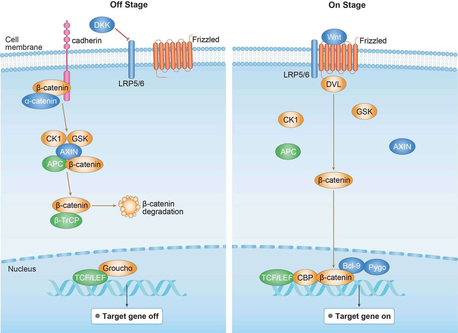 Canonical Wnt Signaling Pathway
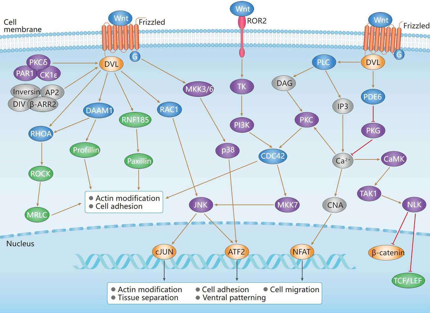 Non-Canonical Wnt Signaling Pathway
Downloadable Resources
Download resources about recombinant antibody development and antibody engineering to boost your research.
Product Notes
This is a product of Creative Biolabs' Hi-Affi™ recombinant antibody portfolio, which has several benefits including:
• Increased sensitivity
• Confirmed specificity
• High repeatability
• Excellent batch-to-batch consistency
• Sustainable supply
• Animal-free production
See more details about Hi-Affi™ recombinant antibody benefits.
Datasheet
MSDS
COA
Certificate of Analysis LookupTo download a Certificate of Analysis, please enter a lot number in the search box below. Note: Certificate of Analysis not available for kit components.
Lot Number:
Protocol & Troubleshooting
We have outlined the assay protocols, covering reagents, solutions, procedures, and troubleshooting tips for common issues in order to better assist clients in conducting experiments with our products. View the full list of Protocol & Troubleshooting.
Isotype Control
- CAT
- Product Name
Secondary Antibody
- CAT
- Product Name
See other products for "CTNNA1"
Select a product category from the dropdown menu below to view related products.
| CAT | Product Name | Application | Type |
|---|---|---|---|
| MOB-2020z | Mouse Anti-CTNNA1 Recombinant Antibody (clone 5G7) | WB, FC, ICC, IF | Mouse IgG1 |
| CAT | Product Name | Application | Type |
|---|---|---|---|
| MOB-0582MZ | Recombinant Mouse Anti-Human CTNNA1 Antibody (clone 36C2) | IHC-Fr, IHC-P, WB | Mouse antibody |
| CAT | Product Name | Application | Type |
|---|---|---|---|
| BRD-0084MZ | Chicken Anti-α-1-Catenin (ab1) Polyclonal IgY | WB | Chicken antibody |
| CAT | Product Name | Application | Type |
|---|---|---|---|
| BRD-0085MZ | Chicken Anti-α-1-Catenin (ab2) Polyclonal IgY | WB | Chicken antibody |
| CAT | Product Name | Application | Type |
|---|---|---|---|
| MOR-0856 | Hi-Affi™ Rabbit Anti-CTNNA1 Recombinant Antibody (clone DS856AB) | WB, FC, ICC | Rabbit IgG |
| CAT | Product Name | Application | Type |
|---|---|---|---|
| MRO-0062-CN | Rabbit Anti-CTNNA1 Recombinant Antibody (clone CBACN-021) | WB, IF, IHC, IP, FC | Rabbit IgG |
| CAT | Product Name | Application | Type |
|---|---|---|---|
| VS3-FY422 | Recombinant Rabbit Anti-CTNNA1 Antibody (clone R07-7D5) | WB, IP | Rabbit IgG |
| CAT | Product Name | Application | Type |
|---|---|---|---|
| VS-1024-XY64 | Rabbit Anti-NHP CTNNA1 Recombinant Antibody (VS-1024-XY64) | WB, IP | Rabbit IgG |
| CAT | Product Name | Application | Type |
|---|---|---|---|
| VS7-HM785 | Mouse Anti-CTNNA1 Recombinant Antibody (clone 8B6C1) | IHC | Mouse IgG1 |
| CAT | Product Name | Application | Type |
|---|---|---|---|
| VS-0325-XY574 | Anti-CTNNA1 Immunohistochemistry Kit | IHC |
| CAT | Product Name | Application | Type |
|---|---|---|---|
| VS-0525-XY1766 | Anti-Human CTNNA1 Immunohistochemistry Kit | IHC |
Specific Inquiry

See Our Custom Production in Action


Popular Products

Application: IF, IP, Neut, FuncS, ELISA, FC, ICC

Application: Neut, ELISA, IF, IP, FuncS, FC, ICC

Application: ELISA, IP, FC, FuncS, Neut, IF, ICC

Application: WB, ELISA, IP, FC, FuncS, Neut, IF

Application: IF, IP, Neut, FuncS, ELISA, FC, WB

Application: FuncS, IF, Neut, ELISA, FC, IP, IHC

Application: FuncS, IF, Neut, ELISA, FC, IP, IHC

Application: WB, FC, IP, ELISA, Neut, FuncS, IF

Application: FC, IP, ELISA, Neut, FuncS, IF, WB

Application: WB, FC, IP, ELISA, Neut, FuncS, IF


Application: IF, IP, Neut, FuncS, ELISA, FC, ICC

Application: FC, IP, ELISA, Neut, FuncS, IF, ICC


For research use only. Not intended for any clinical use. No products from Creative Biolabs may be resold, modified for resale or used to manufacture commercial products without prior written approval from Creative Biolabs.
Send Inquiry
This site is protected by reCAPTCHA and the Google Privacy Policy and Terms of Service apply.