

AbPlus™ Anti-CDC42 Magnetic Beads (VS-0724-YC659)
CAT#: VS-0724-YC659
The AbPlus Anti-CDC42 Magnetic Beads (VS-0724-YC659) is an innovative affinity resin which is bound with anti-CDC42 specific antibody. The beads were designed for small-scale affinity purification and immunoprecipitation (IP) of CDC42 protein under native and denaturing conditions.
Gene Expression

Subcellular Location
Figure 1 IF staining of human cell line U-2 OS
Immunofluorescent staining of human cell line U-2 OS shows localization to microtubules & cytokinetic bridge.
* Image credit: Image credit: Human Protein Atlas v21.proteinatlas.org/images/4360/if_selected.jpg

Normal Tissue
Figure 2 Cerebral cortex
Neuronal cells Staining: Low Intensity: Moderate Quantity: <25% Location: Cytoplasmic/ membranous Neuropil Staining: Medium Intensity: Moderate Quantity:>75% Location: Cytoplasmic/ membranous
* Image credit: Image credit: Human Protein Atlas v21.proteinatlas.org/images/69590/161451_B_8_5.jpg

Normal Tissue
Figure 3 Colon
Endothelial cells Staining: Low Intensity: Weak Quantity: 75%-25% Location: Cytoplasmic/ membranous Glandular cells Staining: Medium Intensity: Moderate Quantity: 75%-25% Location: Cytoplasmic/ membranous Peripheral nerve/ganglion Staining: Medium Intensity: Moderate Quantity: 75%-25% Location: Cytoplasmic/ membranous
* Image credit: Image credit: Human Protein Atlas v21.proteinatlas.org/images/69590/161451_A_7_3.jpg

Normal Tissue
Figure 4 Kidney
Cells in glomeruli Staining: Medium Intensity: Moderate Quantity:>75% Location: Cytoplasmic/ membranous Cells in tubules Staining: Medium Intensity: Moderate Quantity:>75% Location: Cytoplasmic/ membranous
* Image credit: Image credit: Human Protein Atlas v21.proteinatlas.org/images/69590/161451_A_9_5.jpg

Normal Tissue
Figure 5 Testis
Cells in seminiferous ducts Staining: Medium Intensity: Moderate Quantity:>75% Location: Cytoplasmic/ membranous Leydig cells Staining: Low Intensity: Weak Quantity:>75% Location: Cytoplasmic/ membranous
* Image credit: Image credit: Human Protein Atlas v21.proteinatlas.org/images/69590/161451_A_5_6.jpg

Normal Tissue
Figure 6 Lymph node
Germinal center cells Staining: Medium Intensity: Moderate Quantity: 75%-25% Location: Cytoplasmic/ membranous Non-germinal center cells Staining: Medium Intensity: Moderate Quantity:>75% Location: Cytoplasmic/ membranous
* Image credit: Image credit: Human Protein Atlas v21.proteinatlas.org/images/69590/161451_A_9_8.jpg

RNA Expression
Figure 7 RNA cell line category: Low cell line specificity
Cell lines ordered by descending RNA expression order
* Image credit: Image credit: Human Protein Atlas v21.proteinatlas.org/ENSG00000070831-CDC42
❮


 ❯
❯
Specifications
- Applications
- Immunoprecipitation, Protein Purification
- Matrix
- Magnetic bead
- Bead Ligand
- Anti-CDC42 specific antibody
- Target
- CDC42
- Immunogen
- Recombinant human CDC42, aa 1-188.
- Target Species
- Human
- Bead Capacity
- 40 mg/mL
- Bead size
- 25 μm
- Format
- Suspension
- Concentration
- 2 mg/mL
- Buffer
- PBS, pH 7.4
- Preservative
- 0.1% Sodium azide
- Storage
- Stored at 4°C, and is stable for up to 2 years. Do not centrifuge, dry or freeze the magnetic beads.
Applications
- Application Notes
- The beads are in suspension and will settle upon storage. Prior to use, mix the vial gently (do not vortex) to ensure delivery of proper bead volume.
Target
- Introduction
- CDC42 (Cell Division Cycle 42) is a Protein Coding gene. Diseases associated with CDC42 include Takenouchi-Kosaki Syndrome and Temperature-Sensitive Lethal Mutation. Among its related pathways are Development EGFR signaling via small GTPases and RANK Signaling in Osteoclasts. Gene Ontology (GO) annotations related to this gene include identical protein binding and protein kinase binding. An important paralog of this gene is RAC1.
- Alternative Names
- Cell Division Cycle 42; GTP Binding Protein, 25kDa; G25K GTP-Binding Protein; DJ224A6.1.1 (Cell Division Cycle 42 (GTP-Binding Protein, 25kD)); DJ224A6.1.2 (Cell Division Cycle 42 (GTP-Binding Protein, 25kD)); Cell Division Cycle 42 (GTP Binding Protein, 25kDa); Cell Division Cycle 42 (GTP-Binding Protein, 25kD);
- Gene ID
- 998
- UniProt ID
- P60953
REVIEWS AND Q&AS
CITATIONS
RESOURCES
DOWNLOADS
RELATED PRODUCTS
Inquiry
Navs
Customer Review

There are currently no Customer reviews or questions for VS-0724-YC659. Click the button above to contact us or submit your feedback about this product.
Submit Your Publication
Published with our product? Submit your paper and receive a 10% discount on your next order! Share your research to earn exclusive rewards.
Related Signaling Pathways
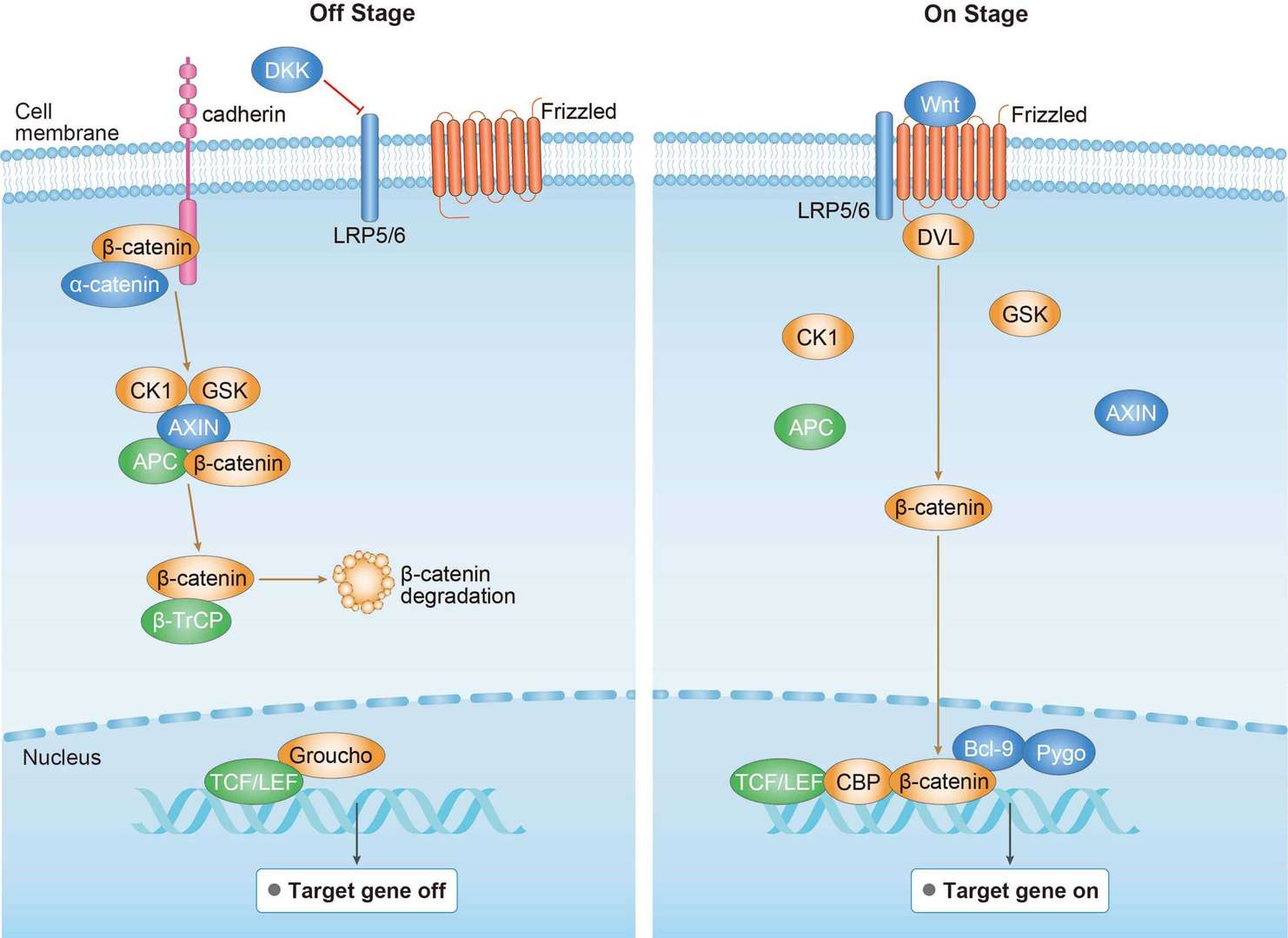 Canonical Wnt Signaling Pathway
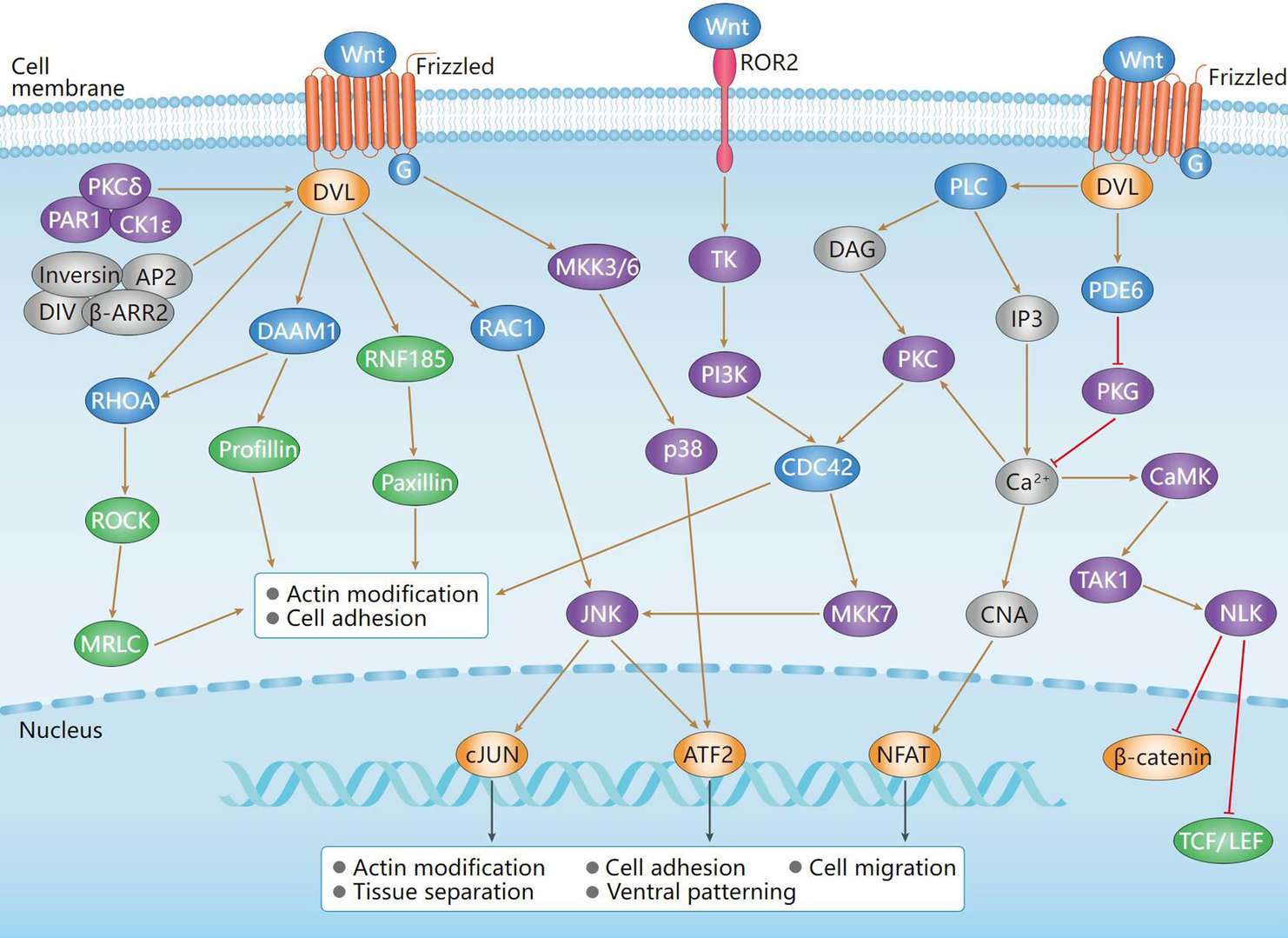 Non-Canonical Wnt Signaling Pathway
Downloadable Resources
Download resources about recombinant antibody development and antibody engineering to boost your research.
Datasheet
MSDS
COA
Certificate of Analysis LookupTo download a Certificate of Analysis, please enter a lot number in the search box below. Note: Certificate of Analysis not available for kit components.
Lot Number:
See other products for "CDC42"
Select a product category from the dropdown menu below to view related products.
| CAT | Product Name | Application | Type |
|---|---|---|---|
| MOB-4073z | Mouse Anti-CDC42 Recombinant Antibody (clone 21B7) | WB, IP, IF, ELISA | Mouse IgG1, κ |
| CAT | Product Name | Application | Type |
|---|---|---|---|
| MOB-2473MZ | Recombinant Mouse Anti-Human CDC42 Antibody (clone 2) | ELISA, WB | Mouse antibody |
| CAT | Product Name | Application | Type |
|---|---|---|---|
| BRD-0921MZ | Chicken Anti-Cdc42 Polyclonal IgY | WB | Chicken antibody |
| CAT | Product Name | Application | Type |
|---|---|---|---|
| MRO-0330-CN | Rabbit Anti-CDC42 Recombinant Antibody (clone CBACN-131) | WB, IHC, IP, FC | Rabbit IgG |
| CAT | Product Name | Application | Type |
|---|---|---|---|
| ZG-0101U | Rabbit Anti-CDC42 Recombinant Antibody (clone 3C3) | ELISA, WB, IHC | Rabbit IgG |
| CAT | Product Name | Application | Type |
|---|---|---|---|
| VS3-CJ1070 | Rabbit Anti-CDC42 Recombinant Antibody (VS3-CJ1070) | WB, IHC, IP, FC | Rabbit IgG |
| CAT | Product Name | Application | Type |
|---|---|---|---|
| VS-0325-XY387 | Anti-CDC42 Immunohistochemistry Kit | IHC |
| CAT | Product Name | Application | Type |
|---|---|---|---|
| VS13-YC187 | CytoStream™ Rabbit Anti-CDC42 Recombinant Antibody (VS13-YC187) | WB, ICC, IF, FC | Rabbit IgG |
| CAT | Product Name | Application | Type |
|---|---|---|---|
| VS-0525-XY1265 | Anti-Human CDC42 Immunohistochemistry Kit | IHC |
Specific Inquiry
See Our Custom Production in Action


Popular Products

Application: Neut, ELISA, IF, IP, FuncS, FC, IHC


Application: FuncS, IF, Neut, ELISA, FC, IP, IHC

Application: IF, IP, Neut, FuncS, ELISA, FC, WB

Application: IF, IP, Neut, FuncS, ELISA, FC, ICC

Application: FC, IP, ELISA, Neut, FuncS, IF, ICC

Application: ELISA, FC, IP, FuncS, IF, Neut, ICC

Application: ELISA, FC, IP, FuncS, IF, Neut, ICC

Application: Neut, ELISA, IF, IP, FuncS, FC, WB

Application: Neut, ELISA, IF, IP, FuncS, FC, ICC

Application: WB, FuncS, IF, Neut, ELISA, FC, IP


Application: Neut, ELISA, FuncS


Application: ELISA, IP, WB, IHC, IF, FuncS


Application: ELISA, SPR, Inhib, FuncS
For research use only. Not intended for any clinical use. No products from Creative Biolabs may be resold, modified for resale or used to manufacture commercial products without prior written approval from Creative Biolabs.
Send Inquiry
This site is protected by reCAPTCHA and the Google Privacy Policy and Terms of Service apply.