

AbPlus™ Anti-MAPK3 Magnetic Beads (CBACN-216)
CAT#: VS-0424-XY181
The AbPlus Anti-MAPK3 Magnetic Beads (CBACN-216) is an innovative affinity resin which is bound with anti-MAPK3 specific antibody (CBACN-216). The beads were designed for small-scale affinity purification and immunoprecipitation (IP) of MAPK3 protein under native and denaturing conditions.
Gene Expression

Subcellular Location
Figure 1 IF staining of human cell line U-2 OS
Immunofluorescent staining of human cell line U-2 OS shows localization to nucleoplasm.
* Image credit: Image credit: Human Protein Atlas v21.proteinatlas.org/images/2683/if_selected.jpg

Normal Tissue
Figure 2 Testis
Cells in seminiferous ducts Staining: Medium Intensity: Moderate Quantity:>75% Location: Cytoplasmic/ membranous Leydig cells Staining: High Intensity: Strong Quantity:>75% Location: Cytoplasmic/ membranous
* Image credit: Image credit: Human Protein Atlas v21.proteinatlas.org/images/5700/20815_A_5_6.jpg

Normal Tissue
Figure 3 Breast
Glandular cells Staining: High Intensity: Strong Quantity:>75% Location: Cytoplasmic/ membranous Myoepithelial cells Staining: Medium Intensity: Moderate Quantity:>75% Location: Cytoplasmic/ membranous
* Image credit: Image credit: Human Protein Atlas v21.proteinatlas.org/images/5700/21384_B_3_4.jpg

Normal Tissue
Figure 4 Lymph node
Germinal center cells Staining: High Intensity: Strong Quantity:>75% Location: Cytoplasmic/ membranous Non-germinal center cells Staining: High Intensity: Strong Quantity:>75% Location: Cytoplasmic/ membranous
* Image credit: Image credit: Human Protein Atlas v21.proteinatlas.org/images/5700/20815_A_9_8.jpg

Normal Tissue
Figure 5 Colon
Endothelial cells Staining: High Intensity: Strong Quantity:>75% Location: Cytoplasmic/ membranous Glandular cells Staining: High Intensity: Strong Quantity:>75% Location: Cytoplasmic/ membranous
* Image credit: Image credit: Human Protein Atlas v21.proteinatlas.org/images/5700/20815_A_9_3.jpg

Normal Tissue
Figure 6 Liver
Cholangiocytes Staining: High Intensity: Strong Quantity:>75% Location: Cytoplasmic/ membranous Hepatocytes Staining: High Intensity: Strong Quantity:>75% Location: Cytoplasmic/ membranous
* Image credit: Image credit: Human Protein Atlas v21.proteinatlas.org/images/5700/20815_A_8_4.jpg

Normal Tissue
Figure 7 Kidney
Cells in glomeruli Staining: Medium Intensity: Moderate Quantity:>75% Location: Cytoplasmic/ membranous Cells in tubules Staining: High Intensity: Strong Quantity:>75% Location: Cytoplasmic/ membranous
* Image credit: Image credit: Human Protein Atlas v21.proteinatlas.org/images/5700/20815_A_9_5.jpg

RNA Expression
Figure 8 RNA cell line category: Low cell line specificity
Cell lines ordered by descending RNA expression order
* Image credit: Image credit: Human Protein Atlas v21.proteinatlas.org/ENSG00000102882-MAPK3
❮


 ❯
❯
Specifications
- Applications
- Immunoprecipitation, Protein Purification
- Matrix
- Magrose bead (> 50 μmol/mL gel)
- Bead Ligand
- Anti-MAPK3 specific antibody (CBACN-216)
- Target
- MAPK3
- Immunogen
- N terminus of human MAPK3
- Target Species
- Human, Mouse
- Antibody Clone
- CBACN-216
- Bead Capacity
- 20-30 mg/mL binding antibody
- Bead size
- 10-37 μm
- Stability
- pH 2-14
- Format
- 20% Suspension
- Buffer
- PBS, pH 7.4, with 1% BSA
- Preservative
- 0.03% Proclin 300
- Storage
- Stored at 4°C, and is stable for up to 2 years. Do not centrifuge, dry or freeze the magnetic beads.
Applications
- Application Notes
- The beads are in suspension and will settle upon storage. Prior to use, mix the vial gently (do not vortex) to ensure delivery of proper bead volume.
Target
- Introduction
- The protein encoded by this gene is a member of the MAP kinase family. MAP kinases, also known as extracellular signal-regulated kinases (ERKs), act in a signaling cascade that regulates various cellular processes such as proliferation, differentiation, and cell cycle progression in response to a variety of extracellular signals. This kinase is activated by upstream kinases, resulting in its translocation to the nucleus where it phosphorylates nuclear targets. Alternatively spliced transcript variants encoding different protein isoforms have been described.
- Alternative Names
- Mitogen-Activated Protein Kinase 3; Extracellular Signal-Regulated Kinase 1; Microtubule-Associated Protein 2 Kinase; Insulin-Stimulated MAP2 Kinase; MAP Kinase Isoform P44; EC 2.7.11.24; P44-ERK1; P44-MAPK; PRKM3; ERK-1; ERK1
- Gene ID
- 5595
- UniProt ID
- P27361
REVIEWS AND Q&AS
CITATIONS
RESOURCES
DOWNLOADS
RELATED PRODUCTS
Inquiry
Navs
Customer Review

There are currently no Customer reviews or questions for VS-0424-XY181. Click the button above to contact us or submit your feedback about this product.
Submit Your Publication
Published with our product? Submit your paper and receive a 10% discount on your next order! Share your research to earn exclusive rewards.
Related Signaling Pathways
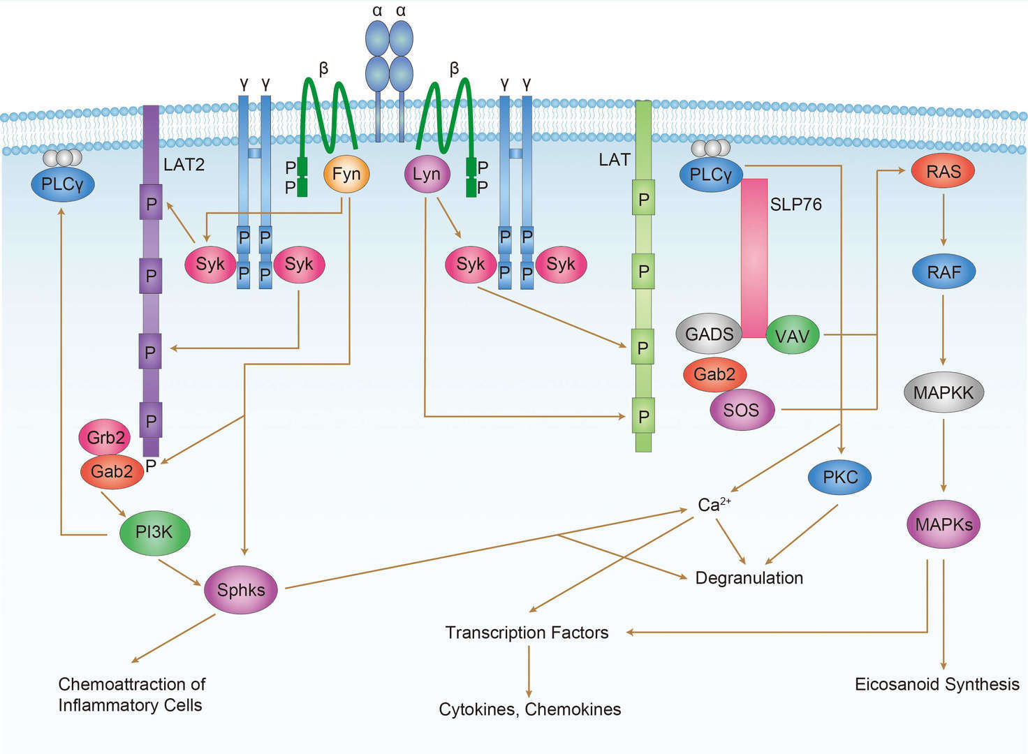 FcεR1 Signaling Pathway
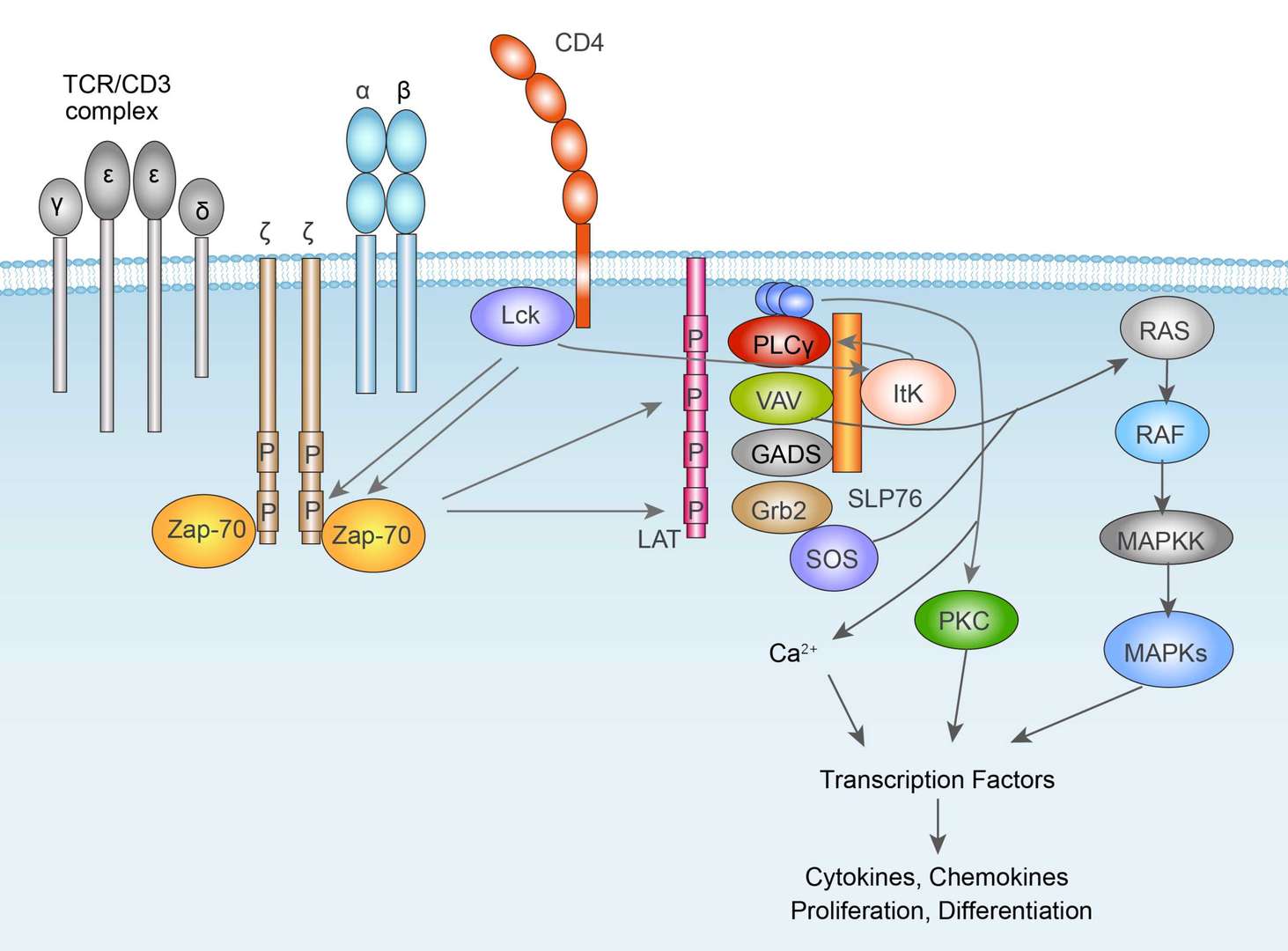 TCR Signaling Pathway
Related Diseases
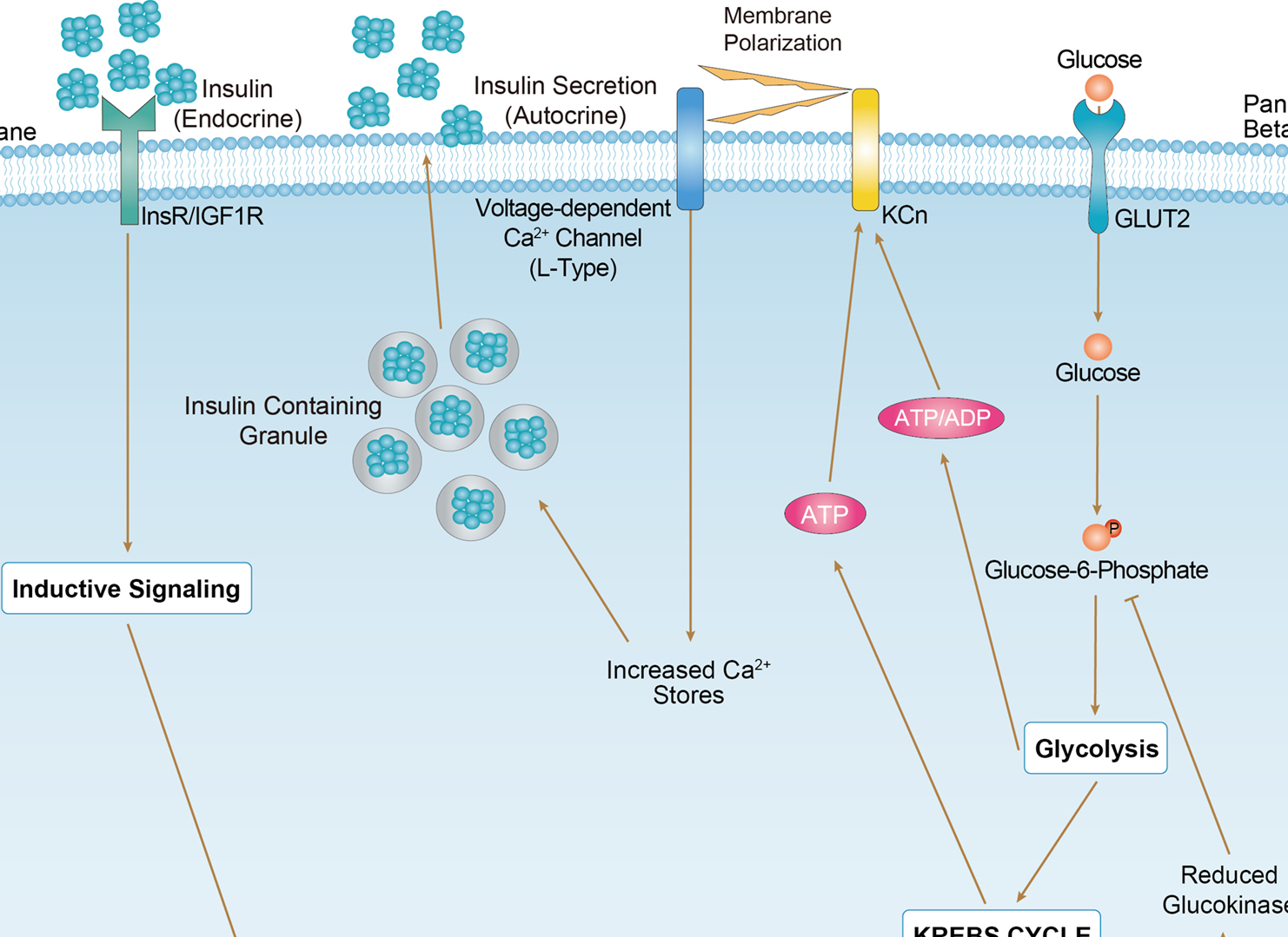 Maturity Onset Diabetes of the Young
Downloadable Resources
Download resources about recombinant antibody development and antibody engineering to boost your research.
Datasheet
MSDS
COA
Certificate of Analysis LookupTo download a Certificate of Analysis, please enter a lot number in the search box below. Note: Certificate of Analysis not available for kit components.
Lot Number:
See other products for "MAPK3"
Select a product category from the dropdown menu below to view related products.
| CAT | Product Name | Application | Type |
|---|---|---|---|
| MOB-2002z | Mouse Anti-MAPK3 Recombinant Antibody (clone 32C11) | WB, ELISA, FC, ICC, IF, IHC | Mouse IgG1 |
| CAT | Product Name | Application | Type |
|---|---|---|---|
| MOB-0572CT | Recombinant Mouse anti-Human MAPK3 Monoclonal antibody (EML1636) | IHC-P, WB |
| CAT | Product Name | Application | Type |
|---|---|---|---|
| BRD-0342MZ | Chicken Anti-MAPK3 Polyclonal IgY | Indirect ELISA, WB | Chicken antibody |
| CAT | Product Name | Application | Type |
|---|---|---|---|
| MOR-2154 | Hi-Affi™ Recombinant Rabbit Anti-MAPK3 Monoclonal Antibody (DS2154AB) | WB, IP | IgG |
| CAT | Product Name | Application | Type |
|---|---|---|---|
| MOR-4659 | Hi-Affi™ Recombinant Rabbit Anti-MAPK3 Monoclonal Antibody (TH173DS) | WB, IF, ICC, FC | IgG |
| CAT | Product Name | Application | Type |
|---|---|---|---|
| MOR-4697 | Hi-Affi™ Recombinant Rabbit Anti-MAPK3 Monoclonal Antibody (TH211DS) | WB, IF, ICC, IHC-P, FC, ChIP, ELISA | IgG |
| CAT | Product Name | Application | Type |
|---|---|---|---|
| MRO-0543-CN | Recombinant Mouse Anti-MAPK3 Monoclonal Antibody (7-D6-E5) | WB, IF, IHC | Mouse IgG1 |
| CAT | Product Name | Application | Type |
|---|---|---|---|
| MRO-0544-CN | Recombinant Rabbit Anti-MAPK3 Monoclonal Antibody (CBACN-215) | WB, IF, IHC | Rabbit IgG |
| CAT | Product Name | Application | Type |
|---|---|---|---|
| MRO-0545-CN | Recombinant Rabbit Anti-MAPK3 Monoclonal Antibody (CBACN-216) | WB, IF, IHC, IP, FC | Rabbit IgG |
| CAT | Product Name | Application | Type |
|---|---|---|---|
| MOR-0040-FY | Rabbit Anti-MAPK3 Recombinant Antibody (clone AFY0011) | IHC-P, IP, WB | Rabbit IgG |
| CAT | Product Name | Application | Type |
|---|---|---|---|
| VS-1024-XY190 | Mouse Anti-NHP MAPK3 Recombinant Antibody (clone 12D11) | WB, IHC, IF | Mouse IgG |
| CAT | Product Name | Application | Type |
|---|---|---|---|
| VS-0525-XY4236 | Anti-MAPK3 Immunohistochemistry Kit | IHC |
| CAT | Product Name | Application | Type |
|---|---|---|---|
| VS-0525-XY4237 | Anti-Mouse MAPK3 Immunohistochemistry Kit | IHC |
| CAT | Product Name | Application | Type |
|---|---|---|---|
| VS-0525-XY4238 | Anti-Monkey MAPK3 Immunohistochemistry Kit | IHC |
Specific Inquiry
See Our Custom Production in Action


Popular Products

Application: Neut, ELISA, IF, IP, FuncS, FC, ICC


Application: WB, ELISA, FC, IP, FuncS, IF, Neut

Application: WB, FuncS, IF, Neut, ELISA, FC, IP

Application: WB, IF, IP, Neut, FuncS, ELISA, FC

Application: FuncS, IF, Neut, ELISA, FC, IP, IHC

Application: ELISA, Neut, IF, IP, FC, FuncS

Application: FC, IP, ELISA, Neut, FuncS, IF, WB
-2-1.png)
Application: IP, IF, FuncS, FC, Neut, ELISA, IHC


For research use only. Not intended for any clinical use. No products from Creative Biolabs may be resold, modified for resale or used to manufacture commercial products without prior written approval from Creative Biolabs.
Send Inquiry
This site is protected by reCAPTCHA and the Google Privacy Policy and Terms of Service apply.