

Anti-Rat BLNK Immunohistochemistry Kit
CAT#: VS-0525-XY782
The BLNK IHC kit provides a complete reagent set for precise and efficient tissue staining. It works well with both paraffin-embedded and frozen tissue sections and demonstrates broad reactivity with multiple species. The user-friendly protocol ensures reproducible, high-quality results for BLNK detection in various tissue types.
Gene Expression

Subcellular Location
Figure 1 IF staining of human cell line REH
Immunofluorescent staining of human cell line REH shows localization to plasma membrane & vesicles.
* Image credit: Image credit: Human Protein Atlas v21.proteinatlas.org/images/38309/1779_G1_1_selected.jpg

Normal Tissue
Figure 2 Tonsil
High expression
* Image credit: Image credit: Human Protein Atlas v21.proteinatlas.org/images/38309/101333_A_4_8_val_selected.jpg

Normal Tissue
Figure 3 Cerebral cortex
Endothelial cells Staining: Low Intensity: Moderate Quantity: <25% Location: Cytoplasmic/ membranous Glial cells Staining: Low Intensity: Moderate Quantity: <25% Location: Cytoplasmic/ membranous
* Image credit: Image credit: Human Protein Atlas v21.proteinatlas.org/images/38309/102126_B_7_5.jpg

Normal Tissue
Figure 4 Colon
Endocrine cells Staining: Medium Intensity: Moderate Enterocytes Staining: Low Intensity: Moderate Quantity: <25% Enterocytes - Gradient Gradient: Intensity: Not representative Quantity: None Enterocytes - Mucosal lymphoid cells Staining: Medium Intensity: Moderate Quantity: 75%-25%
* Image credit: Image credit: Human Protein Atlas v21.proteinatlas.org/images/38309/101333_A_9_3.jpg

Normal Tissue
Figure 5 Kidney
Collecting ducts Staining: Medium Intensity: Moderate Quantity: 75%-25% Distal tubules Staining: Medium Intensity: Moderate Quantity: 75%-25%
* Image credit: Image credit: Human Protein Atlas v21.proteinatlas.org/images/38309/101333_A_7_5.jpg

Normal Tissue
Figure 6 Lymph node
Germinal center cells Staining: Medium Intensity: Moderate Quantity:>75% Location: Cytoplasmic/ membranous Non-germinal center cells Staining: High Intensity: Strong Quantity: 75%-25% Location: Cytoplasmic/ membranous
* Image credit: Image credit: Human Protein Atlas v21.proteinatlas.org/images/38309/101333_A_7_8.jpg

Normal Tissue
Figure 7 Spleen
Cells in red pulp Staining: Medium Intensity: Strong Quantity: <25% Location: Cytoplasmic/ membranous Cells in white pulp Staining: High Intensity: Strong Quantity: 75%-25% Location: Cytoplasmic/ membranous
* Image credit: Image credit: Human Protein Atlas v21.proteinatlas.org/images/38309/102126_B_7_4.jpg

RNA Expression
Figure 8 RNA cell line category: Group enriched (Daudi, REH, RPMI-8226, U-698)
Cell lines ordered by descending RNA expression order
* Image credit: Image credit: Human Protein Atlas v21.proteinatlas.org/ENSG00000095585-BLNK
❮


 ❯
❯
Specifications
- Application
- IHC
- Size
- 50 Tests
- Species Reactivity
- Human, Mouse, Rat
- Target
- BLNK
- Primary Antibody
- Rabbit Anti-BLNK Antibody
- Secondary Antibody
- Goat anti-Rabbit Antibody, HRP
- Sample Type
- FFPE tissue; Frozen section tissue
- Kit Storage
- All reagents should be kept at 2-8°C. The kit remains stable for up to 6 months after arrival.
REVIEWS AND Q&AS
CITATIONS
RESOURCES
DOWNLOADS
RELATED PRODUCTS
Inquiry
Navs
Customer Review

There are currently no Customer reviews or questions for VS-0525-XY782. Click the button above to contact us or submit your feedback about this product.
Submit Your Publication
Published with our product? Submit your paper and receive a 10% discount on your next order! Share your research to earn exclusive rewards.
Related Signaling Pathways
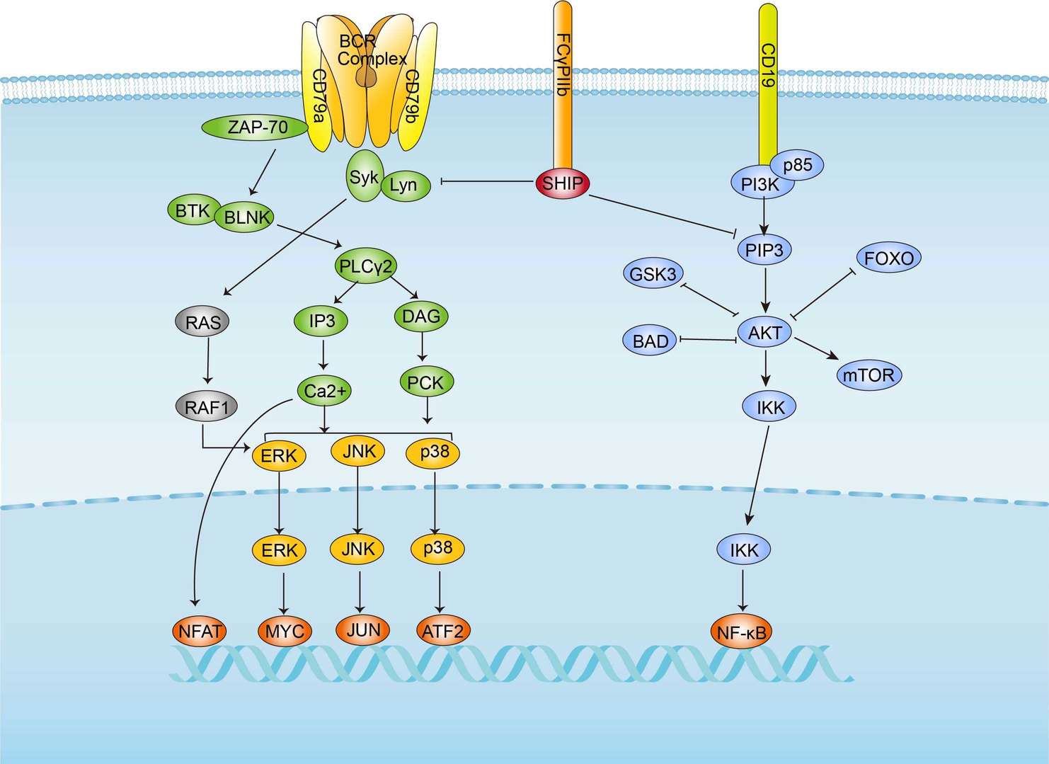 BCR Signaling Pathway
Related Diseases
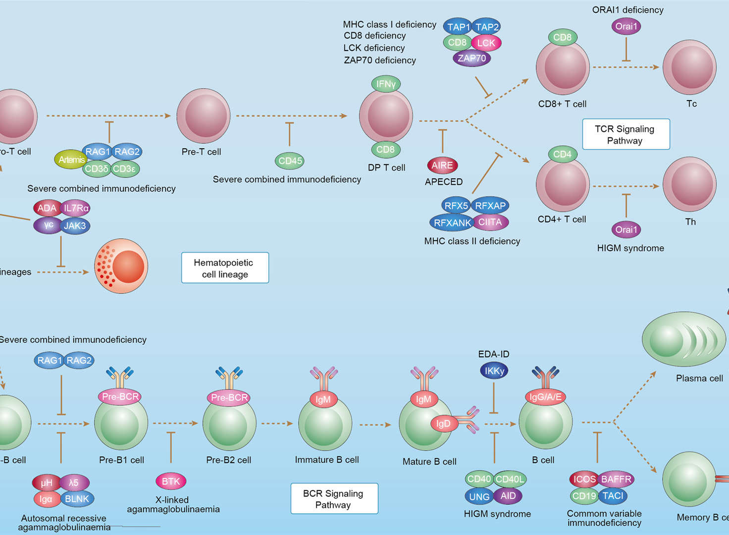 Primary Immunodeficiency
Downloadable Resources
Download resources about recombinant antibody development and antibody engineering to boost your research.
Datasheet
MSDS
COA
Certificate of Analysis LookupTo download a Certificate of Analysis, please enter a lot number in the search box below. Note: Certificate of Analysis not available for kit components.
Lot Number:
Protocol & Troubleshooting
We have outlined the assay protocols, covering reagents, solutions, procedures, and troubleshooting tips for common issues in order to better assist clients in conducting experiments with our products. View the full list of Protocol & Troubleshooting.
See other products for "BLNK"
Select a product category from the dropdown menu below to view related products.
| CAT | Product Name | Application | Type |
|---|---|---|---|
| MOB-1548z | Mouse Anti-BLNK Recombinant Antibody (clone 30G10) | WB, ELISA, FC, ICC, IF, IHC | Mouse IgG1 |
| CAT | Product Name | Application | Type |
|---|---|---|---|
| MOR-0363 | Hi-Affi™ Rabbit Anti-BLNK Recombinant Antibody (clone DS363AB) | ELISA, WB | Rabbit IgG |
| CAT | Product Name | Application | Type |
|---|---|---|---|
| VS3-QX121 | Mouse Anti-BLNK Recombinant Antibody (clone 520CT6.1.1) | WB | Mouse IgM |
| CAT | Product Name | Application | Type |
|---|---|---|---|
| VS3-WK125 | Mouse Anti-BLNK Recombinant Antibody (clone G1-F9) | WB, ELISA | Mouse IgG1 |
| CAT | Product Name | Application | Type |
|---|---|---|---|
| VS3-XY126 | Mouse Anti-BLNK Recombinant Antibody (clone 5G9) | ELISA, WB, IHC | Mouse IgG1 |
| CAT | Product Name | Application | Type |
|---|---|---|---|
| VS3-FY1666 | Rabbit Anti-BLNK Recombinant Antibody (clone R04-7H1) | WB, IP | Rabbit IgG |
| CAT | Product Name | Application | Type |
|---|---|---|---|
| VS-0225-XY5 | CytoStream™ Mouse Anti-BLNK Recombinant Antibody (VS-0225-XY5) | WB, FC | Mouse IgG2a, kappa |
| CAT | Product Name | Application | Type |
|---|---|---|---|
| VS7-HM238 | Mouse Anti-BLNK Recombinant Antibody (clone CBL078HM) | WB, IHC-p, IF, ICC, FCM, ELISA | Mouse IgG |
| CAT | Product Name | Application | Type |
|---|---|---|---|
| VS-0325-XY247 | Anti-BLNK Immunohistochemistry Kit | IHC |
| CAT | Product Name | Application | Type |
|---|---|---|---|
| VS-0525-XY781 | Anti-Mouse BLNK Immunohistochemistry Kit | IHC |
| CAT | Product Name | Application | Type |
|---|---|---|---|
| VS-0525-XY780 | Anti-Human BLNK Immunohistochemistry Kit | IHC |
Specific Inquiry
See Our Custom Production in Action


Popular Products

Application: IP, IF, FuncS, FC, Neut, ELISA, IHC

Application: FC, Cyt, Stim, PP, Agonist

Application: WB, IF, IP, Neut, FuncS, ELISA, FC

Application: ELISA, IP, FC, FuncS, Neut, IF, ICC

Application: ELISA, FC, IP, FuncS, IF, Neut, ICC

Application: IP, IF, FuncS, FC, Neut, ELISA, ICC

Application: WB, FuncS, IF, Neut, ELISA, FC, IP

Application: ELISA, IHC

Application: IF, IP, Neut, FuncS, ELISA, FC, WB

Application: FuncS, IF, Neut, ELISA, FC, IP, IHC

Application: Neut, ELISA, IF, IP, FuncS, FC, ICC

Application: IF, IP, Neut, FuncS, ELISA, FC, WB

Application: ELISA, FC, IP, FuncS, IF, Neut, ICC

Application: IF, IP, Neut, FuncS, ELISA, FC, ICC

Application: ELISA, FC, IP, FuncS, IF, Neut, ICC


Application: Neut, ELISA, FuncS


For research use only. Not intended for any clinical use. No products from Creative Biolabs may be resold, modified for resale or used to manufacture commercial products without prior written approval from Creative Biolabs.
Send Inquiry
This site is protected by reCAPTCHA and the Google Privacy Policy and Terms of Service apply.