

AbPlus™ Anti-STAT6 Magnetic Beads (VS-0724-YC633)
CAT#: VS-0724-YC633
The AbPlus Anti-STAT6 Magnetic Beads (VS-0724-YC633) is an innovative affinity resin which is bound with anti-STAT6 specific antibody. The beads were designed for small-scale affinity purification and immunoprecipitation (IP) of STAT6 protein under native and denaturing conditions.
Gene Expression

Subcellular Location and Protein Expression
Figure 1 IF staining of human cell line A-431
Immunofluorescent staining of human cell line A-431 shows localization to nucleoplasm & cytosol.
* Image credit: Image credit: Human Protein Atlas v21.proteinatlas.org/images/1861/27_A4_1_selected.jpg

Subcellular Location and Protein Expression
Figure 2 IHC staining of human urinary bladder
Immunohistochemical staining of human urinary bladder shows strong cytoplasmic and nuclear positivity in urothelial cells.
* Image credit: Image credit: Human Protein Atlas v21.proteinatlas.org/images/1861/ihc_selected.jpg

Normal Tissue
Figure 3 Cerebral cortex
Endothelial cells
Staining: Medium
Intensity: Moderate
Quantity: 75%-25%
Location: Cytoplasmic/membranous nuclear
* Image credit: Image credit: Human Protein Atlas v21.proteinatlas.org/images/1861/7497_B_8_5.jpg

Normal Tissue
Figure 4 Colon
Endothelial cells
Staining: Medium
Intensity: Moderate
Quantity: 75%-25%
Location: Cytoplasmic/membranous nuclear
Glandular cells
Staining: Medium
Intensity: Moderate
Quantity:>75%
Location: Cytoplasmic/membranous nuclear
Peripheral nerve/ganglion
Staining: Medium
Intensity: Moderate
Quantity: 75%-25%
Location: Cytoplasmic/membranous nuclear
* Image credit: Image credit: Human Protein Atlas v21.proteinatlas.org/images/1861/7497_A_7_3.jpg

Normal Tissue
Figure 5 Kidney
Cells in tubules
Staining: Medium
Intensity: Moderate
Quantity: 75%-25%
Location: Cytoplasmic/membranous
* Image credit: Image credit: Human Protein Atlas v21.proteinatlas.org/images/1861/7497_A_7_5.jpg

Normal Tissue
Figure 6 Testis
Cells in seminiferous ducts
Staining: Medium
Intensity: Moderate
Quantity:>75%
Location: Cytoplasmic/membranous
Leydig cells
Staining: Medium
Intensity: Moderate
Quantity:>75%
Location: Cytoplasmic/membranous nuclear
* Image credit: Image credit: Human Protein Atlas v21.proteinatlas.org/images/1861/7497_A_4_6.jpg

Normal Tissue
Figure 7 Lymph node
Germinal center cells
Staining: High
Intensity: Strong
Quantity:>75%
Location: Cytoplasmic/membranous nuclear
Non-germinal center cells
Staining: Medium
Intensity: Moderate
Quantity: 75%-25%
Location: Cytoplasmic/membranous nuclear
* Image credit: Image credit: Human Protein Atlas v21.proteinatlas.org/images/1861/7497_A_8_8.jpg

RNA Expression
Figure 8 RNA cell line category: Low cell line specificity
Cell lines ordered by descending RNA expression order.
* Image credit: Image credit: Human Protein Atlas v21.proteinatlas.org/ENSG00000166888-STAT6/subcellular
❮


 ❯
❯
Specifications
- Applications
- Immunoprecipitation, Protein Purification
- Matrix
- Magnetic bead
- Bead Ligand
- Anti-STAT6 specific antibody
- Target
- STAT6
- Immunogen
- Recombinant human STAT6 fragment derived from E. coli.
- Target Species
- Human
- Bead Capacity
- 40 mg/mL
- Bead size
- 25 μm
- Format
- Suspension
- Concentration
- 2 mg/mL
- Buffer
- PBS, pH 7.4
- Preservative
- 0.1% Sodium azide
- Storage
- Stored at 4°C, and is stable for up to 2 years. Do not centrifuge, dry or freeze the magnetic beads.
Applications
- Application Notes
- The beads are in suspension and will settle upon storage. Prior to use, mix the vial gently (do not vortex) to ensure delivery of proper bead volume.
Target
- Introduction
- The protein encoded by this gene is a member of the STAT family of transcription factors. In response to cytokines and growth factors, STAT family members are phosphorylated by the receptor associated kinases, and then form homo- or heterodimers that translocate to the cell nucleus where they act as transcription activators. This protein plays a central role in exerting IL4 mediated biological responses. It is found to induce the expression of BCL2L1/BCL-X(L), which is responsible for the anti-apoptotic activity of IL4. Knockout studies in mice suggested the roles of this gene in differentiation of T helper 2 (Th2) cells, expression of cell surface markers, and class switch of immunoglobulins.
- Alternative Names
- HIES6; STAT6B; STAT6C; D12S1644; IL-4-STAT
- Gene ID
- 6778
- UniProt ID
- P42226
REVIEWS AND Q&AS
CITATIONS
RESOURCES
DOWNLOADS
RELATED PRODUCTS
Inquiry
Navs
Customer Review

There are currently no Customer reviews or questions for VS-0724-YC633. Click the button above to contact us or submit your feedback about this product.
Submit Your Publication
Published with our product? Submit your paper and receive a 10% discount on your next order! Share your research to earn exclusive rewards.
Related Diseases
 Endometrial Cancer
 Non-small Cell Lung Cancer
 Pancreatic Cancer
 Throid Cancer
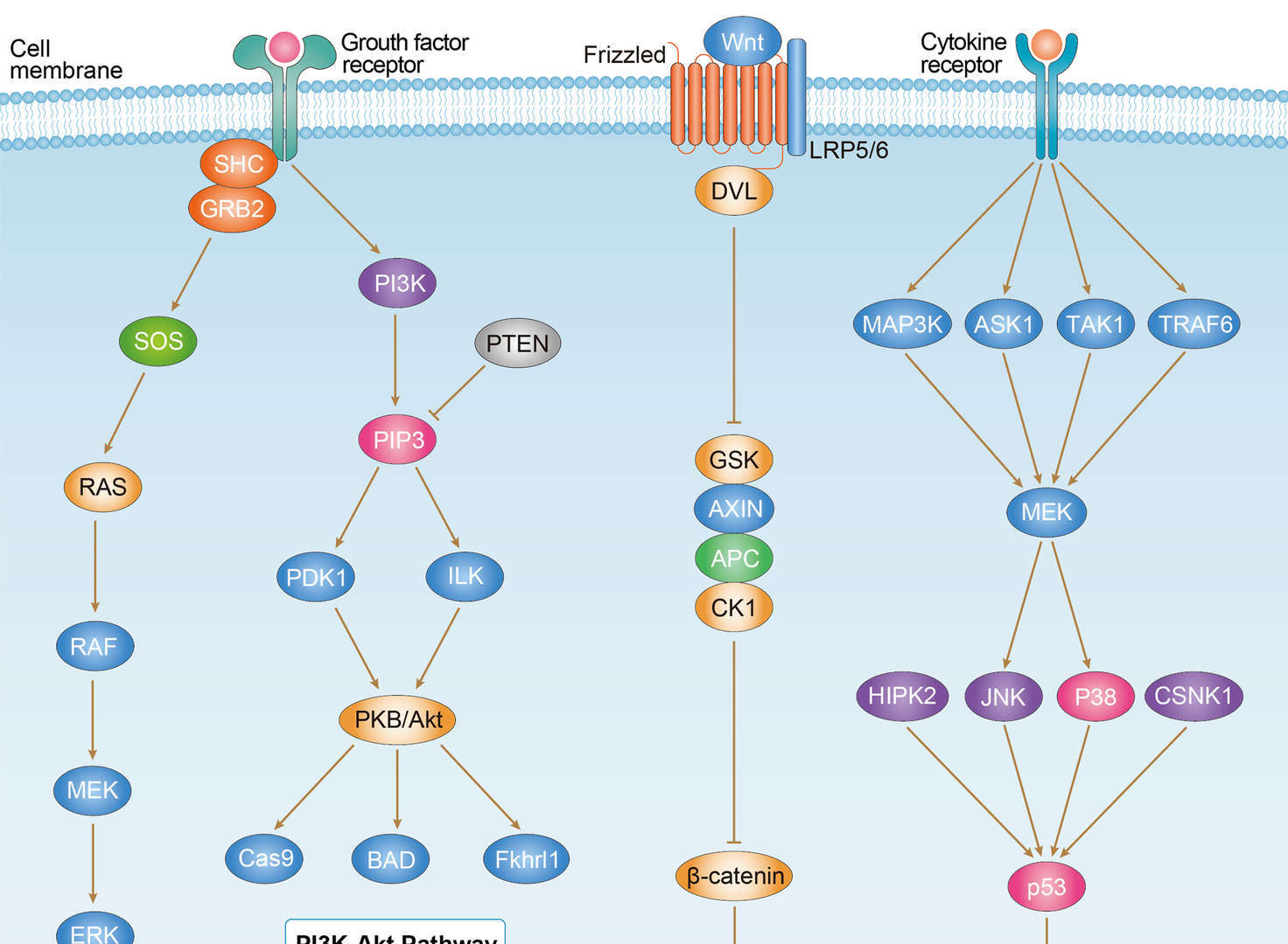
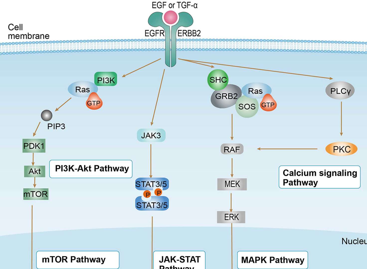
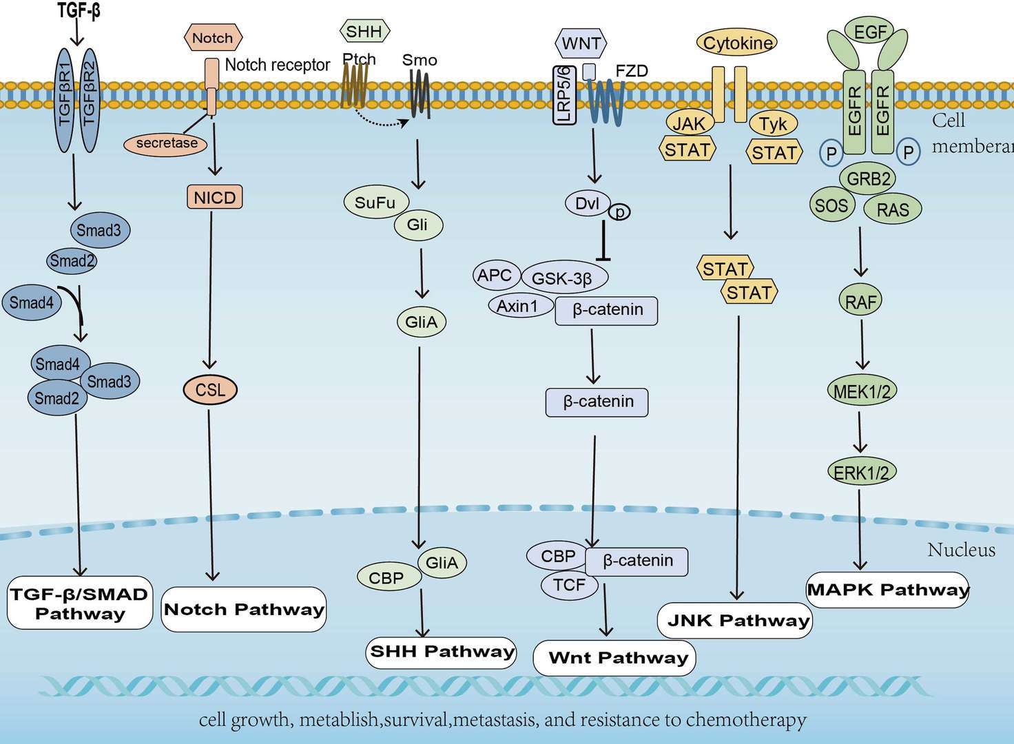
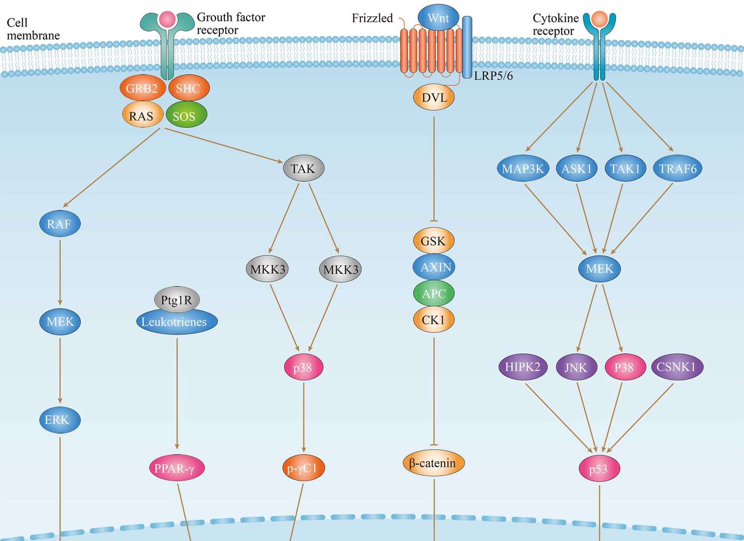
Related Signaling Pathways
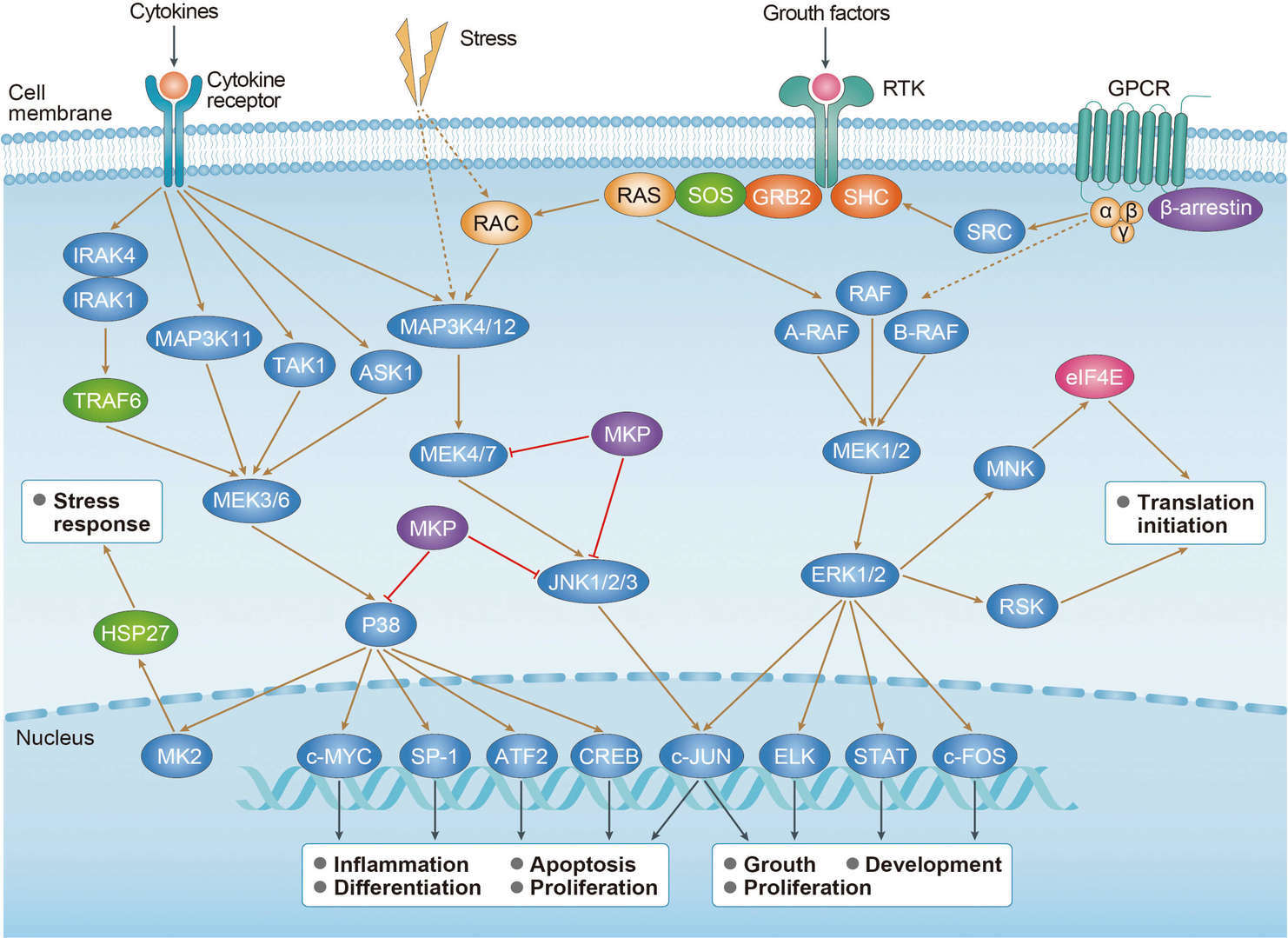 MAPK Signaling Pathway
Downloadable Resources
Download resources about recombinant antibody development and antibody engineering to boost your research.
Datasheet
MSDS
COA
Certificate of Analysis LookupTo download a Certificate of Analysis, please enter a lot number in the search box below. Note: Certificate of Analysis not available for kit components.
Lot Number:
See other products for "STAT6"
Select a product category from the dropdown menu below to view related products.
| CAT | Product Name | Application | Type |
|---|---|---|---|
| MOB-1957CT | Recombinant Mouse anti-Human STAT6 Monoclonal antibody (3H7) | WB |
| CAT | Product Name | Application | Type |
|---|---|---|---|
| BRD-0567MZ | Chicken Anti-STAT6 Polyclonal IgY | WB | Chicken antibody |
| CAT | Product Name | Application | Type |
|---|---|---|---|
| MOR-3430 | Hi-Affi™ Recombinant Rabbit Anti-STAT6 Monoclonal Antibody (DS3430AB) | FC, ICC, IP, WB | IgG |
| CAT | Product Name | Application | Type |
|---|---|---|---|
| MOR-4713 | Hi-Affi™ Recombinant Rabbit Anti-STAT6 Monoclonal Antibody (TH227DS) | WB, IF, ICC, IHC-P, FC, ChIP, ELISA | IgG |
| CAT | Product Name | Application | Type |
|---|---|---|---|
| MOR-4745 | Hi-Affi™ Recombinant Rabbit Anti-STAT6 Monoclonal Antibody (TH259DS) | WB, IF, ICC, IHC-P, FC, ChIP | IgG |
| CAT | Product Name | Application | Type |
|---|---|---|---|
| MRO-1443-CN | Recombinant Rabbit Anti-STAT6 Monoclonal Antibody (CBACN-534) | WB, IF, IHC, IP | Rabbit IgG |
| CAT | Product Name | Application | Type |
|---|---|---|---|
| MRO-1444-CN | Recombinant Rabbit Anti-STAT6 Monoclonal Antibody (CBACN-535) | WB, IF, IHC, IP | Rabbit IgG |
| CAT | Product Name | Application | Type |
|---|---|---|---|
| MRO-2202-CN | Rabbit Anti-STAT6 Polyclonal Antibody (MRO-2202-CN) | WB, IF, IHC, FC | Rabbit IgG |
| CAT | Product Name | Application | Type |
|---|---|---|---|
| ZG-118R | Mouse Anti-STAT6 Recombinant Antibody (ZG-118R) | WB, ELISA, IF | Mouse IgG |
| CAT | Product Name | Application | Type |
|---|---|---|---|
| VS-0325-XY2198 | Anti-STAT6 Immunohistochemistry Kit | IHC |
| CAT | Product Name | Application | Type |
|---|---|---|---|
| VS-0525-XY6961 | Anti-Mouse STAT6 Immunohistochemistry Kit | IHC |
| CAT | Product Name | Application | Type |
|---|---|---|---|
| VS-0525-XY6960 | Anti-Human STAT6 Immunohistochemistry Kit | IHC |
Specific Inquiry
See Our Custom Production in Action


Popular Products

Application: Neut, ELISA, IF, IP, FuncS, FC, IHC

Application: WB, ELISA, IP, FC, FuncS, Neut, IF

Application: FC, IP, ELISA, Neut, FuncS, IF, ICC

Application: ELISA, IP, FC, FuncS, Neut, IF, ICC

Application: WB, ELISA, FC, IP, FuncS, IF, Neut

Application: WB, IF, IP, Neut, FuncS, ELISA, FC

Application: FuncS, IF, Neut, ELISA, FC, IP, IHC

Application: WB, ELISA, FC, IP, FuncS, IF, Neut
-2.png)

Application: FC, IP, ELISA, Neut, FuncS, IF, ICC

Application: IF, IP, Neut, FuncS, ELISA, FC, ICC


Application: Neut, ELISA, FuncS

Application: WB, ELISA, FuncS


For research use only. Not intended for any clinical use. No products from Creative Biolabs may be resold, modified for resale or used to manufacture commercial products without prior written approval from Creative Biolabs.
Send Inquiry
This site is protected by reCAPTCHA and the Google Privacy Policy and Terms of Service apply.