

Recombinant Mouse Anti-HDAC1 Antibody (clone 2A1D3)
CAT#: VS3-FY659
This product is a recombinant mouse antibody that recognizes HDAC1. This antibody has been reported for use in Enzyme-linked Immunosorbent Assay, Western Blot, Immunohistochemistry, Immunocytochemistry, Flow Cytometry. The clone 2A1D3 is specific for HDAC1.
Gene Expression

Subcellular Location
Figure 1 IF staining of human cell line U-2 OS
Immunofluorescent staining of human cell line A-431 shows localization to nucleoplasm.
* Image credit: Image credit: Human Protein Atlas v21.proteinatlas.org/images/5017/if_selected.jpg

Normal Tissue
Figure 2 Cerebral cortex
Glandular cells
Staining: High
Intensity: Strong
Quantity:>75%
Location: Nuclear
* Image credit: Image credit: Human Protein Atlas v21.proteinatlas.org/images/68191/152052_A_7_3.jpg

Normal Tissue
Figure 3 Colon
Glandular cells
Staining: High
Intensity: Strong
Quantity:>75%
Location: Nuclear
* Image credit: Image credit: Human Protein Atlas v21.proteinatlas.org/images/68191/152052_A_7_3.jpg

Normal Tissue
Figure 4 Liver
Cholangiocytes
Staining: Medium
Intensity: Moderate
Quantity:>75%
Location: Nuclear
Hepatocytes
Staining: Medium
Intensity: Moderate
Quantity: 75%-25%
Location: Nuclear
* Image credit: Image credit: Human Protein Atlas v21.proteinatlas.org/images/68191/152052_A_7_4.jpg

Normal Tissue
Figure 5 Kidney
Cells in glomeruli
Staining: Medium
Intensity: Moderate
Quantity:>75%
Location: Nuclear
Cells in tubules
Staining: Medium
Intensity: Moderate
Quantity:>75%
Location: Nuclear
* Image credit: Image credit: Human Protein Atlas v21.proteinatlas.org/images/68191/152052_A_7_5.jpg

Normal Tissue
Figure 6 Testis
Preleptotene spermatocytes
Staining: Medium
Intensity: Strong
Quantity: <25%
* Image credit: Image credit: Human Protein Atlas v21.proteinatlas.org/images/68191/152052_A_6_6.jpg

Normal Tissue
Figure 7 Lymph node
Non-germinal center cells
Staining: Medium
Intensity: Moderate
Quantity: 75%-25%
Location: Nuclear
* Image credit: Image credit: Human Protein Atlas v21.proteinatlas.org/images/68191/152052_A_8_8.jpg

Normal Tissue
Figure 8 Tonsil
Germinal center cells
Staining: High
Intensity: Strong
Quantity:>75%
Location: Nuclear
Non-germinal center cells
Staining: High
Intensity: Strong
Quantity:>75%
Location: Nuclear
Squamous epithelial cells
Staining: High
Intensity: Strong
Quantity:>75%
Location: Nuclear
* Image credit: Image credit: Human Protein Atlas v21.proteinatlas.org/images/68191/152052_A_6_8.jpg

RNA Expression
Figure 9 RNA cell line category: Low cell line specificity
Cell lines ordered by descending RNA expression order.
* Image credit: Image credit: Human Protein Atlas v21.proteinatlas.org/ENSG00000116478-HDAC1
❮


 ❯
❯
Specifications
- Host Species
- Mouse
- Type
- Mouse IgG1
- Clone
- 2A1D3
- Applications
- Enzyme-linked Immunosorbent Assay, Western Blot, Immunohistochemistry, Immunocytochemistry, Flow Cytometry
- Conjugate
- Unconjugated
Product Property
- Purity
- >95% as determined by SDS-PAGE
- Storage
- Centrifuge briefly prior to opening vial. Store at +4°C short term (1-2 weeks). Aliquot and store at -20°C long term. Avoid repeated freeze/thaw cycles.
Applications
- Application Notes
- This antibody has been reported for use in Enzyme-linked Immunosorbent Assay, Western Blot, Immunohistochemistry, Immunocytochemistry, Flow Cytometry.
Target
REVIEWS AND Q&AS
CITATIONS
RESOURCES
DOWNLOADS
RELATED PRODUCTS
Inquiry
Navs
Customer Review

There are currently no Customer reviews or questions for VS3-FY659. Click the button above to contact us or submit your feedback about this product.
Submit Your Publication
Published with our product? Submit your paper and receive a 10% discount on your next order! Share your research to earn exclusive rewards.
Related Diseases
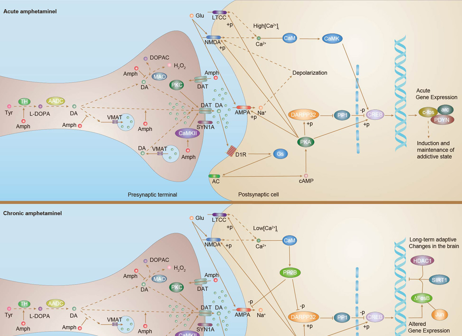 Amphetamine Addiction
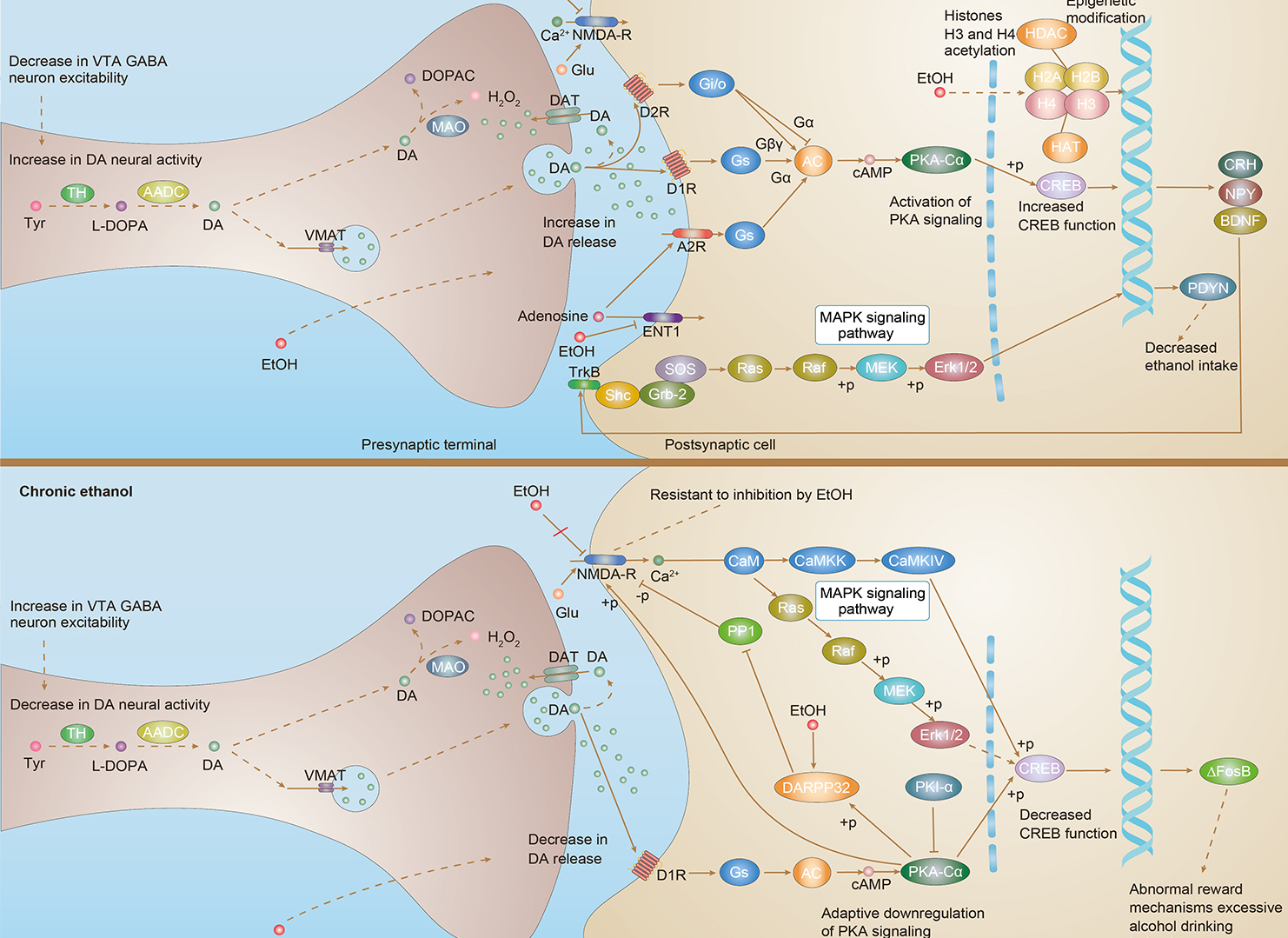 Alcoholism
Downloadable Resources
Download resources about recombinant antibody development and antibody engineering to boost your research.
Product Notes
This is a product of Creative Biolabs' Hi-Affi™ recombinant antibody portfolio, which has several benefits including:
• Increased sensitivity
• Confirmed specificity
• High repeatability
• Excellent batch-to-batch consistency
• Sustainable supply
• Animal-free production
See more details about Hi-Affi™ recombinant antibody benefits.
Datasheet
MSDS
COA
Certificate of Analysis LookupTo download a Certificate of Analysis, please enter a lot number in the search box below. Note: Certificate of Analysis not available for kit components.
Lot Number:
Protocol & Troubleshooting
We have outlined the assay protocols, covering reagents, solutions, procedures, and troubleshooting tips for common issues in order to better assist clients in conducting experiments with our products. View the full list of Protocol & Troubleshooting.
Secondary Antibody
- CAT
- Product Name
Isotype Control
- CAT
- Product Name
See other products for "HDAC1"
Select a product category from the dropdown menu below to view related products.
| CAT | Product Name | Application | Type |
|---|---|---|---|
| MOB-450 | Recombinant Anti-human HDAC1 Antibody | WB, IF, FuncS | IgG |
| CAT | Product Name | Application | Type |
|---|---|---|---|
| MOB-450-F(E) | Recombinant Anti-human HDAC1 Antibody Fab Fragment | WB, Neut, FuncS | Fab |
| CAT | Product Name | Application | Type |
|---|---|---|---|
| MOB-450-S(P) | Recombinant Anti-human HDAC1 Antibody scFv Fragment | FC, WB, FuncS | scFv |
| CAT | Product Name | Application | Type |
|---|---|---|---|
| MHH-450 | Recombinant Human Anti-human HDAC1 Antibody | FC, WB, FuncS | IgG |
| CAT | Product Name | Application | Type |
|---|---|---|---|
| MHH-450-F(E) | Recombinant Human Anti-human HDAC1 Antibody Fab Fragment | ELISA, WB, FC, FuncS | Fab |
| CAT | Product Name | Application | Type |
|---|---|---|---|
| MHH-450-S(P) | Recombinant Human Anti-human HDAC1 Antibody scFv Fragment | IF, Neut, FuncS | scFv |
| CAT | Product Name | Application | Type |
|---|---|---|---|
| MOB-1870MZ | Recombinant Mouse Anti-Human HDAC1 Antibody | IHC-P, WB | Mouse antibody |
| CAT | Product Name | Application | Type |
|---|---|---|---|
| MOR-1571 | Rabbit Anti-HDAC1 Recombinant Antibody (clone DS1571AB) | WB, IHC-P, ICC | Rabbit IgG |
| CAT | Product Name | Application | Type |
|---|---|---|---|
| MHC-LC1937 | PE-A*02:01/Human HDAC1 (RMLPHAPGV) MHC Tetramer | FCM |
| CAT | Product Name | Application | Type |
|---|---|---|---|
| MRO-0716-CN | Rabbit Anti-HDAC1 Recombinant Antibody (clone CBACN-255) | WB, IF, IHC | Rabbit IgG |
| CAT | Product Name | Application | Type |
|---|---|---|---|
| ZG-0096C | Mouse Anti-HDAC1 Recombinant Antibody (clone 4E1) | IF, ICC, IHC-P | Mouse IgG |
| CAT | Product Name | Application | Type |
|---|---|---|---|
| VS-0325-XY997 | Anti-HDAC1 Immunohistochemistry Kit | IHC |
| CAT | Product Name | Application | Type |
|---|---|---|---|
| VS-0525-XY3092 | Anti-Mouse HDAC1 Immunohistochemistry Kit | IHC |
| CAT | Product Name | Application | Type |
|---|---|---|---|
| VS-0525-XY3091 | Anti-Human HDAC1 Immunohistochemistry Kit | IHC |
| CAT | Product Name | Application | Type |
|---|---|---|---|
| VS-0525-XY3093 | Anti-Rat HDAC1 Immunohistochemistry Kit | IHC |
Specific Inquiry

See Our Custom Production in Action


Popular Products

Application: Neut, ELISA, IF, IP, FuncS, FC, IHC

Application: ELISA, IP, FC, FuncS, Neut, IF, ICC

Application: IP, IF, FuncS, FC, Neut, ELISA, ICC

Application: ELISA, IHC

Application: IF, IP, Neut, FuncS, ELISA, FC, WB

Application: FuncS, IF, Neut, ELISA, FC, IP, IHC

-2.png)
Application: ELISA, FC, IP, FuncS, IF, Neut, ICC


Application: WB, Neut, FuncS


Application: WB, ELISA, FuncS


Application: ELISA, IP, WB, IHC, IF, FuncS


For research use only. Not intended for any clinical use. No products from Creative Biolabs may be resold, modified for resale or used to manufacture commercial products without prior written approval from Creative Biolabs.
Send Inquiry
This site is protected by reCAPTCHA and the Google Privacy Policy and Terms of Service apply.