

Recombinant Anti-CD79B Vesicular Antibody, EV Displayed (VS-0425-YC74)
CAT#: VS-0425-YC74
The Recombinant Anti-CD79B Vesicular Antibody, EV Displayed (VS-0425-YC74) is an antibody-displaying extracellular vesicle (Ab-EV). The product combines the benefits of both extracellular vesicle (EV) and antibody (Ab) which can guide the decorated EVs to CD79B-expressed cells or tissues. The CD79B is a transmembrane protein involved in signaling pathways, linked to agammaglobulinemia.
Gene Expression

Subcellular Location and Protein Expression
Figure 1 IF staining of human cell line REH
Immunofluorescent staining of human cell line REH shows localization to plasma membrane.
* Image credit: Image credit: Human Protein Atlas https://v21.proteinatlas.org/images/9178/1889_C4_3_selected.jpg

Subcellular Location and Protein Expression
Figure 2 IHC staining of human lymph node
Immunohistochemical staining of human lymph node shows strong cytoplasmic positivity in lymphoid cells outside reaction centra and weak to moderate in reaction center cells.
* Image credit: Image credit: Human Protein Atlas https://v21.proteinatlas.org/images/9178/ihc_selected.jpg

Subcellular Location and Protein Expression
Figure 3 IHC staining of human lymph node
Immunohistochemical staining of human lymph node shows distinct cytoplasmic positivity in cells of the germinal center corona.
* Image credit: Image credit: Human Protein Atlas https://v21.proteinatlas.org/images/9751/ihc_selected.jpg

Normal Tissue
Figure 4 IF staining of human cell line A-431
Immunofluorescent staining of human cell line A-431 shows localization to Nucleoplasm &
Plasma membrane & Cytosol.
* Image credit: Image credit: Human Protein Atlas https://v21.proteinatlas.org/images/9178/48_C6_1_blue_red_green.jpg

Normal Tissue
Figure 5 IF staining of human cell line U-2 OS
Immunofluorescent staining of human cell line U-2 OS shows localization to Nucleoplasm &
Plasma membrane & Cytosol.
* Image credit: Image credit: Human Protein Atlas https://v21.proteinatlas.org/images/9178/47_C6_1_blue_red_green.jpg

Normal Tissue
Figure 6 Colon
Mucosal lymphoid cells
Staining:Medium
Intensity: Strong
Quantity: <25%
* Image credit: Image credit: Human Protein Atlas https://v21.proteinatlas.org/images/44107/169682_A_8_3.jpg

Normal Tissue
Figure 7 Lymph node
Germinal center cells
Staining:High
Intensity: Strong
Quantity: 75%-25%
Location: Cytoplasmic/membranous
Non-germinal center cells
Staining:Medium
Intensity: Strong
Quantity: <25%
Location: Cytoplasmic/membranous
* Image credit: Image credit: Human Protein Atlas https://v21.proteinatlas.org/images/44107/169682_A_8_8.jpg

RNA Expression
Figure 8 RNA cell line category: Cell line enhanced (Daudi, REH, U-698)
Cell lines ordered by descending RNA expression order.
* Image credit: Image credit: Human Protein Atlas https://v21.proteinatlas.org/ENSG00000007312-CD79B
❮


 ❯
❯
Recombinant Antibody
- Application
- ELISA, FC, Neut, Cell-uptake
- Product Type
- Ab-Fc-EVs
- Antibody Quantification (Ab/EV)
- ~100 Ab/EV
- Target
- CD79B
- Host Animal
- Human
- Antibody Isotype
- IgG
- Species Reactivity
- Human
- Expression Cell
- Mammalian cell
Engineered EVs
- EV-sorting domain
- CD63
- Fc-binding domain
- z domain
- EV Size
- 30~150 nm
- Producing Cell
- HEK293F
- Isolation Method
- Gradient centrifugation
- Purification
- qEV size exclusion chromatography
- Binding Affinity
- Kd = 0.85 µg/mL
- Concentration
- 1 x 10¹⁰
- Size
- 1 mL
- Buffer
- PBS
- Storage
- Store at -80°C for 12 months
Target
- Full Name
- CD79b molecule
- Biological Process
- Adaptive immunity, Immunity
- Cellular Localization
- Plasma membrane, Nucleoplasm, Cytosol
- Introduction
- CD79B (CD79b Molecule) is a Protein Coding gene. Diseases associated with CD79B include Agammaglobulinemia 6, Autosomal Recessive and Agammaglobulinemia, Non-Bruton Type. Among its related pathways are Hematopoietic Stem Cell Differentiation Pathways and Lineage-specific Markers and Innate Immune System. Gene Ontology (GO) annotations related to this gene include transmembrane signaling receptor activity.
- Alternative Names
- B29, Ig-beta, IGB, Igbeta
- Gene ID
- 974
- UniProt ID
- P40259
REVIEWS AND Q&AS
CITATIONS
RESOURCES
DOWNLOADS
RELATED PRODUCTS
Inquiry
Navs
Customer Review

There are currently no Customer reviews or questions for VS-0425-YC74. Click the button above to contact us or submit your feedback about this product.
Submit Your Publication
Published with our product? Submit your paper and receive a 10% discount on your next order! Share your research to earn exclusive rewards.
Related Signaling Pathways
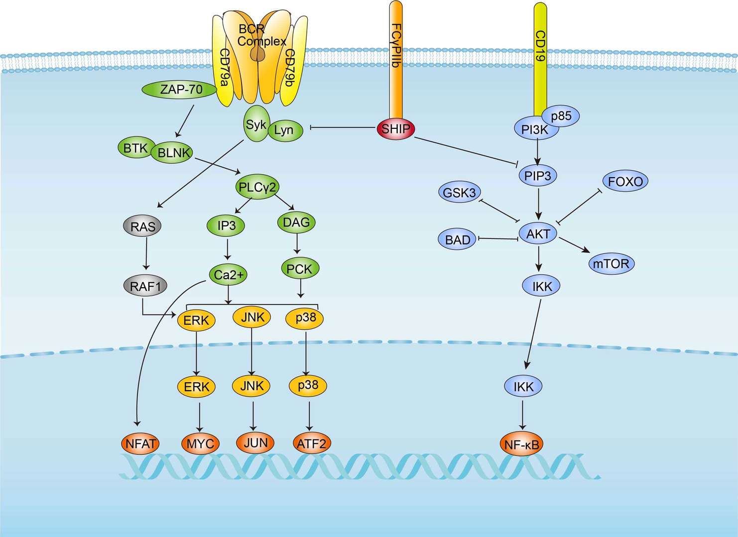 BCR Signaling Pathway
Downloadable Resources
Download resources about recombinant antibody development and antibody engineering to boost your research.
Datasheet
MSDS
COA
Certificate of Analysis LookupTo download a Certificate of Analysis, please enter a lot number in the search box below. Note: Certificate of Analysis not available for kit components.
Lot Number:
See other products for "CD79B"
Select a product category from the dropdown menu below to view related products.
| CAT | Product Name | Application | Type |
|---|---|---|---|
| MOB-1219z | Mouse Anti-CD79B Recombinant Antibody (clone 32B11) | ELISA, WB, FC, FuncS | Mouse IgG1 |
| CAT | Product Name | Application | Type |
|---|---|---|---|
| TAB-897 | Humanized Anti-CD79B Recombinant Antibody (TAB-897) | Neut, ELISA, IF, IP, FuncS, FC, ICC | Humanized (from mouse) IgG1, κ |
| CAT | Product Name | Application | Type |
|---|---|---|---|
| MOB-0360MZ | Recombinant Mouse Anti-Human CD79B CD79b Molecule, Immunoglobulin-associated Beta Antibody (clone TN9) | ELISA | Mouse antibody |
| CAT | Product Name | Application | Type |
|---|---|---|---|
| MOR-0581 | Hi-Affi™ Rabbit Anti-CD79B Recombinant Antibody (clone DS581AB) | WB, IHC, ICC, IP, FC | Rabbit IgG |
| CAT | Product Name | Application | Type |
|---|---|---|---|
| MHC-LC1918 | PE-A*02:01/Human CD79B (LLSAEPVPA) MHC Tetramer | FCM |
| CAT | Product Name | Application | Type |
|---|---|---|---|
| MOB-027LC | Recombinant Mouse Anti-M.fascicularis CD79B Antibody | ELISA, FuncS | Mouse IgG |
| CAT | Product Name | Application | Type |
|---|---|---|---|
| FAMAB-0058YC | Hamster Anti-Cd79b Recombinant Antibody (clone HM79-16) | FuncS | Hamster IgG |
| CAT | Product Name | Application | Type |
|---|---|---|---|
| FAMAB-0058-YC-S(P) | Hamster Anti-Cd79b Recombinant Antibody (clone HM79-16); scFv Fragment | FuncS | Hamster scFv |
| CAT | Product Name | Application | Type |
|---|---|---|---|
| FAMAB-0058-YC-F(E) | Hamster Anti-Cd79b Recombinant Antibody (clone HM79-16); Fab Fragment | FuncS | Hamster Fab |
| CAT | Product Name | Application | Type |
|---|---|---|---|
| HPAB-0364LY | Human Anti-CD79B Recombinant Antibody (HPAB-0364LY) | Inhib, ELISA | Human IgG |
| CAT | Product Name | Application | Type |
|---|---|---|---|
| HPAB-0364LY-S(P) | Human Anti-CD79B Recombinant Antibody; scFv Fragment (HPAB-0364LY-S(P)) | Inhib, ELISA | Human scFv |
| CAT | Product Name | Application | Type |
|---|---|---|---|
| HPAB-0364LY-F(E) | Human Anti-CD79B Recombinant Antibody; Fab Fragment (HPAB-0364LY-F(E)) | Inhib, ELISA | Human Fab |
| CAT | Product Name | Application | Type |
|---|---|---|---|
| HPAB-618-FY-S(P) | Human Anti-CD79B Recombinant Antibody; scFv Fragment (HPAB-618-FY-S(P)) | FC, ELISA | Humanized scFv |
| CAT | Product Name | Application | Type |
|---|---|---|---|
| HPAB-619-FY-S(P) | Human Anti-CD79B Recombinant Antibody; scFv Fragment (HPAB-619-FY-S(P)) | FC, ELISA | Humanized scFv |
| CAT | Product Name | Application | Type |
|---|---|---|---|
| HPAB-618-FY-F(E) | Human Anti-CD79B Recombinant Antibody; Fab Fragment (HPAB-618-FY-F(E)) | FC, ELISA | Humanized Fab |
| CAT | Product Name | Application | Type |
|---|---|---|---|
| HPAB-619-FY-F(E) | Human Anti-CD79B Recombinant Antibody; Fab Fragment (HPAB-619-FY-F(E)) | FC, ELISA | Humanized Fab |
| CAT | Product Name | Application | Type |
|---|---|---|---|
| AFC-TAB-897 | Afuco™ Anti-CD79B ADCC Recombinant Antibody, ADCC Enhanced (AFC-TAB-897) | Neut, ELISA, IF, IP, FuncS, FC | ADCC enhanced antibody |
| CAT | Product Name | Application | Type |
|---|---|---|---|
| HPAB-N0044-YC-S(P) | Human Anti-CD79b Recombinant Antibody; scFv Fragment (HPAB-N0044-YC-S(P)) | FuncS | Humanized scFv |
| CAT | Product Name | Application | Type |
|---|---|---|---|
| HPAB-N0044-YC-F(E) | Human Anti-CD79b Recombinant Antibody; Fab Fragment (HPAB-N0044-YC-F(E)) | FuncS | Humanized Fab |
| CAT | Product Name | Application | Type |
|---|---|---|---|
| VS-0225-XY72 | CytoStream™ Mouse Anti-CD79B Recombinant Antibody (clone CB3-1) | FC | Mouse IgG1, kappa |
| CAT | Product Name | Application | Type |
|---|---|---|---|
| VS-0325-FY74 | Human Anti-CD79B (clone P2C2) scFv-Fc Chimera | IA | Human IgG1, scFv-Fc |
| CAT | Product Name | Application | Type |
|---|---|---|---|
| VS-0525-XY1222 | Anti-Mouse CD79B Immunohistochemistry Kit | IHC |
| CAT | Product Name | Application | Type |
|---|---|---|---|
| VS-0825-YC69 | SmartAb™ Recombinant Anti-CD79B pH-dependent Antibody (VS-0825-YC69) | Neut, ELISA, IF, IP, FC, ICC | Human IgG1 kappa |
| CAT | Product Name | Application | Type |
|---|---|---|---|
| VS-1025-YC210 | Anti-CD79B Antibody Prodrug, Protease Activated (VS-1025-YC210) | ISZ, Cyt, FuncS |
Specific Inquiry
See Our Custom Production in Action


Popular Products

Application: FC, Cyt, Stim, PP, Agonist

Application: WB, IF, IP, Neut, FuncS, ELISA, FC

Application: FuncS, IF, Neut, ELISA, FC, IP, ICC

Application: Neut, ELISA, IF, IP, FuncS, FC, ICC

Application: IP, IF, FuncS, FC, Neut, ELISA, ICC

Application: WB, FuncS, IF, Neut, ELISA, FC, IP


Application: IF, IP, Neut, FuncS, ELISA, FC, ICC

Application: Inhib, Cyt

Application: ELISA, FC, IP, FuncS, IF, Neut, ICC

Application: WB, ELISA, FC, IP, FuncS, IF, Neut

Application: ELISA, WB, BLI, SPR


Application: WB, ELISA, FuncS


Application: WB, IF, FuncS
For research use only. Not intended for any clinical use. No products from Creative Biolabs may be resold, modified for resale or used to manufacture commercial products without prior written approval from Creative Biolabs.
Send Inquiry
This site is protected by reCAPTCHA and the Google Privacy Policy and Terms of Service apply.