

Recombinant Anti-IL18R1 Vesicular Antibody, EV Displayed (VS-0425-YC211)
CAT#: VS-0425-YC211
The Recombinant Anti-IL18R1 Vesicular Antibody, EV Displayed (VS-0425-YC211) is an antibody-displaying extracellular vesicle (Ab-EV). The product combines the benefits of both extracellular vesicle (EV) and antibody (Ab) which can guide the decorated EVs to IL18R1-expressed cells or tissues. The IL18R1 is a receptor binding interleukin 18, crucial for signal transduction in immune responses.
Gene Expression

Subcellular Location
Figure 1 IF staining of human cell line U-2 OS
Immunofluorescent staining of human cell line U-2 OS shows localization to mitochondria.
* Image credit: Image credit: Human Protein Atlas v21.proteinatlas.org/images/7615/if_selected.jpg

Normal Tissue
Figure 2 IHC staining of human stomach
Immunohistochemical staining of human stomach shows strong cytoplasmic positivity in glandular cells.
* Image credit: Image credit: Human Protein Atlas v21.proteinatlas.org/images/7615/ihc_selected.jpg

Normal Tissue
Figure 3 Cerebral cortex
Neuronal cells Staining: High Intensity: Strong Quantity:>75% Location: Cytoplasmic/ membranous Neuropil Staining: Low Intensity: Weak Quantity:>75% Location: Cytoplasmic/ membranous
* Image credit: Image credit: Human Protein Atlas v21.proteinatlas.org/images/7615/28557_B_8_5.jpg

Normal Tissue
Figure 4 Colon
Glandular cells Staining: Medium Intensity: Moderate Quantity:>75% Location: Cytoplasmic/ membranous
* Image credit: Image credit: Human Protein Atlas v21.proteinatlas.org/images/7615/28557_A_9_3.jpg

Normal Tissue
Figure 5 Liver
Cholangiocytes Staining: Medium Intensity: Moderate Quantity:>75% Location: Cytoplasmic/ membranous Hepatocytes Staining: Medium Intensity: Moderate Quantity:>75% Location: Cytoplasmic/ membranous
* Image credit: Image credit: Human Protein Atlas v21.proteinatlas.org/images/7615/28557_A_7_4.jpg

Normal Tissue
Figure 6 Kidney
Cells in tubules Staining: High Intensity: Strong Quantity:>75% Location: Cytoplasmic/ membranous
* Image credit: Image credit: Human Protein Atlas v21.proteinatlas.org/images/7615/28557_A_7_5.jpg

Normal Tissue
Figure 7 Testis
Elongated or late spermatids Staining: Medium Intensity: Moderate Quantity:>75% Leydig cells Staining: High Intensity: Strong Quantity:>75% Pachytene spermatocytes Staining: Medium Intensity: Moderate Quantity: 75%-25% Preleptotene spermatocytes Staining: Low Intensity: Moderate Quantity: <25% Spermatogonia cells Staining: High Intensity: Strong
* Image credit: Image credit: Human Protein Atlas v21.proteinatlas.org/images/7615/28557_A_6_6.jpg

Normal Tissue
Figure 8 Appendix
Glandular cells Staining: High Intensity: Strong Quantity:>75% Location: Cytoplasmic/ membranous Lymphoid tissue Staining: Medium Intensity: Moderate Quantity: 75%-25% Location: Cytoplasmic/ membranous
* Image credit: Image credit: Human Protein Atlas v21.proteinatlas.org/images/7615/28557_A_3_2.jpg

Normal Tissue
Figure 9 Lymph node
Germinal center cells Staining: Medium Intensity: Moderate Quantity: 75%-25% Location: Cytoplasmic/ membranous Non-germinal center cells Staining: Medium Intensity: Strong Quantity: <25% Location: Cytoplasmic/ membranous
* Image credit: Image credit: Human Protein Atlas v21.proteinatlas.org/images/7615/28557_A_9_8.jpg

RNA Expression
Figure 10 RNA cell line category: Cell line enhanced (HDLM-2, HHSteC, U-937)
Cell lines ordered by descending RNA expression order
* Image credit: Image credit: Human Protein Atlas v21.proteinatlas.org/ENSG00000115604-IL18R1
❮


 ❯
❯
Recombinant Antibody
- Application
- ELISA, FC, Cell-uptake
- Product Type
- Ab-Fc-EVs
- Antibody Quantification (Ab/EV)
- ~100 Ab/EV
- Target
- IL18R1
- Host Animal
- Human
- Antibody Isotype
- IgG
- Species Reactivity
- Human, Monkey
- Expression Cell
- Mammalian cell
Engineered EVs
- EV-sorting domain
- CD63
- Fc-binding domain
- z domain
- EV Size
- 30~150 nm
- Producing Cell
- HEK293F
- Isolation Method
- Gradient centrifugation
- Purification
- qEV size exclusion chromatography
- Binding Affinity
- Kd = 0.85 µg/mL
- Concentration
- 1 x 10¹⁰
- Size
- 1 mL
- Buffer
- PBS
- Storage
- Store at -80°C for 12 months
Target
- Full Name
- Interleukin 18 receptor 1
- Biological Process
- Inflammatory response
- Molecular Function
- Hydrolase, Receptor
- Cellular Localization
- Mitochondria
- Introduction
- The protein encoded by this gene is a cytokine receptor that belongs to the interleukin 1 receptor family. This receptor specifically binds interleukin 18 (IL18), and is essential for IL18 mediated signal transduction. IFN-alpha and IL12 are reported to induce the expression of this receptor in NK and T cells. This gene along with four other members of the interleukin 1 receptor family, including IL1R2, IL1R1, ILRL2 (IL-1Rrp2), and IL1RL1 (T1/ST2), form a gene cluster on chromosome 2q. Alternatively spliced transcript variants encoding different isoforms have been found for this gene. [provided by RefSeq, Sep 2013]
- Alternative Names
- CD218a, IL-1Rrp, IL1RRP
- Gene ID
- 8809
- UniProt ID
- Q13478
REVIEWS AND Q&AS
CITATIONS
RESOURCES
DOWNLOADS
RELATED PRODUCTS
Inquiry
Navs
Customer Review

There are currently no Customer reviews or questions for VS-0425-YC211. Click the button above to contact us or submit your feedback about this product.
Submit Your Publication
Published with our product? Submit your paper and receive a 10% discount on your next order! Share your research to earn exclusive rewards.
Related Signaling Pathways
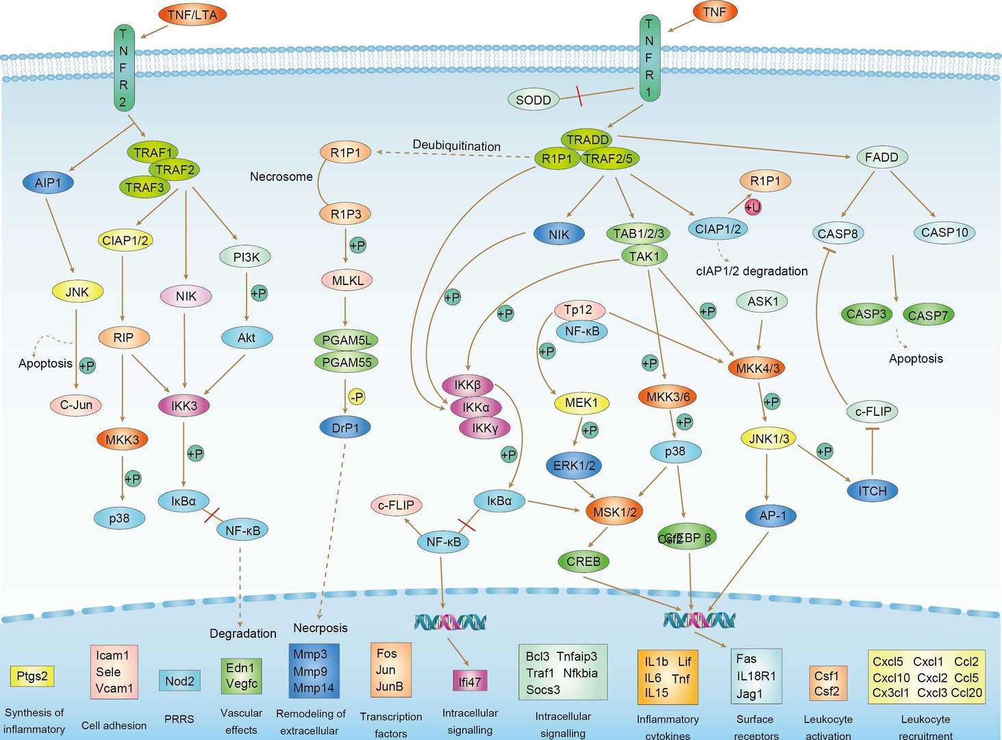 TNF Signaling Pathway
Downloadable Resources
Download resources about recombinant antibody development and antibody engineering to boost your research.
Datasheet
MSDS
COA
Certificate of Analysis LookupTo download a Certificate of Analysis, please enter a lot number in the search box below. Note: Certificate of Analysis not available for kit components.
Lot Number:
See other products for "IL18R1"
Select a product category from the dropdown menu below to view related products.
| CAT | Product Name | Application | Type |
|---|---|---|---|
| MOB-1369z | Mouse Anti-IL18R1 Recombinant Antibody (clone 22B12) | ELISA, WB, IP, FuncS | Mouse IgG1 |
| CAT | Product Name | Application | Type |
|---|---|---|---|
| AGTO-G042E | IL-18-PE immunotoxin | Cytotoxicity assay, Function study |
| CAT | Product Name | Application | Type |
|---|---|---|---|
| AGTO-G042D | IL-18-DT immunotoxin | Cytotoxicity assay, Function study |
| CAT | Product Name | Application | Type |
|---|---|---|---|
| TAB-207CT | Mouse Anti-IL18R1 Recombinant Antibody (TAB-207CT) | Neut, ELISA, Block | Mouse IgG |
| CAT | Product Name | Application | Type |
|---|---|---|---|
| TAB-208CT | Mouse Anti-IL18R1 Recombinant Antibody (TAB-208CT) | Neut, ELISA, Block | Mouse IgG |
| CAT | Product Name | Application | Type |
|---|---|---|---|
| TAB-209CT | Mouse Anti-IL18R1 Recombinant Antibody (TAB-209CT) | FC, ELISA, Ibhibition | Mouse IgG |
| CAT | Product Name | Application | Type |
|---|---|---|---|
| TAB-210CT | Mouse Anti-IL18R1 Recombinant Antibody (TAB-210CT) | FC, ELISA, Ibhibition | Mouse IgG |
| CAT | Product Name | Application | Type |
|---|---|---|---|
| TAB-211CT | Human Anti-IL18R1 Recombinant Antibody (TAB-211CT) | FC, ELISA, Ibhibition | Humanized antibody |
| CAT | Product Name | Application | Type |
|---|---|---|---|
| TAB-207CT-S(P) | Mouse Anti-IL18R1 Recombinant Antibody; scFv Fragment (TAB-207CT-S(P)) | Neut, ELISA, Block | Mouse scFv |
| CAT | Product Name | Application | Type |
|---|---|---|---|
| TAB-211CT-S(P) | Human Anti-IL18R1 Recombinant Antibody; scFv Fragment (TAB-211CT-S(P)) | FC, ELISA, Ibhibition | Humanized scFv |
| CAT | Product Name | Application | Type |
|---|---|---|---|
| TAB-211CT-F(E) | Human Anti-IL18R1 Recombinant Antibody; Fab Fragment (TAB-211CT-F(E)) | FC, ELISA, Ibhibition | Humanized Fab |
| CAT | Product Name | Application | Type |
|---|---|---|---|
| MOB-0235CT | Recombinant Mouse anti-Human IL18R1 Monoclonal antibody (80736.222) | I-ELISA, Neut, WB |
| CAT | Product Name | Application | Type |
|---|---|---|---|
| NEUT-1247CQ | Mouse Anti-IL18R1 Recombinant Antibody (clone CBL489) | FC, IHC, CyTOF, ICC, Neut | Mouse IgG1 |
| CAT | Product Name | Application | Type |
|---|---|---|---|
| NEUT-1248CQ | Mouse Anti-IL18R1 Recombinant Antibody (clone 70625.111) | I-ELISA, Neut, WB | Mouse IgG1 |
| CAT | Product Name | Application | Type |
|---|---|---|---|
| NEUT-1249CQ | Mouse Anti-IL18R1 Recombinant Antibody (clone 8H22) | IHC, Neut, WB | Mouse IgG1 |
| CAT | Product Name | Application | Type |
|---|---|---|---|
| NEUT-1250CQ | Rat Anti-Il18r1 Recombinant Antibody (clone CBL522) | WB, Neut | Rat IgG2a |
| CAT | Product Name | Application | Type |
|---|---|---|---|
| MOR-4250 | Rabbit Anti-Il18r1 Recombinant Antibody (clone SI329DS) | ELISA | Rabbit IgG |
| CAT | Product Name | Application | Type |
|---|---|---|---|
| MOR-4251 | Rabbit Anti-IL18R1 Recombinant Antibody (clone SI330DS) | ELISA | Rabbit IgG |
| CAT | Product Name | Application | Type |
|---|---|---|---|
| HPAB-658-FY | Mouse Anti-IL18R1 Recombinant Antibody (HPAB-658-FY) | FC | Mouse IgG |
| CAT | Product Name | Application | Type |
|---|---|---|---|
| HPAB-659-FY | Mouse Anti-IL18R1 Recombinant Antibody (HPAB-659-FY) | FC | Mouse IgG |
| CAT | Product Name | Application | Type |
|---|---|---|---|
| HPAB-658-FY-S(P) | Mouse Anti-IL18R1 Recombinant Antibody; scFv Fragment (HPAB-658-FY-S(P)) | Inhib, FC, ELISA | Mouse scFv |
| CAT | Product Name | Application | Type |
|---|---|---|---|
| HPAB-659-FY-S(P) | Mouse Anti-IL18R1 Recombinant Antibody; scFv Fragment (HPAB-659-FY-S(P)) | ELISA | Mouse scFv |
| CAT | Product Name | Application | Type |
|---|---|---|---|
| HPAB-658-FY-F(E) | Mouse Anti-IL18R1 Recombinant Antibody; Fab Fragment (HPAB-658-FY-F(E)) | Inhib, FC, ELISA | Mouse Fab |
| CAT | Product Name | Application | Type |
|---|---|---|---|
| HPAB-659-FY-F(E) | Mouse Anti-IL18R1 Recombinant Antibody; Fab Fragment (HPAB-659-FY-F(E)) | ELISA | Mouse Fab |
| CAT | Product Name | Application | Type |
|---|---|---|---|
| FAMAB-0315JF | Mouse Anti-IL18R1 Recombinant Antibody (clone 117-10C) | ELISA, FuncS | Mouse IgM, κ |
| CAT | Product Name | Application | Type |
|---|---|---|---|
| FAMAB-0315JF-F(E) | Mouse Anti-IL18R1 Recombinant Antibody (clone 117-10C); Fab Fragment | ELISA, FuncS | Mouse Fab |
| CAT | Product Name | Application | Type |
|---|---|---|---|
| FAMAB-0315JF-S(P) | Mouse Anti-IL18R1 Recombinant Antibody (clone 117-10C); scFv Fragment | ELISA, FuncS | Mouse scFv |
| CAT | Product Name | Application | Type |
|---|---|---|---|
| VS-0425-FY4 | Mouse Anti-IL18R1 scFv-Fc Chimera (VS-0425-FY4) | Inhib, ELISA | Mouse IgG1, scFv-Fc |
| CAT | Product Name | Application | Type |
|---|---|---|---|
| VS-0525-XY3501 | Anti-IL18R1 Immunohistochemistry Kit | IHC |
Specific Inquiry
See Our Custom Production in Action


Popular Products

Application: WB, FuncS, IF, Neut, ELISA, FC, IP

Application: ELISA, FC, IP, FuncS, IF, Neut, ICC

Application: WB, ELISA, FC, IP, FuncS, IF, Neut

Application: ELISA, FC, IP, FuncS, IF, Neut, ICC

Application: Neut, ELISA, IF, IP, FuncS, FC, IHC


Application: IP, IF, FuncS, FC, Neut, ELISA, ICC

Application: ELISA, Neut, IF, IP, FC, FuncS

Application: ELISA, WB, BLI, SPR


Application: ELISA, Neut, FuncS


For research use only. Not intended for any clinical use. No products from Creative Biolabs may be resold, modified for resale or used to manufacture commercial products without prior written approval from Creative Biolabs.
Send Inquiry
This site is protected by reCAPTCHA and the Google Privacy Policy and Terms of Service apply.