

Afuco™ Anti-Human INSR ADCC Recombinant Antibody (AGT-182), ADCC Enhanced
CAT#: AFC-423CL
Anti-INSR ADCC Enhanced Antibody (AGT-182) is an ADCC enhanced antibody produced by our Afuco™ platform. AGT‑182 is an investigational enzyme replacement therapy (ERT) for the treatment of neurological complications in patients with Hunter syndrome. Currently approved treatments for Hunter syndrome do not penetrate the BBB and therefore do not address the severe and progressive neurological complications of the disease. AGT-182 is a fusion protein of iduronate-2-sulfatase (IDS), engineered to cross the BBB by binding to insulin receptors located on the BBB. AGT‑182 crosses the BBB safely at unprecedented levels.
Gene Expression

Subcellular Location
Figure 1 IF staining of human cell line U-2 OS
Immunofluorescent staining of human cell line U-2 OS shows localization to plasma membrane & vesicles.
* Image credit: Image credit: Human Protein Atlas https://v21.proteinatlas.org/images/36303/737_C10_2_selected.jpg

Subcellular Location
Figure 2 IHC staining of human liver
Immunohistochemical staining of human liver shows moderate cytoplasmic positivity in hepatocytes.
* Image credit: Image credit: Human Protein Atlas https://v21.proteinatlas.org/images/36302/ihc_selected.jpg

Subcellular Location
Figure 3 IF staining of human cell line A-431
Immunofluorescent staining of human cell line A-431 shows localization to vesicles.
* Image credit: Image credit: Human Protein Atlas https://v21.proteinatlas.org/images/36303/836_E10_2_blue_red_green.jpg

Normal Tissue
Figure 4 Colon
landular cells
Staining: Medium
Intensity: Moderate
Quantity:>75%
Location: Cytoplasmic/membranous
* Image credit: Image credit: Human Protein Atlas https://v21.proteinatlas.org/images/36302/112008_A_9_3.jpg

Normal Tissue
Figure 5 Liver
Hepatocytes
Staining: Medium
Intensity: Moderate
Quantity:>75%
Location: Cytoplasmic/membranous
* Image credit: Image credit: Human Protein Atlas https://v21.proteinatlas.org/images/36302/112008_A_8_4.jpg

Normal Tissue
Figure 6 Kidney
Cells in tubules
Staining: Medium
Intensity: Moderate
Quantity:>75%
Location: Cytoplasmic/membranous
* Image credit: Image credit: Human Protein Atlas https://v21.proteinatlas.org/images/36302/112008_A_7_5.jpg

Normal Tissue
Figure 7 Testis
Leydig cells
Staining: Medium
Intensity: Moderate
Quantity:>75%
Location: Cytoplasmic/membranous
* Image credit: Image credit: Human Protein Atlas https://v21.proteinatlas.org/images/36302/112008_A_6_6.jpg

Normal Tissue
Figure 8 Skeletal muscle
Myocytes
Staining: Medium
Intensity: Moderate
Quantity:>75%
Location: Cytoplasmic/membranous
* Image credit: Image credit: Human Protein Atlas https://v21.proteinatlas.org/images/36302/112008_B_3_6.jpg

Normal Tissue
Figure 9 Lymph node
Germinal center cells
Staining: Medium
Intensity: Moderate
Quantity:>75%
Location: Cytoplasmic/membranous
* Image credit: Image credit: Human Protein Atlas https://v21.proteinatlas.org/images/36302/112008_A_8_8.jpg

RNA Expression
Figure 10 RNA cell line category: Cell line enhanced (Karpas-707, U-266/70)
Cell lines ordered by descending RNA expression order.
* Image credit: Image credit: Human Protein Atlas https://v21.proteinatlas.org/ENSG00000171105-INSR
❮


 ❯
❯
Specifications
- Host Species
- Human
- Type
- ADCC enhanced antibody
- Species Reactivity
- Human
- Related Disease
- Hunter Syndrome
Product Property
- Purity
- >95% as determined by analysis by SDS-PAGE
- Storage
- Store at 4°C for up to 3 months. For longer term storage aliquot into small volumes and store at -20°C.
Target
REVIEWS AND Q&AS
CITATIONS
RESOURCES
DOWNLOADS
RELATED PRODUCTS
Inquiry
Navs
Customer Review

There are currently no Customer reviews or questions for AFC-423CL. Click the button above to contact us or submit your feedback about this product.
Submit Your Publication
Published with our product? Submit your paper and receive a 10% discount on your next order! Share your research to earn exclusive rewards.
Related Signaling Pathways
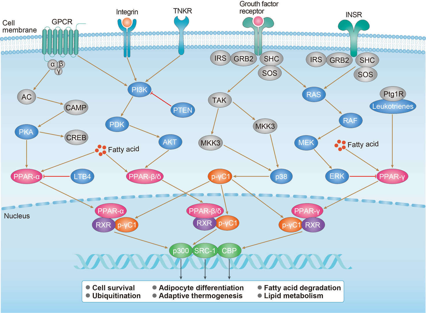 PPAR Signaling Pathway
Related Diseases
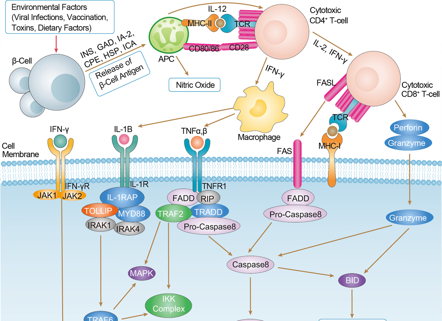 Type I Diabetes Mellitus
Downloadable Resources
Download resources about recombinant antibody development and antibody engineering to boost your research.
Product Notes
This is a product of Creative Biolabs' Hi-Affi™ recombinant antibody portfolio, which has several benefits including:
• Increased sensitivity
• Confirmed specificity
• High repeatability
• Excellent batch-to-batch consistency
• Sustainable supply
• Animal-free production
See more details about Hi-Affi™ recombinant antibody benefits.
Datasheet
MSDS
COA
Certificate of Analysis LookupTo download a Certificate of Analysis, please enter a lot number in the search box below. Note: Certificate of Analysis not available for kit components.
Lot Number:
See other products for "INSR"
Select a product category from the dropdown menu below to view related products.
| CAT | Product Name | Application | Type |
|---|---|---|---|
| MOB-1236z | Mouse Anti-INSR Recombinant Antibody (clone 25F8) | ELISA, FC, ICC, IF, IHC, FuncS | Mouse IgG1, κ |
| CAT | Product Name | Application | Type |
|---|---|---|---|
| TAB-441CL | Human Anti-INSR Recombinant Antibody (TAB-441CL) | ELISA | Human IgG |
| CAT | Product Name | Application | Type |
|---|---|---|---|
| PABL-246 | Mouse Anti-INSR Recombinant Antibody (clone 83-7) | WB, ELISA, FuncS | Mouse IgG |
| CAT | Product Name | Application | Type |
|---|---|---|---|
| PSBL-246 | Mouse Anti-INSR Recombinant Antibody (clone 83-7); scFv Fragment | WB, ELISA, FuncS | Mouse scFv |
| CAT | Product Name | Application | Type |
|---|---|---|---|
| PFBL-246 | Mouse Anti-INSR Recombinant Antibody (clone 83-7); Fab Fragment | WB, ELISA, FuncS | Mouse Fab |
| CAT | Product Name | Application | Type |
|---|---|---|---|
| PABZ-083 | Mouse Anti-INSR Recombinant Antibody (clone 83-14) | WB, FuncS | Mouse IgG |
| CAT | Product Name | Application | Type |
|---|---|---|---|
| PFBZ-083 | Mouse Anti-INSR Recombinant Antibody (clone 83-14); Fab Fragment | WB, FuncS | Mouse Fab |
| CAT | Product Name | Application | Type |
|---|---|---|---|
| PSBZ-083 | Mouse Anti-INSR Recombinant Antibody (clone 83-14); scFv Fragment | WB, FuncS | Mouse scFv |
| CAT | Product Name | Application | Type |
|---|---|---|---|
| TAB-711LC | Human Anti-INSR Recombinant Antibody (TAB-711LC) | ELISA, FC, FuncS | Human IgG |
| CAT | Product Name | Application | Type |
|---|---|---|---|
| TAB-712LC | Human Anti-INSR Recombinant Antibody (TAB-712LC) | ELISA, FC, Agonist, FuncS | Human IgG |
| CAT | Product Name | Application | Type |
|---|---|---|---|
| TAB-711LC-S(P) | Human Anti-INSR Recombinant Antibody; scFv Fragment (TAB-711LC-S(P)) | ELISA, FC | Human scFv |
| CAT | Product Name | Application | Type |
|---|---|---|---|
| TAB-712LC-S(P) | Human Anti-INSR Recombinant Antibody; scFv Fragment (TAB-712LC-S(P)) | ELISA, FC | Human scFv |
| CAT | Product Name | Application | Type |
|---|---|---|---|
| TAB-711LC-F(E) | Human Anti-INSR Recombinant Antibody; Fab Fragment (TAB-711LC-F(E)) | ELISA, FC | Human Fab |
| CAT | Product Name | Application | Type |
|---|---|---|---|
| MOB-0984CT | Recombinant Mouse anti-Human INSR Monoclonal antibody (94-8) | ELISA, FC, IHC-P, IP |
| CAT | Product Name | Application | Type |
|---|---|---|---|
| PABX-124 | Recombinant Mouse Anti-IR Antibody (Fab83-14) | Neut, FuncS | IgG |
| CAT | Product Name | Application | Type |
|---|---|---|---|
| PABX-125 | Recombinant Mouse Anti-IR Antibody (Fab83-7) | WB, ELISA, FuncS | IgG |
| CAT | Product Name | Application | Type |
|---|---|---|---|
| PABX-124-F (E) | Recombinant Mouse Anti-IR Antibody Fab Fragment (Fab83-14 ) | Neut, FuncS | Fab |
| CAT | Product Name | Application | Type |
|---|---|---|---|
| PABX-125-F (E) | Recombinant Mouse Anti-IR Antibody Fab Fragment (Fab83-7 ) | WB, ELISA, FuncS | Fab |
| CAT | Product Name | Application | Type |
|---|---|---|---|
| MHC-LC1205 | PE-H-2Kb/Mouse Insr (GNYSFYAL) MHC Tetramer | FCM |
| CAT | Product Name | Application | Type |
|---|---|---|---|
| MHC-LC1206 | APC-H-2Kb/Mouse Insr (GNYSFYAL) MHC Tetramer | FCM |
| CAT | Product Name | Application | Type |
|---|---|---|---|
| MHC-LC1207 | BV421-H-2Kb/Mouse Insr (GNYSFYAL) MHC Tetramer | FCM |
| CAT | Product Name | Application | Type |
|---|---|---|---|
| NEUT-1533CQ | Mouse Anti-INSR Recombinant Antibody (NEUT-1533CQ) | Inhib, IP | Mouse IgG2a |
| CAT | Product Name | Application | Type |
|---|---|---|---|
| NEUT-1534CQ | Mouse Anti-INSR Recombinant Antibody (clone 29B4) | IP, Neut | Mouse IgG1 |
| CAT | Product Name | Application | Type |
|---|---|---|---|
| NEUT-1535CQ | Mouse Anti-INSR Recombinant Antibody (clone 47-9) | FC, Block, WB | Mouse IgG1 |
| CAT | Product Name | Application | Type |
|---|---|---|---|
| NEUT-1536CQ | Mouse Anti-INSR Recombinant Antibody (clone 18-44) | FC, ICC, IF, Block, FuncS, IP, WB | Mouse IgG2b |
| CAT | Product Name | Application | Type |
|---|---|---|---|
| MOR-1844 | Rabbit Anti-INSR Recombinant Antibody (clone DS1844AB) | ICC, IP, WB | Rabbit IgG |
| CAT | Product Name | Application | Type |
|---|---|---|---|
| MOR-4630 | Rabbit Anti-INSR Recombinant Antibody (clone TH143DS) | WB | Rabbit IgG |
| CAT | Product Name | Application | Type |
|---|---|---|---|
| MOR-4688 | Rabbit Anti-INSR Recombinant Antibody (clone TH202DS) | WB, FC | Rabbit IgG |
| CAT | Product Name | Application | Type |
|---|---|---|---|
| HPAB-0129-YC | Mouse Anti-INSR Recombinant Antibody (HPAB-0129-YC) | ELISA, FC | Mouse IgG |
| CAT | Product Name | Application | Type |
|---|---|---|---|
| HPAB-0129-YC-S(P) | Mouse Anti-INSR Recombinant Antibody; scFv Fragment (HPAB-0129-YC-S(P)) | ELISA, FC | Mouse scFv |
| CAT | Product Name | Application | Type |
|---|---|---|---|
| HPAB-0129-YC-F(E) | Mouse Anti-INSR Recombinant Antibody; Fab Fragment (HPAB-0129-YC-F(E)) | ELISA, FC | Mouse Fab |
| CAT | Product Name | Application | Type |
|---|---|---|---|
| HPAB-2351LY-F(E) | Human Anti-INSR Recombinant Antibody; Fab Fragment (HPAB-2351LY-F(E)) | ELISA, WB | Humanized Fab |
| CAT | Product Name | Application | Type |
|---|---|---|---|
| HPAB-2352LY-F(E) | Human Anti-INSR Recombinant Antibody; Fab Fragment (HPAB-2352LY-F(E)) | ELISA, WB | Humanized Fab |
| CAT | Product Name | Application | Type |
|---|---|---|---|
| HPAB-2351LY-S(P) | Human Anti-INSR Recombinant Antibody; scFv Fragment (HPAB-2351LY-S(P)) | ELISA, WB | Humanized scfv |
| CAT | Product Name | Application | Type |
|---|---|---|---|
| HPAB-2352LY-S(P) | Human Anti-INSR Recombinant Antibody; scFv Fragment (HPAB-2352LY-S(P)) | ELISA, WB | Humanized scfv |
| CAT | Product Name | Application | Type |
|---|---|---|---|
| VS-0424-XY155 | AbPlus™ Anti-INSR Magnetic Beads (1.B.109) | IP, Protein Purification |
| CAT | Product Name | Application | Type |
|---|---|---|---|
| VS-0225-XY148 | CytoStream™ Mouse Anti-INSR Recombinant Antibody (VS-0225-XY148) | FC | Mouse IgG1, kappa |
| CAT | Product Name | Application | Type |
|---|---|---|---|
| VS-0325-FY78 | Mouse Anti-INSR scFv-Fc Chimera (VS-0325-FY78) | IA | Mouse IgG1, scFv-Fc |
| CAT | Product Name | Application | Type |
|---|---|---|---|
| VS-0425-YC382 | Recombinant Anti-INSR Vesicular Antibody, EV Displayed (VS-0425-YC382) | ELISA, FC, Neut, Cell-uptake |
| CAT | Product Name | Application | Type |
|---|---|---|---|
| VS-0525-XY3571 | Anti-INSR Immunohistochemistry Kit | IHC |
| CAT | Product Name | Application | Type |
|---|---|---|---|
| VS-0525-XY3572 | Anti-Human INSR Immunohistochemistry Kit | IHC |
| CAT | Product Name | Application | Type |
|---|---|---|---|
| VS-0525-YC114 | Recombinant Anti-INSR (AA 191-290 x AA 485-592) Biparatopic Antibody, Tandem scFv (Clone IR 18-146 x Clone IR 47-46) | ELISA | Tandem scFv |
| CAT | Product Name | Application | Type |
|---|---|---|---|
| VS-1025-YC208 | Anti-INSR Antibody Prodrug, Protease Activated (VS-1025-YC208) | ISZ, Cyt, FuncS |
Specific Inquiry

See Our Custom Production in Action


Popular Products

Application: Neut, ELISA, IF, IP, FuncS, FC, ICC

Application: WB, FC, IP, ELISA, Neut, FuncS, IF

Application: WB, ELISA, IP, FC, FuncS, Neut, IF

Application: FC, IP, ELISA, Neut, FuncS, IF, ICC

Application: IF, IP, Neut, FuncS, ELISA, FC, ICC

Application: ELISA, Neut, IF, IP, FC, FuncS

Application: IP, IF, FuncS, FC, Neut, ELISA, ICC

Application: IP, IF, FuncS, FC, Neut, ELISA, ICC

Application: IP, IF, FuncS, FC, Neut, ELISA, ICC

Application: FuncS, IF, Neut, ELISA, FC, IP, ICC


Application: Block, Cyt, FuncS, Inhib


Application: WB, ELISA, FuncS

Application: Neut, ELISA, Inhib, ICC, WB


For research use only. Not intended for any clinical use. No products from Creative Biolabs may be resold, modified for resale or used to manufacture commercial products without prior written approval from Creative Biolabs.
Send Inquiry
This site is protected by reCAPTCHA and the Google Privacy Policy and Terms of Service apply.