

Mouse Anti-MDM2 scFv-Fc Chimera (VS-0625-FY8)
CAT#: VS-0625-FY8
This anti-MDM2 scFv-FC is an innovative recombinant fusion protein, meticulously developed through a cutting-edge in vitro selection process combining mRNA display and a microfluidic system. This advanced methodology enabled the isolation of this single-chain variable fragment (scFv) with remarkably high affinity and specificity for human MDM2, a critical negative regulator of the p53 tumor suppressor. Isolated after just one round of selection from a naive mouse scFv library, its development showcases ultra-high enrichment efficiency. By specifically binding to MDM2, the scFv interferes with MDM2's ubiquitination and degradation of p53, thereby stabilizing p53 and potentially restoring its vital tumor-suppressing functions. The inclusion of a mouse IgG1 Fc domain further enhances its therapeutic potential.
Gene Expression

Subcellular Location and Protein Expression
Figure 1 IF staining of human cell line U-251 MG
Immunofluorescent staining of human cell line U-251 MG shows localization to nucleoplasm.
* Image credit: Image credit: Human Protein Atlas v21.proteinatlas.org/images/16303/if_selected.jpg

Subcellular Location and Protein Expression
Figure 2 IF staining of human cell line U-2 OS
Immunofluorescent staining of human cell line U-2 OS shows localization to nucleoplasm & cytosol.
* Image credit: Image credit: Human Protein Atlas v21.proteinatlas.org/images/79977/1999_F7_3_selected.jpg

Subcellular Location and Protein Expression
Figure 3 IHC staining of human cerebellum
Immunohistochemical staining of human cerebellum shows strong nuclear positivity.
* Image credit: Image credit: Human Protein Atlas v21.proteinatlas.org/images/86/ihc_selected.jpg

Subcellular Location and Protein Expression
Figure 4 IHC staining of human ovary
Immunohistochemical staining of human ovary shows strong nuclear positivity in ovarian stroma cells.
* Image credit: Image credit: Human Protein Atlas v21.proteinatlas.org/images/16303/37938_A_6_7_selected.jpg

Subcellular Location and Protein Expression
Figure 5 IHC staining of human placenta
Immunohistochemical staining of human placenta shows strong nuclear and cytoplasmic positivity in trophoblastic cells.
* Image credit: Image credit: Human Protein Atlas v21.proteinatlas.org/images/79977/170698_A_2_7_selected.jpg

Normal Tissue
Figure 6 Cerebral cortex
Endothelial cells
Staining: Medium
Intensity: Moderate
Quantity:>75%
Location: Nuclear
Glial cells
Staining: High
Intensity: Strong
Quantity:>75%
Location: Nuclear
Neuronal cells
Staining: High
Intensity: Strong
Quantity:>75%
Location: Nuclear
* Image credit: Image credit: Human Protein Atlas v21.proteinatlas.org/images/86/33293_B_8_5.jpg

Normal Tissue
Figure 7 Colon
Endothelial cells
Staining: High
Intensity: Strong
Quantity:>75%
Location: Nuclear
Glandular cells
Staining: High
Intensity: Strong
Quantity: 75%-25%
Location: Nuclear
Peripheral nerve/ganglion
Staining: High
Intensity: Strong
Quantity:>75%
Location: Nuclear
* Image credit: Image credit: Human Protein Atlas v21.proteinatlas.org/images/86/33293_A_9_3.jpg

Normal Tissue
Figure 8 Liver
Cholangiocytes
Staining: High
Intensity: Strong
Quantity:>75%
Location: Nuclear
Hepatocytes
Staining: High
Intensity: Strong
Quantity:>75%
Location: Nuclear
* Image credit: Image credit: Human Protein Atlas v21.proteinatlas.org/images/86/33293_A_8_4.jpg

Normal Tissue
Figure 9 Pancreas
Exocrine glandular cells
Staining: High
Intensity: Strong
Quantity:>75%
Location: Nuclear
Pancreatic endocrine cells
Staining: High
Intensity: Strong
Quantity:>75%
Location: Nuclear
* Image credit: Image credit: Human Protein Atlas v21.proteinatlas.org/images/86/33293_A_2_3.jpg

Normal Tissue
Figure 10 Kidney
Cells in glomeruli
Staining: High
Intensity: Strong
Quantity: 75%-25%
Location: Nuclear
Cells in tubules
Staining: High
Intensity: Strong
Quantity:>75%
Location: Nuclear
* Image credit: Image credit: Human Protein Atlas v21.proteinatlas.org/images/86/33293_A_8_5.jpg

Normal Tissue
Figure 11 Testis
Pachytene spermatocytes
Staining: High
Intensity: Strong
Quantity:>75%
Peritubular cells
Staining: High
Intensity: Strong
Quantity:>75%
Preleptotene spermatocytes
Staining: High
Intensity: Strong
Quantity:>75%
Round or early spermatids
Staining: High
Intensity: Strong
Quantity:>75%
Spermatogonia cells
Staining: High
Intensity: Strong
Quantity:>75%
* Image credit: Image credit: Human Protein Atlas v21.proteinatlas.org/images/86/33293_A_5_6.jpg

Normal Tissue
Figure 12 Lymph node
Germinal center cells
Staining: Medium
Intensity: Moderate
Quantity:>75%
Location: Nuclear
Non-germinal center cells
Staining: Medium
Intensity: Moderate
Quantity: 75%-25%
Location: Nuclear
* Image credit: Image credit: Human Protein Atlas v21.proteinatlas.org/images/86/33293_A_9_8.jpg

RNA Expression
Figure 13 RNA cell line category: Cell line enhanced (HBEC3-KT, U-698)
Cell lines ordered by descending RNA expression order.
* Image credit: Image credit: Human Protein Atlas v21.proteinatlas.org/ENSG00000135679-MDM2
❮


 ❯
❯
Specifications
- Immunogen
- The human MDM2 protein.
- Host Species
- Mouse
- Type
- Mouse IgG1, scFv-Fc
- Antibody Isotype
- scFv-Fc
- Specificity
- Human MDM2
- Species Reactivity
- Human
- Applications
- ELISA, Western Blot
- Related Disease
- Cancer
Product Property
- Purity
- >95% as determined by SDS-PAGE
- Concentration
- Please refer to the vial label for the specific concentration.
- Storage
- Centrifuge briefly prior to opening vial. Store at +4°C short term (1-2 weeks). Aliquot and store at -20°C long term. Avoid repeated freeze/thaw cycles.
- Shipping
- Ice packs
Applications
- Application Notes
- The optimal dilution concentration is determined by the experiment.
Target
- Alternative Names
- MDM2 Proto-Oncogene; MDM2 Proto-Oncogene, E3 Ubiquitin Protein Ligase; Oncoprotein Mdm2; Hdm2; Mdm2, Transformed 3T3 Cell Double Minute 2, P53 Binding Protein (Mouse); Mdm2, Transformed 3T3 Cell Double Minute 2, P53 Binding Protein; Mouse Double Minute 2, Human Homolog Of; P53-Binding Protein; Double Minute 2, Human Homolog Of; P53-Binding Protein; Mdm2, P53 E3 Ubiquitin Protein Ligase Homolog; MDM2 Oncogene, E3 Ubiquitin Protein Ligase
- Gene ID
- 4193
- UniProt ID
- Q00987
- Long Name
- MDM2 Proto-Oncogene
- Sequence Similarities
- Belongs to the MDM2/MDM4 family.
- Cellular Localization
- Nucleus, nucleoplasm; Cytoplasm; Nucleus
- Post Translation Modifications
- Phosphorylation on Ser-166 by SGK1 activates ubiquitination of p53/TP53.
Phosphorylated at multiple sites near the RING domain by ATM upon DNA damage; this prevents oligomerization and E3 ligase processivity and impedes constitutive p53/TP53 degradation.
- Protein Refseq
- XP_047284809.1; XP_054228034.1; XP_054228035.1; NP_001138809.1; NP_001138811.1
- Function
- E3 ubiquitin-protein ligase that mediates ubiquitination of p53/TP53, leading to its degradation by the proteasome (PubMed:29681526).
Inhibits p53/TP53- and p73/TP73-mediated cell cycle arrest and apoptosis by binding its transcriptional activation domain.
Also acts as a ubiquitin ligase E3 toward itself and ARRB1.
Permits the nuclear export of p53/TP53.
REVIEWS AND Q&AS
CITATIONS
RESOURCES
DOWNLOADS
RELATED PRODUCTS
Inquiry
Navs
Customer Review

There are currently no Customer reviews or questions for VS-0625-FY8. Click the button above to contact us or submit your feedback about this product.
Submit Your Publication
Published with our product? Submit your paper and receive a 10% discount on your next order! Share your research to earn exclusive rewards.
Related Diseases
 Bladder Cancer
 EGFR Tyrosine Kinase Inhibitor Resistance
 Gastric Cancer
 Prostate Cancer
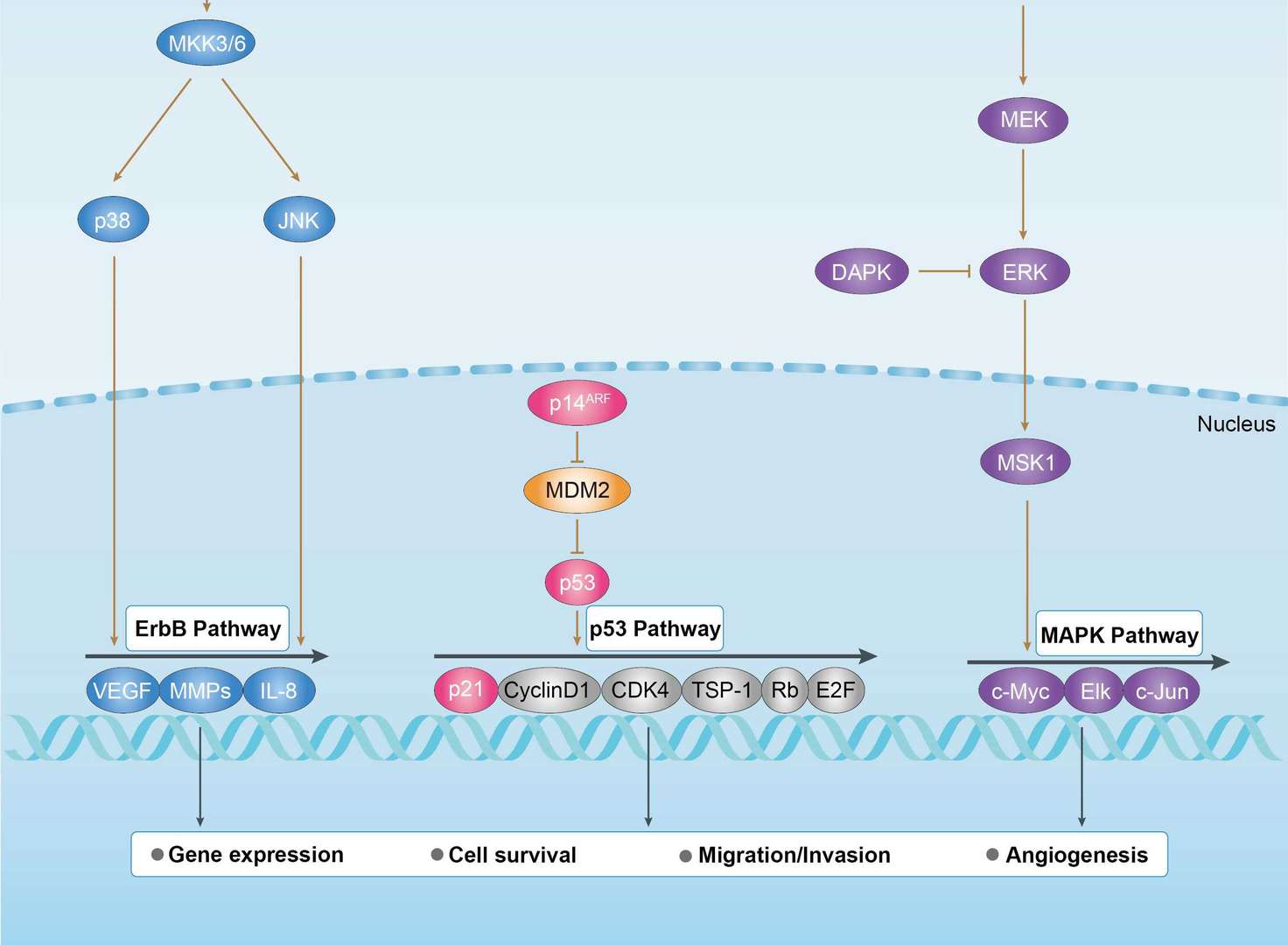
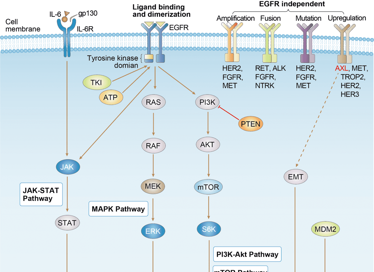
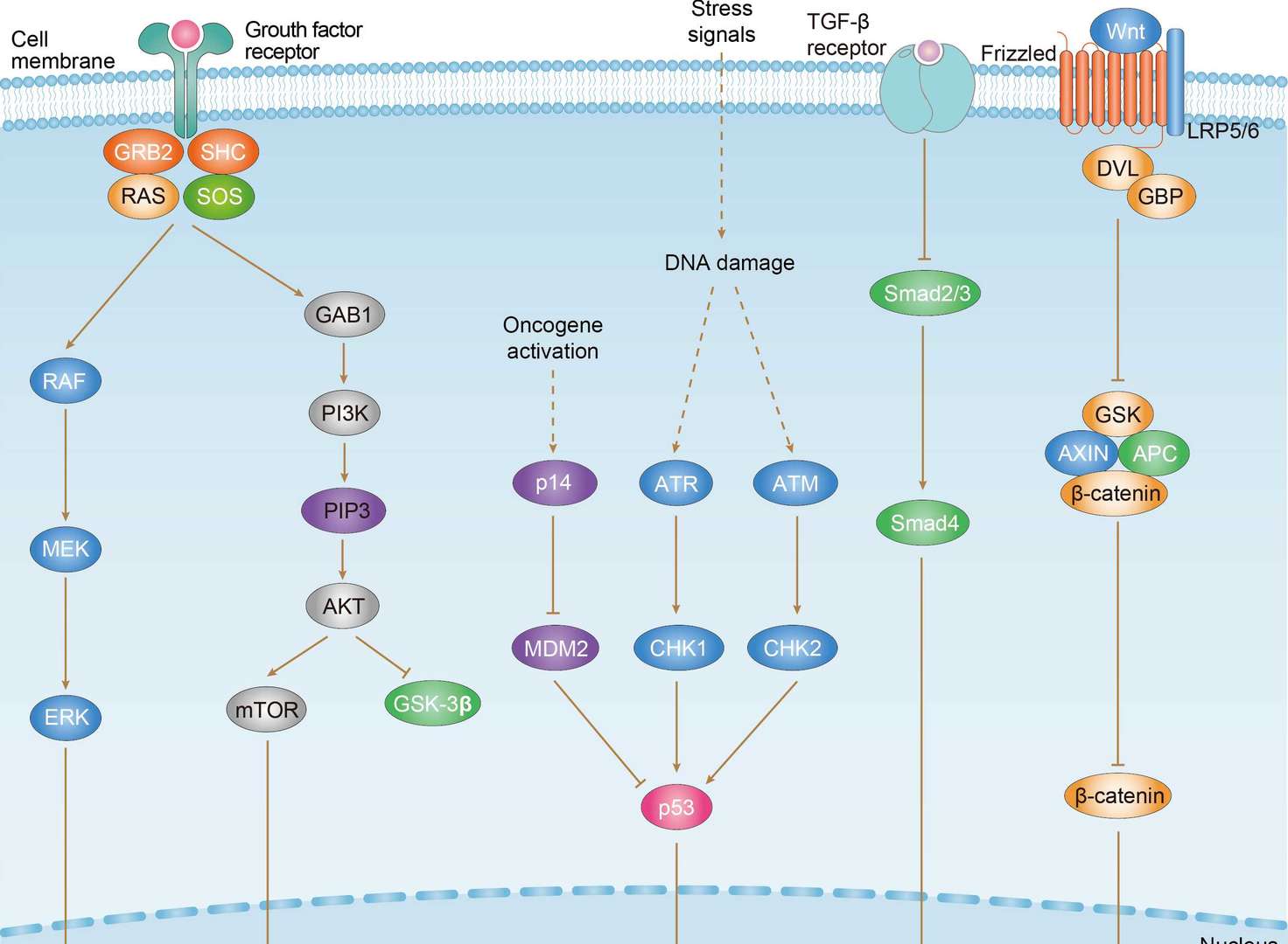
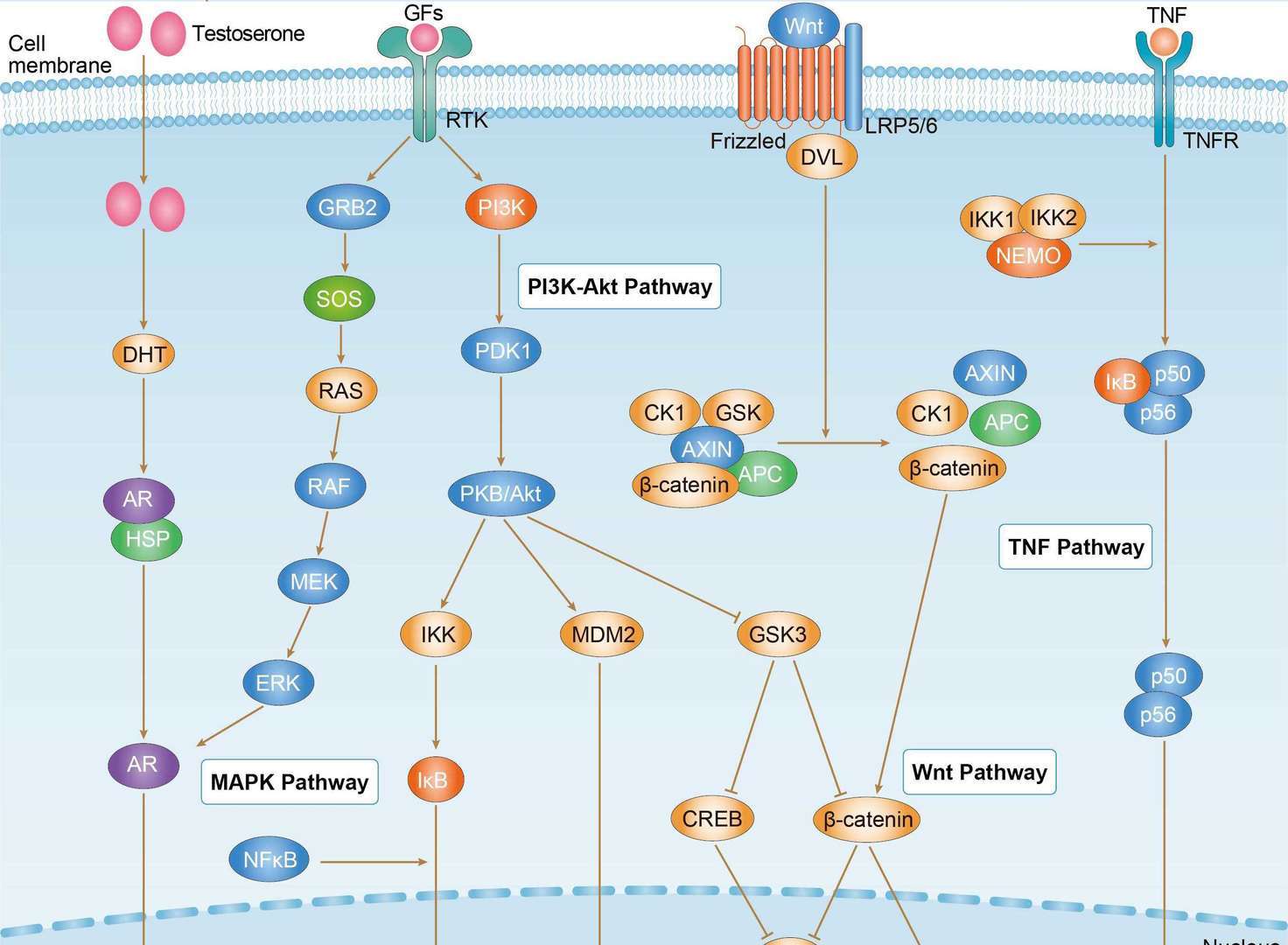
Related Signaling Pathways
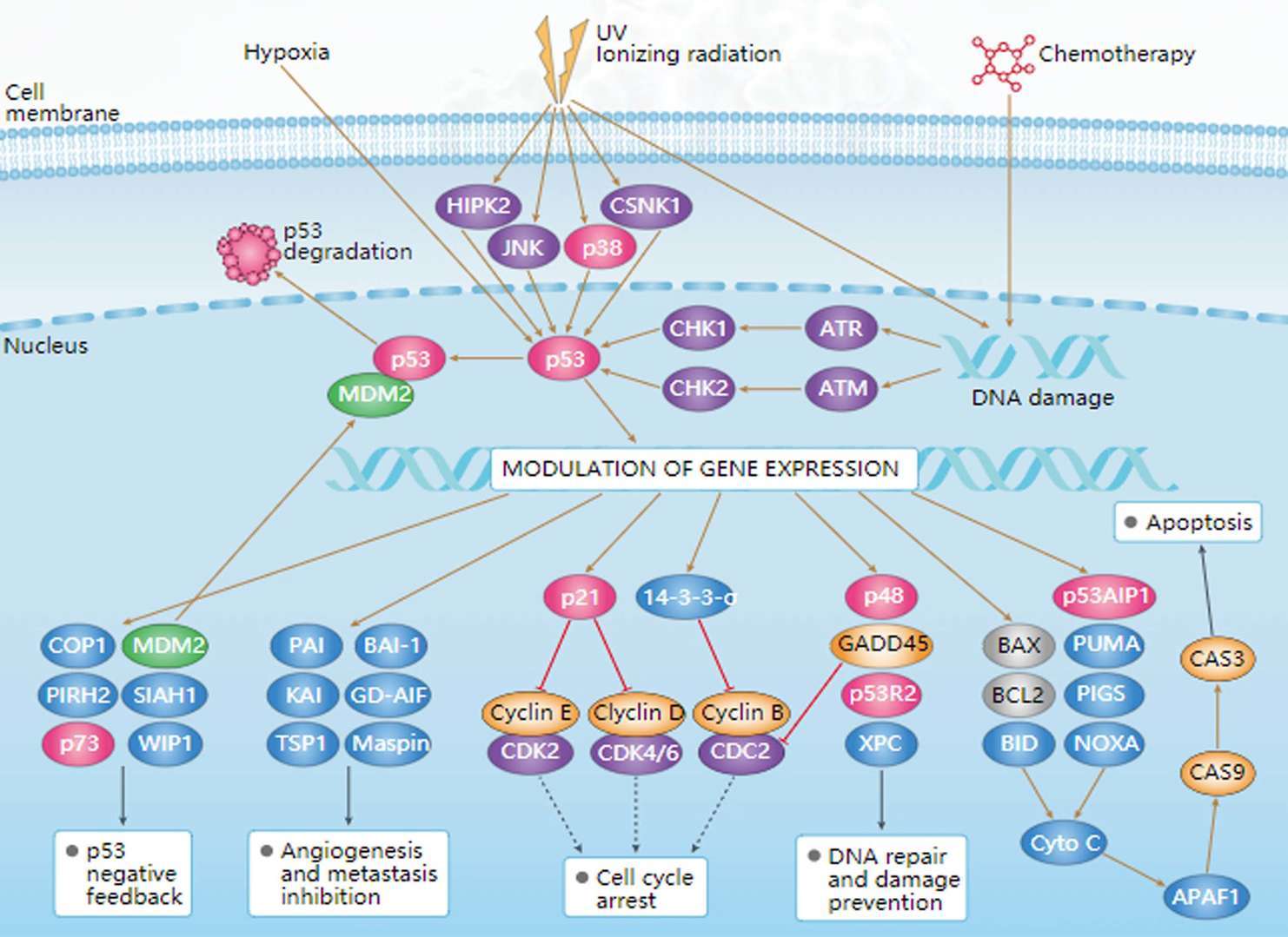 p53 Signaling Pathway
Downloadable Resources
Download resources about recombinant antibody development and antibody engineering to boost your research.
Product Notes
This is a product of Creative Biolabs' Hi-Affi™ recombinant antibody portfolio, which has several benefits including:
• Increased sensitivity
• Confirmed specificity
• High repeatability
• Excellent batch-to-batch consistency
• Sustainable supply
• Animal-free production
See more details about Hi-Affi™ recombinant antibody benefits.
Datasheet
MSDS
COA
Certificate of Analysis LookupTo download a Certificate of Analysis, please enter a lot number in the search box below. Note: Certificate of Analysis not available for kit components.
Lot Number:
See other products for "MDM2"
Select a product category from the dropdown menu below to view related products.
| CAT | Product Name | Application | Type |
|---|---|---|---|
| MOB-648 | Recombinant Anti-human MDM2 Antibody | ELISA, WB, IF, FuncS | IgG |
| CAT | Product Name | Application | Type |
|---|---|---|---|
| MOB-648-F(E) | Recombinant Anti-human MDM2 Antibody Fab Fragment | WB, FACS, FuncS | Fab |
| CAT | Product Name | Application | Type |
|---|---|---|---|
| MOB-648-S(P) | Recombinant Anti-human MDM2 Antibody scFv Fragment | ELISA, WB, FuncS | scFv |
| CAT | Product Name | Application | Type |
|---|---|---|---|
| MHH-648 | Recombinant Human Anti-human MDM2 Antibody | IP, RIA, FuncS | IgG |
| CAT | Product Name | Application | Type |
|---|---|---|---|
| MHH-648-F(E) | Recombinant Human Anti-human MDM2 Antibody Fab Fragment | ELISA, WB, FC, FuncS | Fab |
| CAT | Product Name | Application | Type |
|---|---|---|---|
| MHH-648-S(P) | Recombinant Human Anti-human MDM2 Antibody scFv Fragment | ELISA, WB, FC, FuncS | scFv |
| CAT | Product Name | Application | Type |
|---|---|---|---|
| MOB-1012CT | Recombinant Mouse anti-Human MDM2 Monoclonal antibody (3H3) | WB |
| CAT | Product Name | Application | Type |
|---|---|---|---|
| BRD-0353MZ | Chicken Anti-MDM2 Polyclonal IgY | WB | Chicken antibody |
| CAT | Product Name | Application | Type |
|---|---|---|---|
| MOR-4642 | Hi-Affi™ Recombinant Rabbit Anti-MDM2 Monoclonal Antibody (TH155DS) | WB, IF, ICC | IgG |
| CAT | Product Name | Application | Type |
|---|---|---|---|
| MHC-CN1420 | PE-A*02:01/Human MDM2 (LLGDLFGV) MHC Tetramer | FCM |
| CAT | Product Name | Application | Type |
|---|---|---|---|
| MHC-YF287 | A*02:01/Human MDM2 (LLGDLFGV) MHC Monomer | MHC Multimer |
| CAT | Product Name | Application | Type |
|---|---|---|---|
| ZG-0396U | Rabbit Anti-MDM2 Recombinant Antibody (clone E22-L) | IHC-P | Rabbit IgG |
| CAT | Product Name | Application | Type |
|---|---|---|---|
| VS3-XY1056 | Mouse Anti-MDM2 Recombinant Antibody (clone 3G2) | WB, IP, IF, IHC | Mouse IgG1 |
| CAT | Product Name | Application | Type |
|---|---|---|---|
| VS-0525-XY4322 | Anti-MDM2 Immunohistochemistry Kit | IHC |
| CAT | Product Name | Application | Type |
|---|---|---|---|
| VS-0525-XY4323 | Anti-Human MDM2 Immunohistochemistry Kit | IHC |
| CAT | Product Name | Application | Type |
|---|---|---|---|
| VS-0126-XL257 | Rabbit Anti-MDM2 (phospho S166) Monoclonal Antibody | Phospho Antibodies for Cell Signaling Research |
Specific Inquiry

See Our Custom Production in Action


Popular Products

Application: FC, Cyt, Stim, PP, Agonist


Application: WB, FuncS, IF, Neut, ELISA, FC, IP

Application: IF, IP, Neut, FuncS, ELISA, FC, ICC

Application: FC, IP, ELISA, Neut, FuncS, IF, ICC

Application: IF, IP, Neut, FuncS, ELISA, FC, WB

Application: ELISA, Neut, IF, IP, FC, FuncS

Application: ELISA, IP, FC, FuncS, Neut, IF, ICC


Application: FuncS, IF, Neut, ELISA, FC, IP, IHC


Application: FC, IP, ELISA, Neut, FuncS, IF, ICC

Application: ELISA, FC, IP, FuncS, IF, Neut, ICC

Application: WB, Neut, ELISA, IF, IP, FuncS, FC


For research use only. Not intended for any clinical use. No products from Creative Biolabs may be resold, modified for resale or used to manufacture commercial products without prior written approval from Creative Biolabs.
Send Inquiry
This site is protected by reCAPTCHA and the Google Privacy Policy and Terms of Service apply.