

Recombinant Anti-NOTCH1 Vesicular Antibody, EV Displayed (VS-0425-YC381)
CAT#: VS-0425-YC381
The Recombinant Anti-NOTCH1 Vesicular Antibody, EV Displayed (VS-0425-YC381) is an antibody-displaying extracellular vesicle (Ab-EV). The product combines the benefits of both extracellular vesicle (EV) and antibody (Ab) which can guide the decorated EVs to NOTCH1-expressed cells or tissues. The NOTCH1 is a transmembrane receptor that is part of the Notch signaling pathway, influencing cell development with mutations linked to several conditions, including cancers.
Gene Expression

Subcellular Location
Figure 1 IF staining of human cell line HaCaT
Immunofluorescent staining of human cell line HaCaT shows localization to nucleoplasm.
* Image credit: Image credit: Human Protein Atlas v21.proteinatlas.org/images/67168/1315_F7_4_selected.jpg

Normal Tissue
Figure 2 IHC staining of human pancreas
Immunohistochemical staining of human pancreas shows strong cytoplasmic positivity in exocrine glandular cells.
* Image credit: Image credit: Human Protein Atlas v21.proteinatlas.org/images/22466/61564_A_3_3_selected.jpg

Normal Tissue
Figure 3 Cerebral cortex
Neuronal cells Staining: Medium Intensity: Moderate Quantity:>75% Location: Cytoplasmic/ membranous nuclear
* Image credit: Image credit: Human Protein Atlas v21.proteinatlas.org/images/8112/61559_B_8_5.jpg

Normal Tissue
Figure 4 Colon
Endocrine cells Staining: Medium Intensity: Moderate Endothelial cells Staining: Medium Intensity: Moderate Quantity:>75% Enterocytes Staining: Medium Intensity: Moderate Quantity:>75% Enterocytes - Microvilli Staining: Medium Intensity: Moderate Fibroblasts Staining: High Intensity: Strong Mucosal lymphoid cells Staining: Medium Intensity: Moderate Quantity: 75%-25% Peripheral nerve/ganglion Staining: Low Intensity: Weak
* Image credit: Image credit: Human Protein Atlas v21.proteinatlas.org/images/8112/61559_A_8_3.jpg

Normal Tissue
Figure 5 Liver
Hepatocytes Staining: Medium Intensity: Moderate Quantity:>75% Location: Cytoplasmic/ membranous nuclear
* Image credit: Image credit: Human Protein Atlas v21.proteinatlas.org/images/8112/61559_A_9_4.jpg

Normal Tissue
Figure 6 Kidney
Cells in glomeruli Staining: Medium Intensity: Moderate Quantity:>75% Location: Cytoplasmic/ membranous Nuclear Cells in tubules Staining: Medium Intensity: Moderate Quantity:>75% Location: Cytoplasmic/ membranous nuclear
* Image credit: Image credit: Human Protein Atlas v21.proteinatlas.org/images/8112/61559_A_8_5.jpg

Normal Tissue
Figure 7 Testis
Elongated or late spermatids Staining: Medium Intensity: Moderate Quantity:>75% Leydig cells Staining: Medium Intensity: Moderate Quantity:>75% Pachytene spermatocytes Staining: Medium Intensity: Moderate Quantity:>75% Preleptotene spermatocytes Staining: Medium Intensity: Moderate Quantity:>75% Round or early spermatids Staining: Medium Intensity: Moderate Quantity:>75% Sertoli cells Staining: Low Intensity: Weak Quantity:>75% Spermatogonia cells Staining: High Intensity: Strong
* Image credit: Image credit: Human Protein Atlas v21.proteinatlas.org/images/8112/61559_A_6_6.jpg

Normal Tissue
Figure 8 Lymph node
Non-germinal center cells Staining: Medium Intensity: Moderate Quantity: 75%-25% Location: Cytoplasmic/ membranous nuclear
* Image credit: Image credit: Human Protein Atlas v21.proteinatlas.org/images/8112/61559_A_9_8.jpg

RNA Expression
Figure 9 RNA cell line category: Cell line enhanced (HaCaT, HDLM-2, HMC-1, MOLT-4)
Cell lines ordered by descending RNA expression order
* Image credit: Image credit: Human Protein Atlas v21.proteinatlas.org/ENSG00000148400-NOTCH1
❮


 ❯
❯
Recombinant Antibody
- Application
- ELISA, FC, Neut, Cell-uptake
- Product Type
- Ab-Fc-EVs
- Antibody Quantification (Ab/EV)
- ~100 Ab/EV
- Target
- NOTCH1
- Host Animal
- Human
- Antibody Isotype
- IgG1
- Species Reactivity
- Human
- Expression Cell
- Mammalian cell
Engineered EVs
- EV-sorting domain
- CD63
- Fc-binding domain
- z domain
- EV Size
- 30~150 nm
- Producing Cell
- HEK293F
- Isolation Method
- Gradient centrifugation
- Purification
- qEV size exclusion chromatography
- Binding Affinity
- Kd = 0.85 µg/mL
- Concentration
- 1 x 10¹⁰
- Size
- 1 mL
- Buffer
- PBS
- Storage
- Store at -80°C for 12 months
Target
- Full Name
- Notch receptor 1
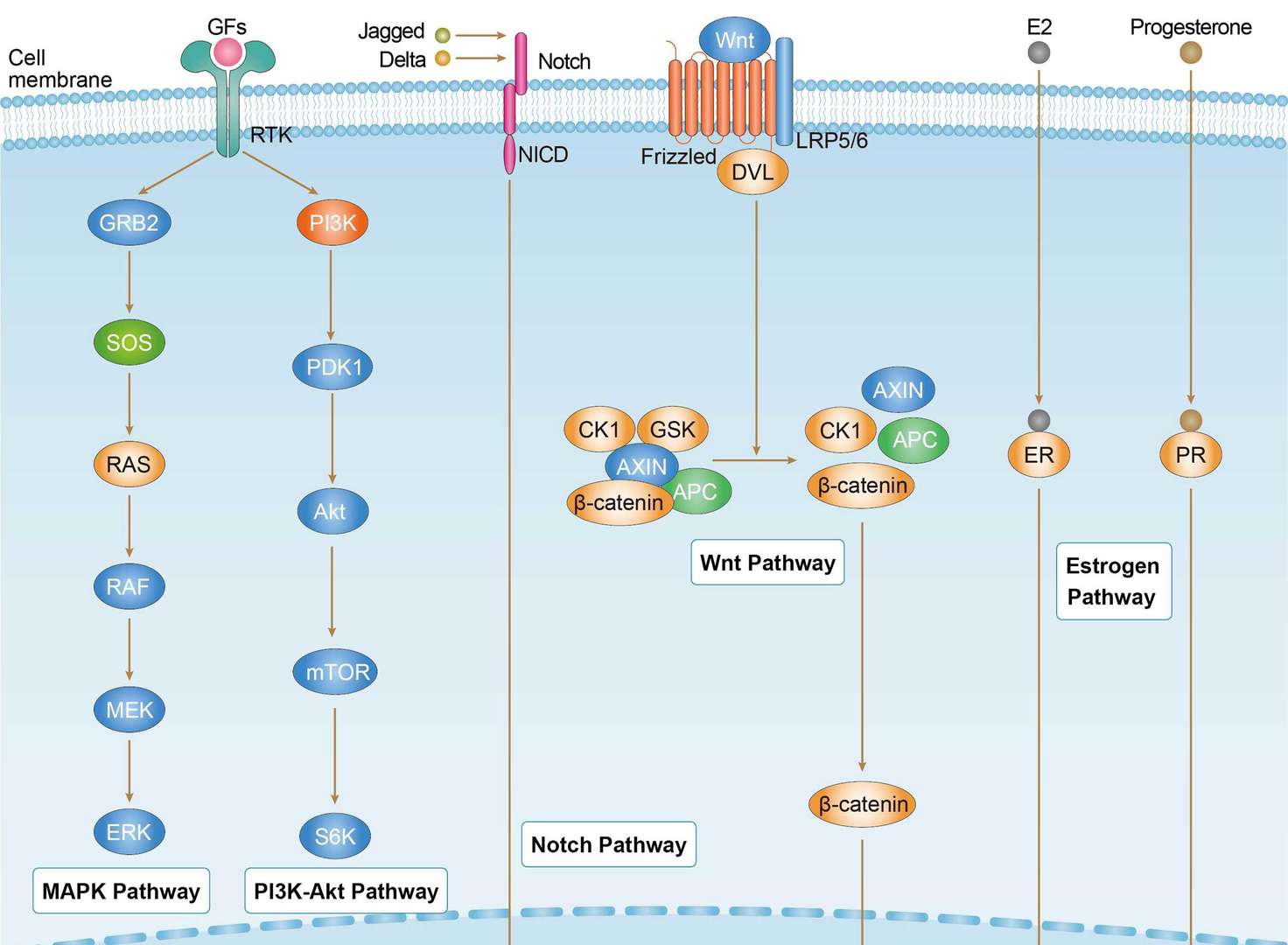
- Biological Process
- Angiogenesis, Differentiation, Notch signaling pathway, Transcription, Transcription regulation
- Molecular Function
- Activator, Developmental protein, Receptor
- Cellular Localization
- Nucleoplasm
- Introduction
- This gene encodes a member of the NOTCH family of proteins. Members of this Type I transmembrane protein family share structural characteristics including an extracellular domain consisting of multiple epidermal growth factor-like (EGF) repeats, and an intracellular domain consisting of multiple different domain types. Notch signaling is an evolutionarily conserved intercellular signaling pathway that regulates interactions between physically adjacent cells through binding of Notch family receptors to their cognate ligands. The encoded preproprotein is proteolytically processed in the trans-Golgi network to generate two polypeptide chains that heterodimerize to form the mature cell-surface receptor. This receptor plays a role in the development of numerous cell and tissue types. Mutations in this gene are associated with aortic valve disease, Adams-Oliver syndrome, T-cell acute lymphoblastic leukemia, chronic lymphocytic leukemia, and head and neck squamous cell carcinoma. [provided by RefSeq, Jan 2016]
- Alternative Names
- TAN1
- Gene ID
- 4851
- UniProt ID
- P46531
REVIEWS AND Q&AS
CITATIONS
RESOURCES
DOWNLOADS
RELATED PRODUCTS
Inquiry
Navs
Customer Review

There are currently no Customer reviews or questions for VS-0425-YC381. Click the button above to contact us or submit your feedback about this product.
Submit Your Publication
Published with our product? Submit your paper and receive a 10% discount on your next order! Share your research to earn exclusive rewards.
Related Diseases
 Breast Cancer
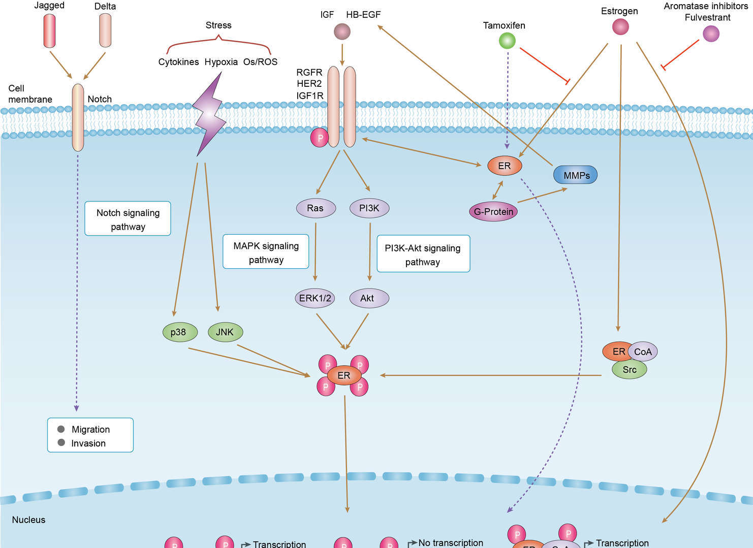 Endocrine Resistance
Downloadable Resources
Download resources about recombinant antibody development and antibody engineering to boost your research.
Datasheet
MSDS
COA
Certificate of Analysis LookupTo download a Certificate of Analysis, please enter a lot number in the search box below. Note: Certificate of Analysis not available for kit components.
Lot Number:
See other products for "NOTCH1"
Select a product category from the dropdown menu below to view related products.
| CAT | Product Name | Application | Type |
|---|---|---|---|
| MOB-1747z | Mouse Anti-NOTCH1 Recombinant Antibody (clone 21H9) | WB, ICC, IF, IHC | Mouse IgG2b |
| CAT | Product Name | Application | Type |
|---|---|---|---|
| TAB-H11 | Anti-Human NOTCH1 Recombinant Antibody (Brontictuzumab) | Inhib | IgG2 - lambda |
| CAT | Product Name | Application | Type |
|---|---|---|---|
| PABL-666 | Human Anti-NOTCH1 Recombinant Antibody | ELISA | Human IgG |
| CAT | Product Name | Application | Type |
|---|---|---|---|
| PFBL-660 | Human Anti-NOTCH1 Recombinant Antibody; Fab Fragment | ELISA | Human Fab |
| CAT | Product Name | Application | Type |
|---|---|---|---|
| PSBL-660 | Human Anti-NOTCH1 Recombinant Antibody; scFv Fragment | ELISA | Human scFv |
| CAT | Product Name | Application | Type |
|---|---|---|---|
| TAB-539MZ | Rat Anti-NOTCH1 Recombinant Antibody (TAB-539MZ) | WB, Competition ELISA, Neut | Rat IgG1 |
| CAT | Product Name | Application | Type |
|---|---|---|---|
| TAB-540MZ | Rat Anti-NOTCH1 Recombinant Antibody (TAB-540MZ) | WB, Competition ELISA, Neut | Rat IgG1 |
| CAT | Product Name | Application | Type |
|---|---|---|---|
| TAB-541MZ | Human Anti-NOTCH1 Recombinant Antibody (TAB-541MZ) | Neut, WB | Humanized antibody |
| CAT | Product Name | Application | Type |
|---|---|---|---|
| TAB-542MZ | Human Anti-NOTCH1 Recombinant Antibody (TAB-542MZ) | Neut, WB | Humanized antibody |
| CAT | Product Name | Application | Type |
|---|---|---|---|
| TAB-543MZ | Anti-Human NOTCH1 Recombinant Antibody (mAb N248A) | ELISA, WB, Luciferase reporter assay |
| CAT | Product Name | Application | Type |
|---|---|---|---|
| TAB-544MZ | Anti-Human NOTCH1 Recombinant Antibody (A12.2) | ELISA, FuncS, Inhib | Humanized antibody |
| CAT | Product Name | Application | Type |
|---|---|---|---|
| TAB-545MZ | Anti-Human NOTCH1 Recombinant Antibody (23814) | ELISA, FuncS, Inhib, WB, FACS | Human antibody |
| CAT | Product Name | Application | Type |
|---|---|---|---|
| TAB-546MZ | Anti-Human NOTCH1 Recombinant Antibody (23418.8) | FuncS, Inhib | Human antibody |
| CAT | Product Name | Application | Type |
|---|---|---|---|
| TAB-547MZ | Human Anti-NOTCH1 Recombinant Antibody (TAB-547MZ) | Inhib | Human IgG1 |
| CAT | Product Name | Application | Type |
|---|---|---|---|
| TAB-548MZ | Human Anti-NOTCH1 Recombinant Antibody (TAB-548MZ) | FuncS, Inhib | Human IgG1 |
| CAT | Product Name | Application | Type |
|---|---|---|---|
| TAB-549MZ | Human Anti-NOTCH1 Recombinant Antibody (TAB-549MZ) | Inhib | Human IgG1 |
| CAT | Product Name | Application | Type |
|---|---|---|---|
| TAB-539MZ-S(P) | Rat Anti-NOTCH1 Recombinant Antibody; scFv Fragment (TAB-539MZ-S(P)) | WB, Competition ELISA | Rat scFv |
| CAT | Product Name | Application | Type |
|---|---|---|---|
| TAB-540MZ-S(P) | Rat Anti-NOTCH1 Recombinant Antibody; scFv Fragment (TAB-540MZ-S(P)) | WB, Competition ELISA | Rat scFv |
| CAT | Product Name | Application | Type |
|---|---|---|---|
| TAB-541MZ-S(P) | Human Anti-NOTCH1 Recombinant Antibody; scFv Fragment (TAB-541MZ-S(P)) | WB, Competition ELISA | Humanized scFv |
| CAT | Product Name | Application | Type |
|---|---|---|---|
| MOB-1438CT | Recombinant Mouse anti-Mouse NOTCH1 Monoclonal antibody (3E12) | FC |
| CAT | Product Name | Application | Type |
|---|---|---|---|
| NEUT-1786CQ | Recombinant Mouse Anti-NOTCH1 Antibody (MHN1-519) | FC, BL | IgG1, κ |
| CAT | Product Name | Application | Type |
|---|---|---|---|
| MOR-2473 | Hi-Affi™ Recombinant Rabbit Anti-NOTCH1 Monoclonal Antibody (DS2473AB) | WB, IHC | IgG |
| CAT | Product Name | Application | Type |
|---|---|---|---|
| HPAB-0078-YC | Mouse Anti-NOTCH1 Recombinant Antibody (clone OMP-52M51) | IHC | Mouse IgG |
| CAT | Product Name | Application | Type |
|---|---|---|---|
| HPAB-0078-YC-S(P) | Mouse Anti-NOTCH1 Recombinant Antibody (clone OMP-52M51); scFv Fragment | IHC | Mouse scFv |
| CAT | Product Name | Application | Type |
|---|---|---|---|
| HPAB-0078-YC-F(E) | Mouse Anti-NOTCH1 Recombinant Antibody (clone OMP-52M51); Fab Fragment | IHC | Mouse Fab |
| CAT | Product Name | Application | Type |
|---|---|---|---|
| NS-060CN | Mouse Anti-NOTCH1 Recombinant Antibody (clone 2E6) | FC, IF, ELISA, Inhib, FuncS | Mouse IgG1 |
| CAT | Product Name | Application | Type |
|---|---|---|---|
| NS-061CN | Mouse Anti-NOTCH1 Recombinant Antibody (clone 2G10) | FC, IF, ELISA | Mouse IgG1 |
| CAT | Product Name | Application | Type |
|---|---|---|---|
| NS-062CN | Mouse Anti-NOTCH1 Recombinant Antibody (clone 2A11) | FC, IF, ELISA, Inhib | Mouse IgG2b |
| CAT | Product Name | Application | Type |
|---|---|---|---|
| NS-063CN | Mouse Anti-NOTCH1 Recombinant Antibody (clone 2D11) | FC, IF, ELISA | Mouse IgG1 |
| CAT | Product Name | Application | Type |
|---|---|---|---|
| NS-060CN-F(E) | Mouse Anti-NOTCH1 Recombinant Antibody (clone 2E6); Fab Fragment | FC, IF, ELISA | Mouse Fab |
| CAT | Product Name | Application | Type |
|---|---|---|---|
| NS-061CN-F(E) | Mouse Anti-NOTCH1 Recombinant Antibody (clone 2G10); Fab Fragment | FC, IF, ELISA | Mouse Fab |
| CAT | Product Name | Application | Type |
|---|---|---|---|
| NS-062CN-F(E) | Mouse Anti-NOTCH1 Recombinant Antibody (clone 2A11); Fab Fragment | FC, IF, ELISA | Mouse Fab |
| CAT | Product Name | Application | Type |
|---|---|---|---|
| NS-063CN-F(E) | Mouse Anti-NOTCH1 Recombinant Antibody (clone 2D11); Fab Fragment | FC, IF, ELISA | Mouse Fab |
| CAT | Product Name | Application | Type |
|---|---|---|---|
| NS-060CN-S(P) | Mouse Anti-NOTCH1 Recombinant Antibody (clone 2E6); scFv Fragment | FC, IF, ELISA | Mouse scFv |
| CAT | Product Name | Application | Type |
|---|---|---|---|
| NS-061CN-S(P) | Mouse Anti-NOTCH1 Recombinant Antibody (clone 2G10); scFv Fragment | FC, IF, ELISA | Mouse scFv |
| CAT | Product Name | Application | Type |
|---|---|---|---|
| NS-062CN-S(P) | Mouse Anti-NOTCH1 Recombinant Antibody (clone 2A11); scFv Fragment | FC, IF, ELISA | Mouse scFv |
| CAT | Product Name | Application | Type |
|---|---|---|---|
| HPAB-0044LY | Mouse Anti-NOTCH1 Recombinant Antibody (HPAB-0044LY) | WB | Mouse IgG1 |
| CAT | Product Name | Application | Type |
|---|---|---|---|
| AFC-TAB-H11 | Afuco™ Anti-NOTCH1 ADCC Recombinant Antibody, ADCC Enhanced (AFC-TAB-H11) | FC, IP, ELISA, Neut, FuncS, IF | ADCC enhanced antibody |
| CAT | Product Name | Application | Type |
|---|---|---|---|
| VS-0924-YC79 | Rabbit Anti-NOTCH1 Antibody (VS-0924-YC79) - Cancer Stem Cell Marker | IF | Rabbit IgG |
| CAT | Product Name | Application | Type |
|---|---|---|---|
| VS-1024-XY345 | Mouse Anti-NHP NOTCH1 Recombinant Antibody (clone mN1A) | ELISA | Mouse IgG1 |
| CAT | Product Name | Application | Type |
|---|---|---|---|
| VS13-YC828 | CytoStream™ Rabbit Anti-NOTCH1 Recombinant Antibody (VS13-YC828) | WB, ICC, IF, IHC-P, IP, FC | Rabbit IgG |
| CAT | Product Name | Application | Type |
|---|---|---|---|
| VS13-YC829 | CytoStream™ Rabbit Anti-NOTCH1 Polyclonal Antibody (VS13-YC829) | WB, IHC, IF, FC | Rabbit IgG |
| CAT | Product Name | Application | Type |
|---|---|---|---|
| VS-0425-FY159 | Human Anti-NOTCH1 scFv-Fc Chimera (VS-0425-FY159) | ELISA, Inhib, WB, FC | Human IgG1, scFv-Fc |
| CAT | Product Name | Application | Type |
|---|---|---|---|
| VS-0325-XY1506 | Anti-NOTCH1 Immunohistochemistry Kit | IHC |
| CAT | Product Name | Application | Type |
|---|---|---|---|
| VS-0525-XY4907 | Anti-Mouse NOTCH1 Immunohistochemistry Kit | IHC |
| CAT | Product Name | Application | Type |
|---|---|---|---|
| VS-0525-YC139 | Recombinant Anti-NOTCH1 (LBD domain x HD-LNR domain) Biparatopic Antibody, Tandem scFv (Clone hN1wc49 x Clone N1wc104) | ELISA | Tandem scFv |
| CAT | Product Name | Application | Type |
|---|---|---|---|
| VS-0525-XY4906 | Anti-Human NOTCH1 Immunohistochemistry Kit | IHC |
| CAT | Product Name | Application | Type |
|---|---|---|---|
| VS-0825-YC267 | SmartAb™ Recombinant Anti-NOTCH1 pH-dependent Antibody (Clone Brontictuzumab) | Inhibition | Human IgG2 lambda |
| CAT | Product Name | Application | Type |
|---|---|---|---|
| VS-1025-YC28 | Anti-NOTCH1 Antibody Prodrug, Protease Activated (OMP-52M51) | ISZ, Cyt, FuncS |
Specific Inquiry
See Our Custom Production in Action


Popular Products

Application: WB, FC, IP, ELISA, Neut, FuncS, IF

Application: ELISA, FC, IP, FuncS, IF, Neut, ICC

Application: ELISA, FC, IP, FuncS, IF, Neut, ICC

Application: FC, IP, ELISA, Neut, FuncS, IF, ICC

Application: Neut, ELISA, IF, IP, FuncS, FC, ICC

Application: Neut, ELISA, IF, IP, FuncS, FC, WB

Application: ELISA, FC, IP, FuncS, IF, Neut, ICC

Application: IF, IP, Neut, FuncS, ELISA, FC, ICC

Application: ELISA, IP, FC, FuncS, Neut, IF, ICC


Application: Neut, ELISA, FuncS

Application: WB, Neut, FuncS


Application: FuncS, Inhib, IP, ELISA


For research use only. Not intended for any clinical use. No products from Creative Biolabs may be resold, modified for resale or used to manufacture commercial products without prior written approval from Creative Biolabs.
Send Inquiry
This site is protected by reCAPTCHA and the Google Privacy Policy and Terms of Service apply.