

Rabbit Anti-AKT1 Recombinant Antibody (clone 4H12)
CAT#: ZG-0539J
This product is a rabbit antibody that recognizes AKT1.
Tested Data
Gene Expression

WB
Figure 1 Rabbit Anti-AKT1 Antibody (ZG-0539J) in WB
Western Blot
Positive WB detected in 293 whole cell lysate(treated with Calyculin A or not)
All lanes Phospho-AKT1 antibody at 1.08μg/ml
Secondary
Goat polyclonal to rabbit IgG at 1/50000 dilution
Predicted band size: 60 KDa
Observed band size: 60 KDa

IHC
Figure 2 Rabbit Anti-AKT1 Antibody (ZG-0539J) in IHC
IHC image of ZG-0539J diluted at 1:100 and staining in paraffin-embedded human breast cancer performed on a Leica BondTM system. After dewaxing and hydration, antigen retrieval was mediated by high pressure in a citrate buffer (pH 6.0). Section was blocked with 10% normal goat serum 30min at RT. Then primary antibody (1% BSA) was incubated at 4°C overnight. The primary is detected by a biotinylated secondary antibody and visualized using an HRP conjugated SP system.

IHC
Figure 3 Rabbit Anti-AKT1 Antibody (ZG-0539J) in IHC
IHC image of ZG-0539J diluted at 1:100 and staining in paraffin-embedded human lung cancer performed on a Leica BondTM system. After dewaxing and hydration, antigen retrieval was mediated by high pressure in a citrate buffer (pH 6.0). Section was blocked with 10% normal goat serum 30min at RT. Then primary antibody (1% BSA) was incubated at 4°C overnight. The primary is detected by a biotinylated secondary antibody and visualized using an HRP conjugated SP system.

IF
Figure 4 Rabbit Anti-AKT1 Antibody (ZG-0539J) in IF
Immunofluorescence staining of Hela cells(treated with 100mM EGF for 20min) with ZG-0539J at 1:68,counter-stained with DAPI. The cells were fixed in 4% formaldehyde, permeabilized using 0.2% Triton X-100 and blocked in 10% normal Goat Serum. The cells were then incubated with the antibody overnight at 4°C. The secondary antibody was Alexa Fluor 488-congugated Goat Anti-Rabbit IgG (H+L).

IP
Figure 5 Rabbit Anti-AKT1 Antibody (ZG-0539J) in IP
Immunoprecipitating Phospho-AKT1 in 293 whole cell lysate treated with Calyculin A
Lane 1: Rabbit control IgG(1μg)instead of ZG-0539J in 293 whole cell lysate treated with Calyculin A. For western blotting,a HRP-conjugated Protein G antibody was used as the secondary antibody (1/2000)
Lane 2: ZG-0539J(3μg)+ 293 whole cell lysate treated with Calyculin A(1mg)
Lane 3: 293 whole cell lysate treated with Calyculin A (20μg)
❮


 ❯
❯

Subcellular Location and Protein Expression
Figure 1 IF staining of human cell line A-431
Immunofluorescent staining of human cell line A-431 shows localization to nucleoplasm & microtubules.
* Image credit: Image credit: Human Protein Atlas https://v21.proteinatlas.org/images/2891/39_F7_2_selected.jpg

Subcellular Location and Protein Expression
Figure 2 IHC staining of human gall bladder
Immunohistochemical staining of human gall bladder shows strong nuclear positivity in glandular cells.
* Image credit: Image credit: Human Protein Atlas https://v21.proteinatlas.org/images/2891/ihc_selected.jpg

Subcellular Location and Protein Expression
Figure 3 IHC staining of human cerebellum
Immunohistochemical staining of human cerebellum shows strong cytoplasmic and nuclear positivity in Purkinje cells and cells in molecular layer.
* Image credit: Image credit: Human Protein Atlas https://v21.proteinatlas.org/images/3765/ihc_selected.jpg

Subcellular Location and Protein Expression
Figure 4 IF staining of human cell line U-2 OS
Immunofluorescent staining of human cell line U-2 OS shows localization to nucleoplasm & microtubules.
* Image credit: Image credit: Human Protein Atlas https://v21.proteinatlas.org/images/2891/38_F7_1_red_green.jpg

Subcellular Location and Protein Expression
Figure 5 IF staining of human cell line U-251 MG
Immunofluorescent staining of human cell line U-251 MG shows localization to nucleoplasm & microtubules.
* Image credit: Image credit: Human Protein Atlas https://v21.proteinatlas.org/images/2891/40_F7_1_red_green.jpg

Normal Tissue
Figure 6 Cerebral cortex
Endothelial cells
Staining:Medium
Intensity: Moderate
Quantity:>75%
Location: Nuclear
Glial cells
Staining:Medium
Intensity: Moderate
Quantity:>75%
Location: Nuclear
Neuronal cells
Staining:High
Intensity: Strong
Quantity:>75%
Location: Cytoplasmic/membranous nuclear
* Image credit: Image credit: Human Protein Atlas https://v21.proteinatlas.org/images/2891/7680_B_8_5.jpg

Normal Tissue
Figure 7 Colon
Endothelial cells
Staining:Medium
Intensity: Moderate
Quantity:>75%
Location: Nuclear
Glandular cells
Staining:High
Intensity: Strong
Quantity:>75%
Location: Nuclear
Peripheral nerve/ganglion
Staining:Medium
Intensity: Moderate
Quantity:>75%
Location: Nuclear
* Image credit: Image credit: Human Protein Atlas https://v21.proteinatlas.org/images/2891/7680_A_7_3.jpg

Normal Tissue
Figure 8 Liver
Cholangiocytes
Staining:High
Intensity: Strong
Quantity:>75%
Location: Nuclear
* Image credit: Image credit: Human Protein Atlas https://v21.proteinatlas.org/images/2891/7680_A_8_4.jpg

Normal Tissue
Figure 9 Kidney
Cells in glomeruli
Staining:Medium
Intensity: Moderate
Quantity:>75%
Location: Nuclear
Cells in tubules
Staining:Medium
Intensity: Moderate
Quantity:>75%
Location: Nuclear
* Image credit: Image credit: Human Protein Atlas https://v21.proteinatlas.org/images/2891/7680_A_9_5.jpg

Normal Tissue
Figure 10 Testis
Elongated or late spermatids
Staining:High
Intensity: Strong
Quantity:>75%
Leydig cells
Staining:Medium
Intensity: Moderate
Quantity:>75%
Pachytene spermatocytes
Staining:Medium
Intensity: Moderate
Quantity:>75%
Peritubular cells
Staining:Medium
Intensity: Moderate
Quantity: 75%-25%
Preleptotene spermatocytes
Staining:Medium
Intensity: Moderate
Quantity:>75%
Round or early spermatids
Staining:High
Intensity: Strong
Quantity:>75%
Sertoli cells
Staining:High
Intensity: Strong
Quantity:>75%
Spermatogonia cells
Staining:Medium
Intensity: Moderate
Quantity:>75%
* Image credit: Image credit: Human Protein Atlas https://v21.proteinatlas.org/images/2891/7680_A_4_6.jpg

Normal Tissue
Figure 11 Lymph node
Germinal center cells
Staining:High
Intensity: Strong
Quantity:>75%
Location: Nuclear
Non-germinal center cells
Staining:High
Intensity: Strong
Quantity:>75%
Location: Nuclear
* Image credit: Image credit: Human Protein Atlas https://v21.proteinatlas.org/images/2891/7680_A_8_8.jpg

RNA Expression
Figure 12 RNA cell line category: Cell line enhanced (MCF7)
Cell lines ordered by descending RNA expression order.
* Image credit: Image credit: Human Protein Atlas https://v21.proteinatlas.org/ENSG00000142208-AKT1
❮


 ❯
❯
Specifications
- Immunogen
- A synthesized peptide derived from human Phospho-AKT1 (Ser473)
- Host Species
- Rabbit
- Type
- Rabbit IgG
- Specificity
- Human AKT1
- Species Reactivity
- Human
- Clone
- 4H12
- Applications
- ELISA, WB, IHC, IF, IP
- Conjugate
- Uconjugated
Product Property
- Purification
- Affinity purified
- Format
- Liquid
- Concentration
- See the COA
- Buffer
- PBS, pH 7.4, 150mM NaCl, 0.02% sodium azide and 50% glycerol.
- Preservative
- 0.02% sodium azide
- Storage
- Centrifuge briefly prior to opening vial. Store at +4°C for short term (1-2 weeks). Aliquot and store at -20°C for long term. Avoid repeated freeze/thaw cycles.
Applications
- Application Notes
- The antibody was validated for Enzyme-linked Immunosorbent Assay, Western Blot, Immunohistochemistry, Immunofluorescence, Immunoprecipitation.
WB:1:500-1:5000, IHC:1:50-1:200, IF:1:20-1:200, IP:1:200-1:1000
Target
- Alternative Names
- AKT; PKB; RAC; PRKBA; PKB-ALPHA; RAC-ALPHA; RAC-alpha serine/threonine-protein kinase; AKT1m; PKB alpha; RAC-PK-alpha; protein kinase B alpha; proto-oncogene c-Akt; rac protein kinase alpha; serine-threonine protein kinase; v-akt murine thymoma viral oncogene homolog 1; v-akt murine thymoma viral oncogene-like protein 1
- Gene ID
- 207
- UniProt ID
- P31749
REVIEWS AND Q&AS
CITATIONS
RESOURCES
DOWNLOADS
RELATED PRODUCTS
Inquiry
Navs
Customer Review

There are currently no Customer reviews or questions for ZG-0539J. Click the button above to contact us or submit your feedback about this product.
Submit Your Publication
Published with our product? Submit your paper and receive a 10% discount on your next order! Share your research to earn exclusive rewards.
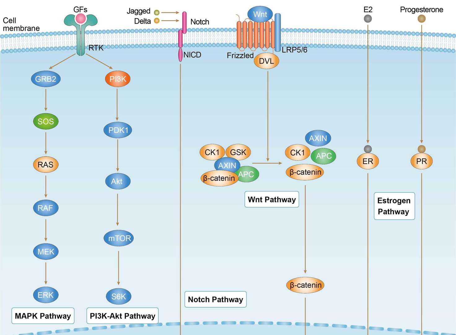
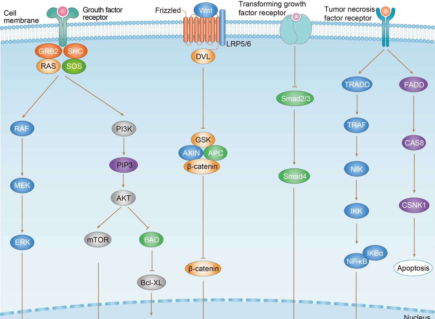
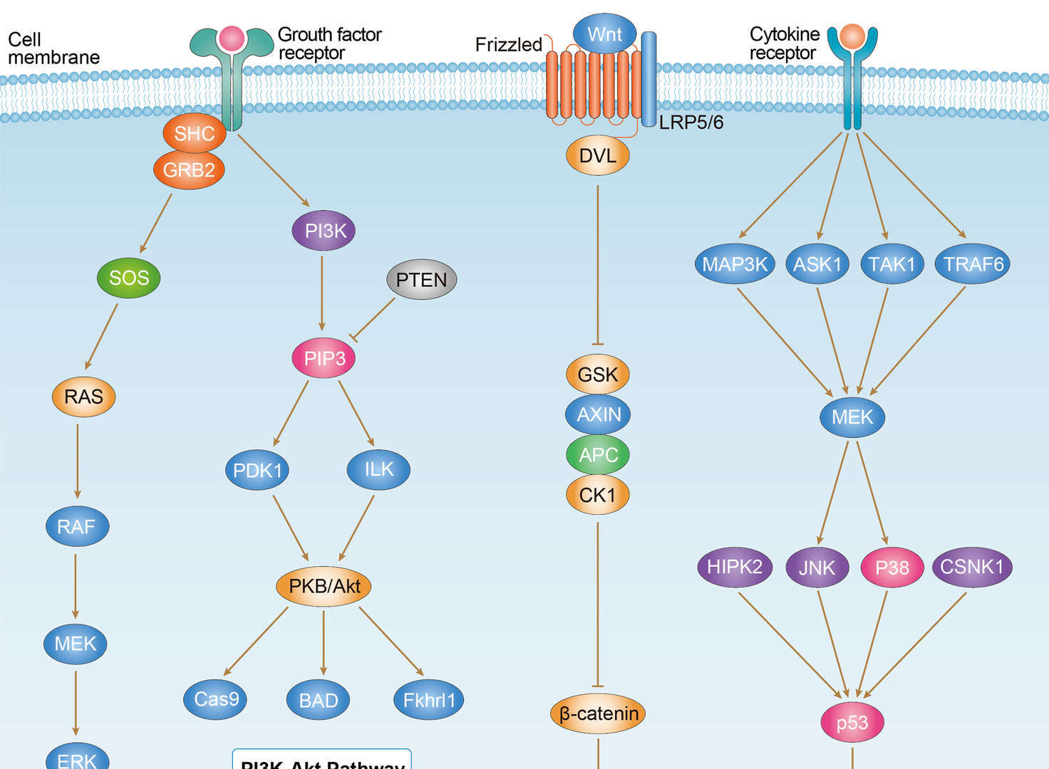
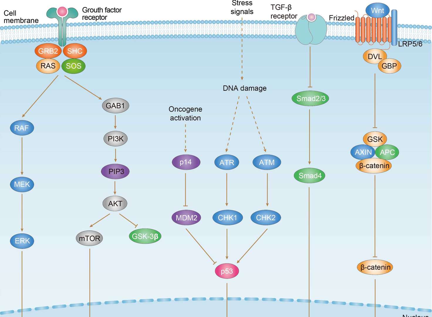
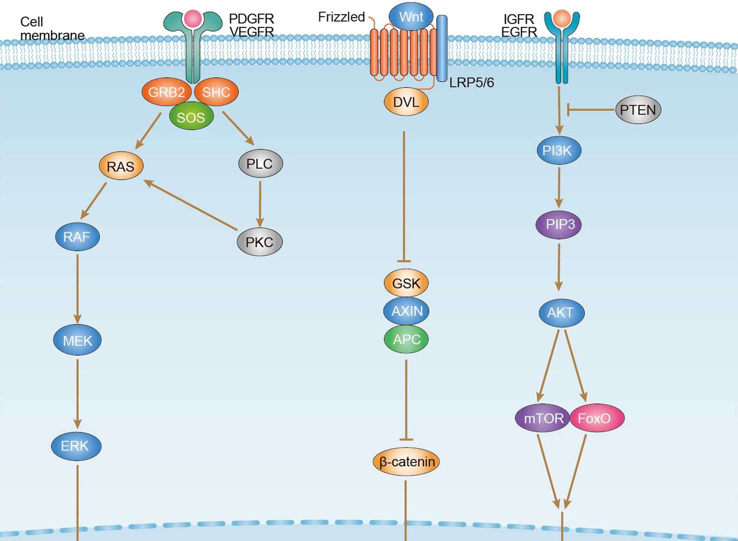
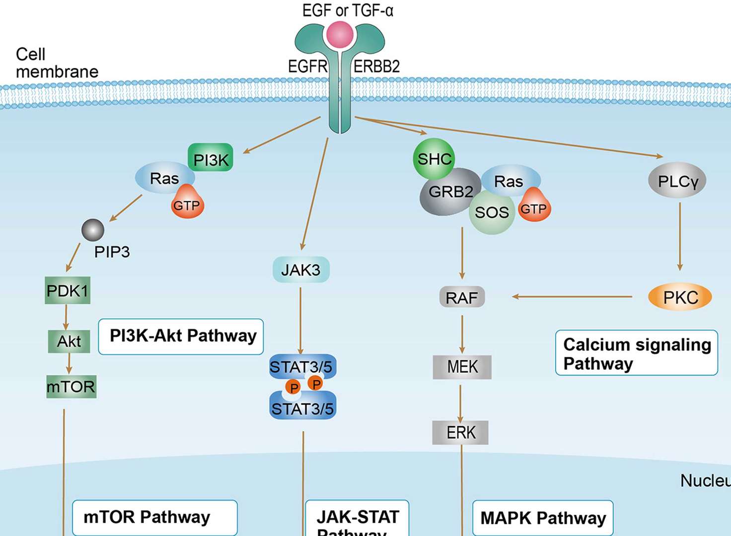
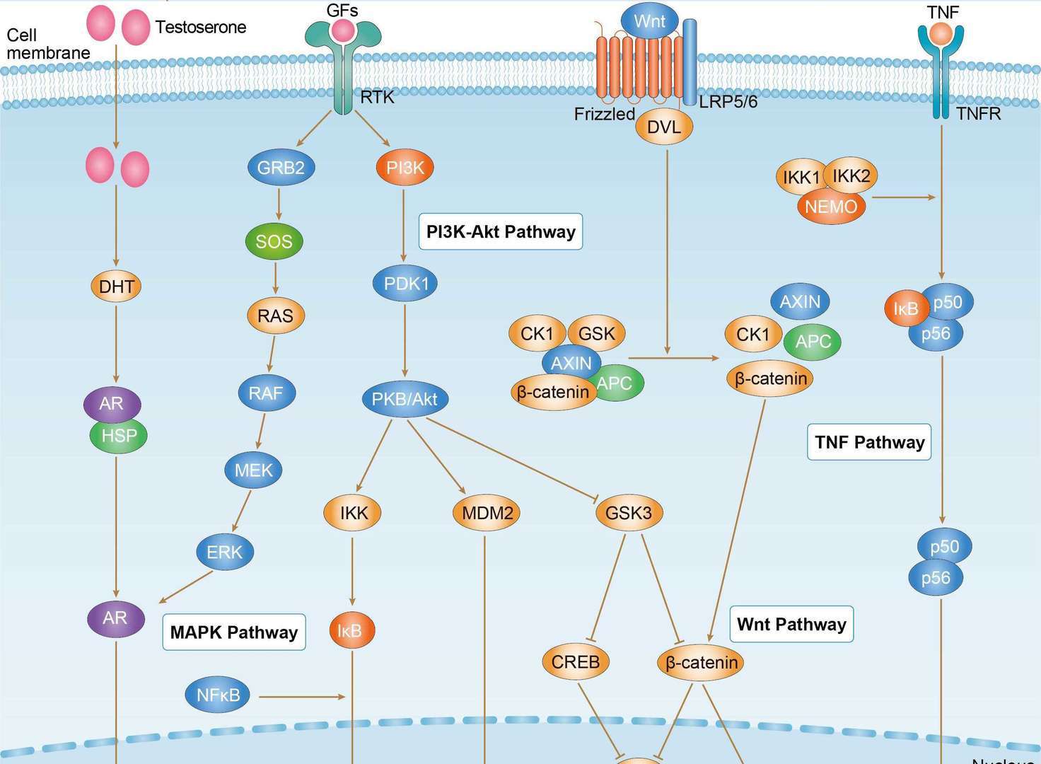
Related Signaling Pathways
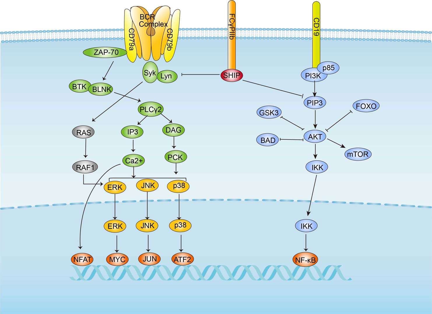 BCR Signaling Pathway
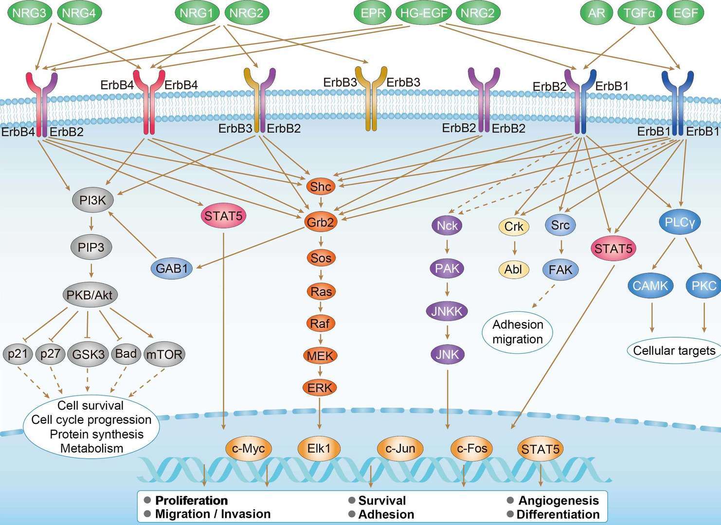 ErbB Signaling Pathway
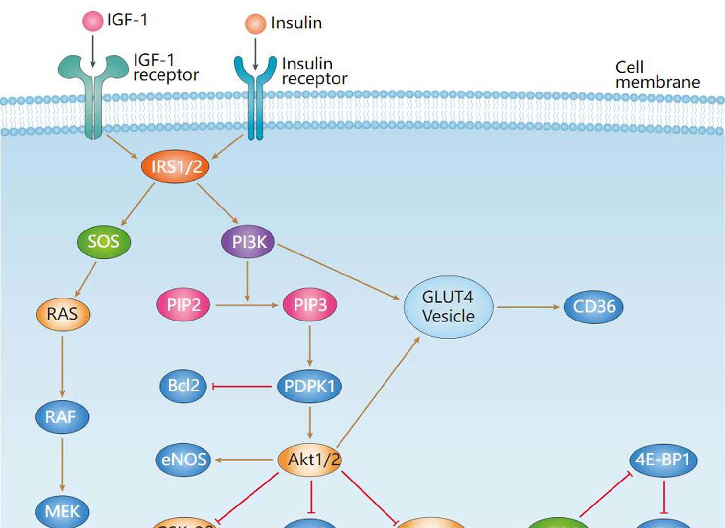 Insulin Signaling Pathway
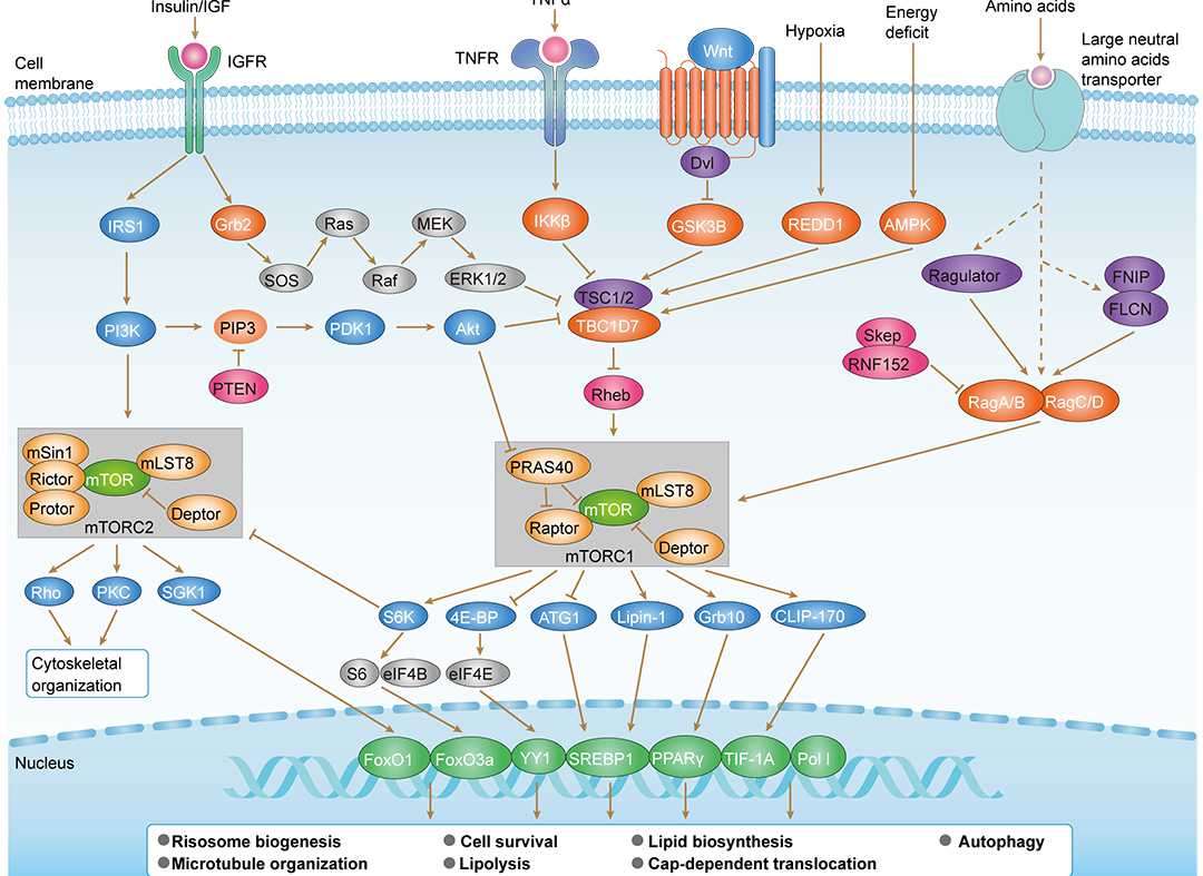 mTOR Signaling Pathway
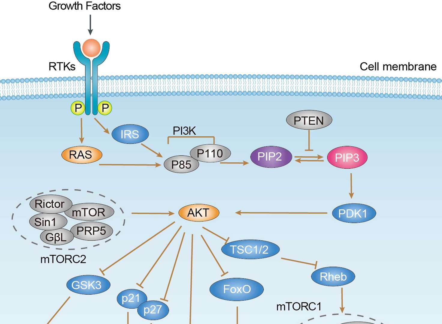 PI3K-Akt Signaling Pathway
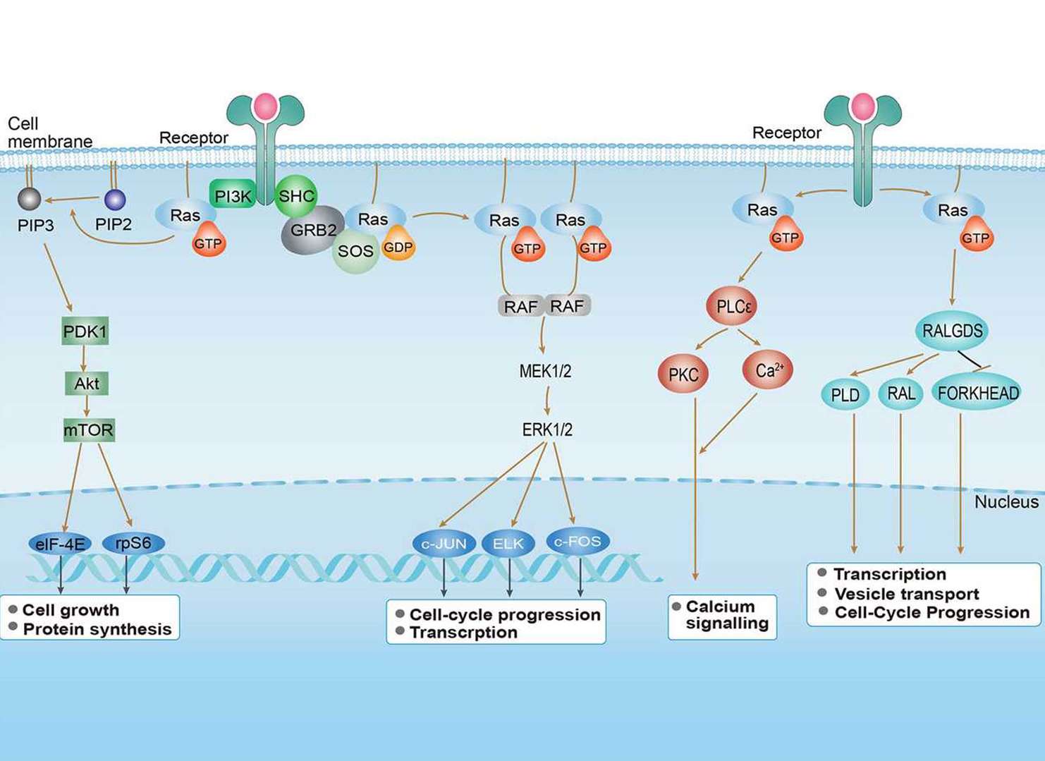 Ras Signaling Pathway
Related Diseases
 Breast Cancer
 Colorectal Cancer
 Endometrial Cancer
 Gastric Cancer
 Hepatocellular Carcinoma
 Non-small Cell Lung Cancer
 Prostate Cancer
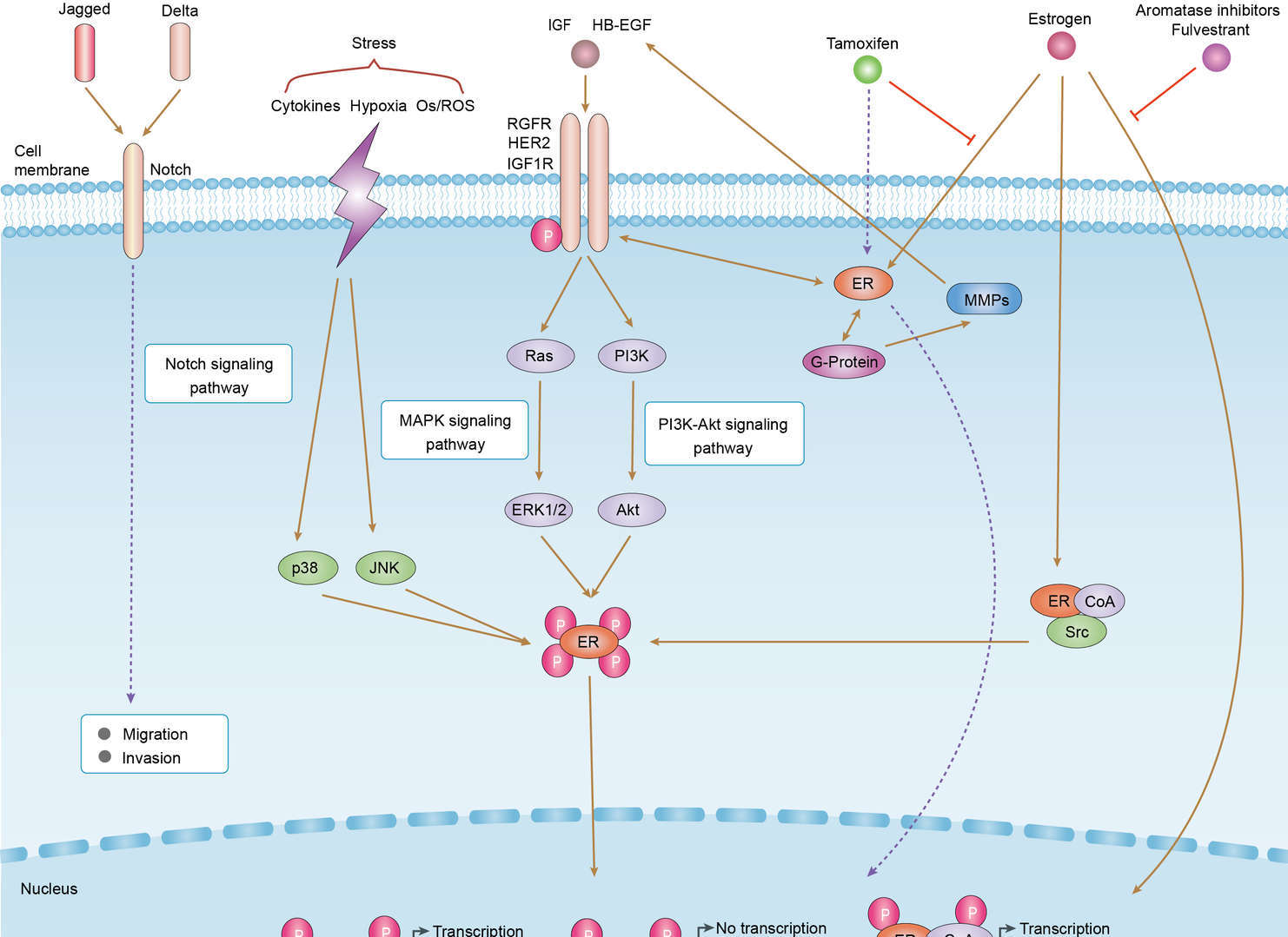 Endocrine Resistance
Downloadable Resources
Download resources about recombinant antibody development and antibody engineering to boost your research.
Product Notes
This is a product of Creative Biolabs' Hi-Affi™ recombinant antibody portfolio, which has several benefits including:
• Increased sensitivity
• Confirmed specificity
• High repeatability
• Excellent batch-to-batch consistency
• Sustainable supply
• Animal-free production
See more details about Hi-Affi™ recombinant antibody benefits.
Datasheet
MSDS
COA
Certificate of Analysis LookupTo download a Certificate of Analysis, please enter a lot number in the search box below. Note: Certificate of Analysis not available for kit components.
Lot Number:
Protocol & Troubleshooting
We have outlined the assay protocols, covering reagents, solutions, procedures, and troubleshooting tips for common issues in order to better assist clients in conducting experiments with our products. View the full list of Protocol & Troubleshooting.
See other products for "AKT1"
Select a product category from the dropdown menu below to view related products.
| CAT | Product Name | Application | Type |
|---|---|---|---|
| NAB-33-VHH | Recombinant Anti-human AKT1 VHH Single Domain Antibody | WB, IP, ChiP, Neut, ELISA | Llama VHH |
| CAT | Product Name | Application | Type |
|---|---|---|---|
| MOB-1676z | Mouse Anti-AKT1 Recombinant Antibody (clone 29A11) | WB, ELISA, IHC | Mouse IgG1 |
| CAT | Product Name | Application | Type |
|---|---|---|---|
| MOB-0566MZ | Mouse Anti-AKT1 Recombinant Antibody (clone 205B393) | WB | Mouse IgG1 |
| CAT | Product Name | Application | Type |
|---|---|---|---|
| BRD-0031MZ | Chicken Anti-AKT1 (ab1) Polyclonal IgY | WB | Chicken antibody |
| CAT | Product Name | Application | Type |
|---|---|---|---|
| BRD-0032MZ | Chicken Anti-AKT1 (ab2) Polyclonal IgY | Indirect ELISA, WB | Chicken antibody |
| CAT | Product Name | Application | Type |
|---|---|---|---|
| BRD-0683MZ | Chicken Anti-AKT1 Polyclonal IgY | WB | Chicken antibody |
| CAT | Product Name | Application | Type |
|---|---|---|---|
| MOR-0126 | Hi-Affi™ Rabbit Anti-AKT1 Recombinant Antibody (clone DS126AB) | WB | Rabbit IgG |
| CAT | Product Name | Application | Type |
|---|---|---|---|
| MOR-4668 | Hi-Affi™ Rabbit Anti-AKT1 Recombinant Antibody (clone TH182DS) | WB, IF, ICC, IHC-P, FC, ELISA | Rabbit IgG |
| CAT | Product Name | Application | Type |
|---|---|---|---|
| MOR-4669 | Hi-Affi™ Rabbit Anti-AKT1 Recombinant Antibody (clone TH183DS) | WB, IF, ICC, FC, ELISA | Rabbit IgG |
| CAT | Product Name | Application | Type |
|---|---|---|---|
| MOR-4670 | Hi-Affi™ Rabbit Anti-AKT1 Recombinant Antibody (clone TH184DS) | WB, IF, ICC, FC | Rabbit IgG |
| CAT | Product Name | Application | Type |
|---|---|---|---|
| MRO-0043-CN | Rabbit Anti-AKT1 Recombinant Antibody (clone CBACN-016) | WB, IF, IHC, IP, FC | Rabbit IgG |
| CAT | Product Name | Application | Type |
|---|---|---|---|
| MRO-0044-CN | Mouse Anti-AKT1 Recombinant Antibody (clone D9-9-C9) | WB, IF, IHC, FC | Mouse IgG2b |
| CAT | Product Name | Application | Type |
|---|---|---|---|
| MRO-2295-CN | Rabbit Anti-AKT1 Recombinant Antibody (clone CBACN-590) | WB, IF, IHC, IP | Rabbit IgG |
| CAT | Product Name | Application | Type |
|---|---|---|---|
| VS-0424-XY10 | AbPlus™ Anti-AKT1 Magnetic Beads (CBACN-016) | IP, Protein Purification |
| CAT | Product Name | Application | Type |
|---|---|---|---|
| VS-1024-XY15 | Rabbit Anti-NHP AKT1 Recombinant Antibody (VS-1024-XY15) | WB, IP | Rabbit IgG |
| CAT | Product Name | Application | Type |
|---|---|---|---|
| VS13-YC39 | CytoStream™ Mouse Anti-AKT1 Recombinant Antibody (VS13-YC39) | WB, ICC, IF, IHC-P, FC | Mouse IgG2b |
| CAT | Product Name | Application | Type |
|---|---|---|---|
| VS-0525-XY278 | Anti-AKT1 Immunohistochemistry Kit | IHC |
| CAT | Product Name | Application | Type |
|---|---|---|---|
| VS-0525-XY279 | Anti-Mouse AKT1 Immunohistochemistry Kit | IHC |
| CAT | Product Name | Application | Type |
|---|---|---|---|
| VS-0525-XY281 | Anti-Zebrafish AKT1 Immunohistochemistry Kit | IHC |
| CAT | Product Name | Application | Type |
|---|---|---|---|
| VS-0525-XY280 | Anti-Rat AKT1 Immunohistochemistry Kit | IHC |
Specific Inquiry

See Our Custom Production in Action


Popular Products

Application: WB, IF, IP, Neut, FuncS, ELISA, FC

Application: Neut, ELISA, IF, IP, FuncS, FC, IHC

Application: Neut, ELISA, IF, IP, FuncS, FC, ICC


Application: WB, IP, IF, FuncS, FC, Neut, ELISA

Application: Neut, ELISA, IF, IP, FuncS, FC, IHC

Application: ELISA, Neut, IF, IP, FC, FuncS

Application: FC, IHC, FuncS, Inhib, Cyt

Application: ELISA, FC, IP, FuncS, IF, Neut, ICC

Application: Neut, ELISA, IF, IP, FuncS, FC, ICC

Application: FuncS, IF, Neut, ELISA, FC, IP, WB
-2.png)
Application: ELISA, FC, IP, FuncS, IF, Neut, ICC

Application: Neut, ELISA, IF, IP, FuncS, FC, ICC

Application: ELISA, WB, BLI, SPR

Application: Block, Cyt, FuncS, Inhib

Application: ELISA, Neut, FuncS


For research use only. Not intended for any clinical use. No products from Creative Biolabs may be resold, modified for resale or used to manufacture commercial products without prior written approval from Creative Biolabs.
Send Inquiry
This site is protected by reCAPTCHA and the Google Privacy Policy and Terms of Service apply.