

Anti-ATP2C1 Immunohistochemistry Kit (CAT#: VS-0325-XY204)
This ATP2C1 IHC Kit comes with a comprehensive set of reagents. The user - friendly protocol is easy to follow, making it suitable for exploring ATP2C1's functions in calcium - dependent signaling pathways and its potential role in cellular processes such as exocytosis and cell - cell communication. This kit can be applied to paraffin sections and frozen sections.
Specific Inquiry
Subcellular Location
 Normal Tissue


 RNA Expression

Figure 1 IF staining of human cell line HaCaT
(Immunofluorescent staining of human cell line HaCaT shows localization to the Golgi apparatus.)
* Image credit: Human Protein Atlas v21.proteinatlas.org/images/69684/1346_A10_2_selected.jpg
(Immunofluorescent staining of human cell line HaCaT shows localization to the Golgi apparatus.)
* Image credit: Human Protein Atlas v21.proteinatlas.org/images/69684/1346_A10_2_selected.jpg
Figure 2 Cerebral cortex
(Endothelial cells Staining: Medium Intensity: Moderate Quantity:>75% Location: Cytoplasmic/ membranous Glial cells Staining: High Intensity: Strong Quantity:>75% Location: Cytoplasmic/ membranous Neuronal cells Staining: High Intensity: Strong Quantity:>75% Location: Cytoplasmic/ membranous nuclear Neuropil Staining: Low Intensity: Weak Quantity:>75% Location: Cytoplasmic/ membranous)
* Image credit: Human Protein Atlas v21.proteinatlas.org/images/35116/84724_B_8_5.jpg
Figure 3 Colon
(Endothelial cells Staining: Medium Intensity: Moderate Quantity:>75% Location: Cytoplasmic/ membranous Glandular cells Staining: High Intensity: Strong Quantity:>75% Location: Cytoplasmic/ membranous Peripheral nerve/ganglion Staining: Medium Intensity: Moderate Quantity:>75% Location: Cytoplasmic/ membranous)
* Image credit: Human Protein Atlas v21.proteinatlas.org/images/35116/84724_A_9_3.jpg
Figure 4 Liver
(Cholangiocytes Staining: Medium Intensity: Moderate Quantity:>75% Location: Cytoplasmic/ membranous Nuclear Hepatocytes Staining: High Intensity: Strong Quantity:>75% Location: Cytoplasmic/ membranous)
* Image credit: Human Protein Atlas v21.proteinatlas.org/images/35116/84724_A_8_4.jpg
(Cholangiocytes Staining: Medium Intensity: Moderate Quantity:>75% Location: Cytoplasmic/ membranous Nuclear Hepatocytes Staining: High Intensity: Strong Quantity:>75% Location: Cytoplasmic/ membranous)
* Image credit: Human Protein Atlas v21.proteinatlas.org/images/35116/84724_A_8_4.jpg
Figure 5 Kidney
(Cells in glomeruli Staining: Medium Intensity: Moderate Quantity: 75%-25% Location: Cytoplasmic/ membranous Cells in tubules Staining: High Intensity: Strong Quantity:>75% Location: Cytoplasmic/ membranous)
* Image credit: Human Protein Atlas v21.proteinatlas.org/images/35116/84724_A_9_5.jpg
(Cells in glomeruli Staining: Medium Intensity: Moderate Quantity: 75%-25% Location: Cytoplasmic/ membranous Cells in tubules Staining: High Intensity: Strong Quantity:>75% Location: Cytoplasmic/ membranous)
* Image credit: Human Protein Atlas v21.proteinatlas.org/images/35116/84724_A_9_5.jpg
Figure 6 Testis
(Leydig cells Staining: High Intensity: Strong Quantity:>75% Pachytene spermatocytes Staining: Medium Intensity: Moderate Quantity: 75%-25% Sertoli cells Staining: Medium Intensity: Moderate Quantity:>75% Spermatogonia cells Staining: High Intensity: Strong)
* Image credit: Human Protein Atlas v21.proteinatlas.org/images/35116/84724_A_6_6.jpg
Figure 7 Lymph node
(Germinal center cells Staining: High Intensity: Strong Quantity:>75% Location: Cytoplasmic/ membranous Non-germinal center cells Staining: High Intensity: Strong Quantity:>75% Location: Cytoplasmic/ membranous)
* Image credit: Human Protein Atlas v21.proteinatlas.org/images/35116/84724_A_7_8.jpg
(Germinal center cells Staining: High Intensity: Strong Quantity:>75% Location: Cytoplasmic/ membranous Non-germinal center cells Staining: High Intensity: Strong Quantity:>75% Location: Cytoplasmic/ membranous)
* Image credit: Human Protein Atlas v21.proteinatlas.org/images/35116/84724_A_7_8.jpg
Figure 8 RNA cell line category: Low cell line specificity
(Cell lines ordered by descending RNA expression order)
* Image credit: Human Protein Atlas v21.proteinatlas.org/ENSG00000017260-ATP2C1
(Cell lines ordered by descending RNA expression order)
* Image credit: Human Protein Atlas v21.proteinatlas.org/ENSG00000017260-ATP2C1
Specifications
- Application
- IHC
- Size
- 50 Tests
- Species Reactivity
- Human, Mouse, Rat
- Target
- ATP2C1
- Primary Antibody
- Rabbit Anti-ATP2C1 Antibody
- Secondary Antibody
- Goat anti-Rabbit Antibody, HRP
- Sample Type
- FFPE tissue; Frozen section tissue
- Kit Storage
- All reagents should be kept at 2-8°C. The kit remains stable for up to 6 months after arrival.
Related Resources
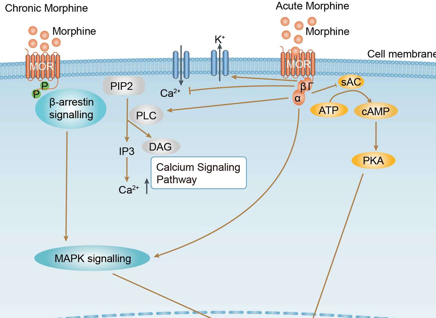 Morphine Addiction
Downloads
Download resources about recombinant antibody development and antibody engineering to boost your research.
See other products for "ATP2C1"
Select a product category from the dropdown menu below to view related products.
Please select product type
IgG Antibody Products
Non-Human Primate (NHP) Antibody Products
| CAT | Product Name | Application | Type |
|---|---|---|---|
| MOB-2938z | Mouse Anti-ATP2C1 Recombinant Antibody (clone 21G12) | WB, ELISA, IHC | Mouse IgG1 |
| MOB-0483MZ | Mouse Anti-ATP2C1 Recombinant Antibody (clone CBLS-047) | WB | Mouse IgG1 |
| ZG-0383C | Mouse Anti-ATP2C1 Recombinant Antibody (ZG-0383C) | WB, IHC, ELISA | Mouse IgG |
| VS3-XY103 | Mouse Anti-ATP2C1 Recombinant Antibody (clone 4G12) | ELISA, WB, IHC | Mouse IgG1 |
| VS7-HM172 | Mouse Anti-ATP2C1 Recombinant Antibody (clone CBL058HM) | WB, IHC-p, IF, ELISA | Mouse IgG |
Customer Reviews and Q&As

There are currently no Customer reviews or questions for VS-0325-XY204. Click the button above to contact us or submit your feedback about this product.
Popular products with customers


Application: FC, IP, ELISA, Neut, FuncS, IF, ICC

Application: IF, IP, Neut, FuncS, ELISA, FC, WB

Application: IP, IF, FuncS, FC, Neut, ELISA, IHC


Application: WB, ELISA, FC, IHC, IP

Application: ELISA, FC


Application: ELISA, FC, IF, IHC, WB


For Research Use Only. Not For Clinical Use.
For research use only. Not intended for any clinical use. No products from Creative Biolabs may be resold, modified for resale or used to manufacture commercial products without prior written approval from Creative Biolabs.
Send Inquiry
This site is protected by reCAPTCHA and the Google Privacy Policy and Terms of Service apply.