

Caspases
Product List
+ Filter
 Loading...
- AbPlus™ Anti-CASP7 Magnetic Beads (VS-0724-YC1356) (VS-0724-YC1356)
-
- Target: CASP7
- Target Species: Human
- Application: IP, Protein Purification
 Compare
- Rabbit Anti-CASP8 Recombinant Antibody (VS3-WK1583) (VS3-WK1583)
-
- Species Reactivity: Human, Mouse, Rat
- Type: Rabbit IgG
- Application: WB, ELISA
Compare
-
- Derivation: Mouse
- Species Reactivity: Human, Mouse, Rat
- Type: Mouse IgG
- Application: ELISA, WB, IHC
Compare
-
- Derivation: Mouse
- Species Reactivity: Human, Mouse, Rat
- Type: Mouse IgG
- Application: WB, IHC
Compare
- Rabbit Anti-CASP7 Recombinant Antibody (VS3-CJ1043) (VS3-CJ1043)
-
- Species Reactivity: Human
- Type: Rabbit IgG
- Application: WB, ICC, IF, IHC
Compare
-
- Species Reactivity: Human, Mouse, Rat
- Type: Rabbit IgG
- Application: WB, ICC, IF, IHC, IP, FC
Compare
- Recombinant Human Anti-CASP8 Soluble TCR (121) (KIH) (VS-0622-YF5432)
-
- Species Reactivity: Human
- Epitope: FPSDSWCYF
- MHC: HLA-B*3503
Compare
- Recombinant Human Anti-CASP8 Soluble TCR (121) (C-Cys) (VS-0622-YF2724)
-
- Species Reactivity: Human
- Epitope: FPSDSWCYF
- MHC: HLA-B*3503
Compare
- Rabbit Anti-CASP1 Polyclonal Antibody (MRO-1719-CN) (MRO-1719-CN)
-
- Species Reactivity: Human
- Type: Rabbit IgG
- Application: IF, IHC, FC
Compare
- Rabbit Anti-CASP1 Polyclonal Antibody (MRO-1718-CN) (MRO-1718-CN)
-
- Species Reactivity: Human
- Type: Rabbit IgG
- Application: WB, IHC
Compare
- Recombinant Human Anti-CASP8 Soluble TCR (clone 121) (scTCR-016CQ-S(P))
-
- Species Reactivity: Human
- Epitope: FPSDSWCYF
- MHC: HLA-B*3503
Compare
- Mouse Anti-CASP1 Recombinant Antibody (clone B0-6) (MRO-0236-CN)
-
- Species Reactivity: Human, Mouse, Rat
- Type: Mouse IgG1
- Application: WB, IF, IHC
Compare
- Rabbit Anti-CASP1 Recombinant Antibody (clone CBACN-086) (MRO-0237-CN)
-
- Species Reactivity: Human, Mouse, Rat
- Type: Rabbit IgG
- Application: WB, IHC, FC
Compare
- Rabbit Anti-CASP2 Recombinant Antibody (clone CBACN-089) (MRO-0240-CN)
-
- Species Reactivity: Human, Mouse, Rat
- Type: Rabbit IgG
- Application: WB, IF, IHC, FC
Compare
- Rabbit Anti-CASP8 Recombinant Antibody (clone CBACN-093) (MRO-0244-CN)
-
- Species Reactivity: Human
- Type: Rabbit IgG
- Application: WB, IF, IHC
Compare
- Rabbit Anti-CASP8 Recombinant Antibody (clone CBACN-094) (MRO-0245-CN)
-
- Species Reactivity: Human
- Type: Rabbit IgG
- Application: WB, IF, IHC
Compare
- Rabbit Anti-CASP7 Recombinant Antibody (clone CBACN-460) (MRO-1271-CN)
-
- Species Reactivity: Human
- Type: Rabbit IgG
- Application: WB, IF, IHC
Compare
- Mouse Anti-CASP1 Recombinant Antibody (clone EML275) (MOB-0323CT)
-
- Species Reactivity: Human
- Type: Mouse IgG2a
- Application: IHC-P, WB
Compare
- Mouse Anti-CASP2 Recombinant Antibody (clone 5j24) (MOB-0326CT)
-
- Species Reactivity: Human
- Type: Mouse IgG1
- Application: WB
Compare
- Mouse Anti-CASP8 Recombinant Antibody (clone 1H11) (MOB-0330CT)
-
- Species Reactivity: Human
- Type: Mouse IgG1
- Application: WB
Compare
- Mouse Anti-CASP7 Recombinant Antibody (clone 8E2) (MOB-2355CT)
-
- Species Reactivity: Human
- Type: Mouse IgG2b
- Application: IP, WB
Compare
-
- Derivation: Mouse
- Species Reactivity: Human
- Type: Mouse IgG1, κ
- Application: WB, IP, IF, ELISA
Compare
-
- Derivation: Mouse
- Species Reactivity: Human
- Type: Mouse IgG1
- Application: WB, ELISA, FC, IHC
Compare
-
- Derivation: Mouse
- Species Reactivity: Human
- Type: Mouse IgG1, κ
- Application: WB, IF, IHC, IP
Compare
- Recombinant Anti-Mouse Casp1 VHH Single Domain Antibody (NAB-209-sdAb)
-
- Species Reactivity: Mouse
- Type: Llama VHH
- Application: FA, FC, ICC, ELISA
Compare
-
- Derivation: Mouse
- Species Reactivity: Human, Mouse, Rat, Monkey
- Type: Mouse IgG
- Application: WB, IHC-p, IF, FCM, ELISA
Compare
- Anti-CASP7 Immunohistochemistry Kit (VS-0325-XY320)
-
- Species Reactivity: Human, Mouse, Rat
- Target: CASP7
- Application: IHC
Compare
- Anti-CASP8 Immunohistochemistry Kit (VS-0325-XY321)
-
- Species Reactivity: Human
- Target: CASP8
- Application: IHC
Compare
-
- Species Reactivity: Human, Mouse, Rat
- Type: Rabbit IgG
- Application: WB, ELISA, IHC-P, IHC-Fr, FC, ICC, IF
Compare
- Rabbit Anti-Casp12 Recombinant Antibody (VS13-YC125) (VS13-YC125)
-
- Species Reactivity: Human, Mouse, Rat
- Type: Rabbit IgG
- Application: WB, IHC-P
Compare
- Rabbit Anti-Casp7 Recombinant Antibody (VS13-YC131) (VS13-YC131)
-
- Species Reactivity: Mouse, Rat
- Type: Rabbit IgG
- Application: WB, IF, IP
Compare
- Rabbit Anti-CASP8 Recombinant Antibody (VS13-YC132) (VS13-YC132)
-
- Species Reactivity: Human
- Type: Rabbit IgG
- Application: WB, ICC, IF, IHC-P
Compare
- Anti-CASP1 Immunohistochemistry Kit (VS-0525-XY993)
-
- Species Reactivity: Human
- Target: CASP1
- Application: IHC
Compare
- Anti-Mouse CASP12 Immunohistochemistry Kit (VS-0525-XY995)
-
- Species Reactivity: Mouse, Rat
- Target: CASP12
- Application: IHC
Compare
- Anti-CASP14 Immunohistochemistry Kit (VS-0525-XY996)
-
- Species Reactivity: Human
- Target: CASP14
- Application: IHC
Compare
- Anti-Mouse CASP14 Immunohistochemistry Kit (VS-0525-XY997)
-
- Species Reactivity: Human, Mouse
- Target: CASP14
- Application: IHC
Compare
- Anti-CASP2 Immunohistochemistry Kit (VS-0525-XY998)
-
- Species Reactivity: Human
- Target: CASP2
- Application: IHC
Compare
- Anti-Mouse CASP2 Immunohistochemistry Kit (VS-0525-XY999)
-
- Species Reactivity: Human, Mouse, Rat, Canine, Monkey
- Target: CASP2
- Application: IHC
Compare
- Anti-Mouse CASP7 Immunohistochemistry Kit (VS-0525-XY1007)
-
- Species Reactivity: Human, Mouse, Rat, Porcine, Canine, Rabbit
- Target: CASP7
- Application: IHC
Compare
- Anti-Mouse CASP8 Immunohistochemistry Kit (VS-0525-XY1009)
-
- Species Reactivity: Mouse
- Target: CASP8
- Application: IHC
Compare
- Anti-Human CASP8 Immunohistochemistry Kit (VS-0525-XY1012)
-
- Species Reactivity: Human, Monkey
- Target: CASP8
- Application: IHC
Compare
- Anti-Rat CASP2 Immunohistochemistry Kit (VS-0525-XY1000)
-
- Species Reactivity: Human, Mouse, Rat
- Target: CASP2
- Application: IHC
Compare
- Anti-Human CASP7 Immunohistochemistry Kit (VS-0525-XY1006)
-
- Species Reactivity: Human
- Target: CASP7
- Application: IHC
Compare
- Anti-Rat CASP7 Immunohistochemistry Kit (VS-0525-XY1008)
-
- Species Reactivity: Mouse, Rat
- Target: CASP7
- Application: IHC
Compare
- Anti-Monkey CASP8 Immunohistochemistry Kit (VS-0525-XY1010)
-
- Species Reactivity: Human, Mouse, Rat, Monkey
- Target: CASP8
- Application: IHC
Compare
- Rabbit Anti-CASP8 Polyclonal Antibody (VS-0725-YC72)
-
- Species Reactivity: Human, Mouse
- Type: Rabbit IgG
- Application: ICC, IF, WB
Compare
- Recombinant Anti-human CASP7 Intrabody [(D-Arg)9] (IAB-B072(A))
-
- Species Reactivity: human
- Type: scFv-(D-Arg)9
- Application: WB, ICC, FuncS
Compare
- Recombinant Anti-human CASP7 Intrabody [+36 GFP] (IAB-B072(G))
-
- Species Reactivity: human
- Type: scFv-(+36GFP)
- Application: WB, ICC, Neut, FuncS
Compare
- Recombinant Anti-human CASP7 Intrabody [Tat] (IAB-B072(T))
-
- Species Reactivity: human
- Type: scFv-Tat
- Application: IF, FC, FuncS
Compare
- Chicken Anti-Caspase 8 Polyclonal IgY (BRD-0082MZ)
-
- Species Reactivity: Human
- Type: Chicken antibody
- Application: WB
Compare
-
- Derivation: Phage display library screening
- Species Reactivity: Human
- Type: Rabbit IgG
- Application: WB
Compare
-
- Derivation: Phage display library screening
- Species Reactivity: Human
- Type: Rabbit IgG
- Application: ELISA
Compare
-
- Derivation: Phage display library screening
- Species Reactivity: Human
- Type: Rabbit IgG
- Application: IP, WB
Compare
-
- Derivation: Phage display library screening
- Species Reactivity: Human
- Type: Rabbit IgG
- Application: IF, IHC-P, IP, WB
Compare
View More Products
Our customer service representatives are available 24 hours a day, from Monday to Sunday. Contact Us
Can't find the products you're looking for? Try to filter in the left sidebar.Filter By Tag
Representative Targets of Caspases Full List of Targets of Caspases Tested Data-Supported Products Targeting Caspases
Caspases, essential in mediating cellular apoptosis and inflammation, are pivotal to cellular integrity and organismal health. Structurally, they are synthesized as inactive proenzymes (zymogens) comprising a pro-domain and a large and small subunit. Upon receiving apoptotic signals, caspases are cleaved at specific aspartic acid residues, a process that separates the pro-domain from the subunits, which then form the active enzyme. This activation mechanism is finely tuned and can initiate through intrinsic (mitochondrial) or extrinsic (death receptor) pathways.
In the intrinsic pathway, internal stressors like DNA damage or oxidative stress lead to mitochondrial outer membrane permeabilization (MOMP), resulting in the release of cytochrome c. Cytochrome c then binds with Apaf-1 and ATP to form the apoptosome, a platform that facilitates the auto-activation of caspase-9. Caspase-9, an initiator caspase, then cleaves and activates executioner caspases such as caspase-3 and -7, which dismantle the cell by cleaving key structural and regulatory proteins.
Conversely, the extrinsic pathway begins at the cell surface through the engagement of death receptors like Fas or TNF receptor with their respective ligands. This interaction recruits adaptor proteins such as FADD or TRADD, which in turn bind and activate caspase-8 or -10. These caspases can directly activate executioner caspases or cleave the BH3-only protein Bid, linking extrinsic signaling to the intrinsic pathway and enhancing the apoptotic response.
Regulation of caspases is crucial to prevent inappropriate activation and cellular damage, involving inhibitors such as the IAP family, which directly bind and inhibit caspases, and FLIP, which competes with caspase-8 for binding to FADD, thus blocking the extrinsic pathway. Dysregulation in caspase activity is implicated in numerous diseases; for instance, excessive caspase activation can result in excessive cell death, contributing to diseases like Alzheimer's and Huntington's, while reduced activity can lead to cancer due to inadequate apoptosis.
Therapeutically, caspases present a dual target. Inhibitors are researched for conditions marked by excessive apoptosis, while activators are investigated for their potential to kill cancer cells. Research continues to explore caspases' nuanced roles in cell death and survival, aiming to harness their mechanisms for targeted disease intervention, demonstrating their significance beyond mere executioners of cell death to being central figures in the maintenance of cellular and organismal health.
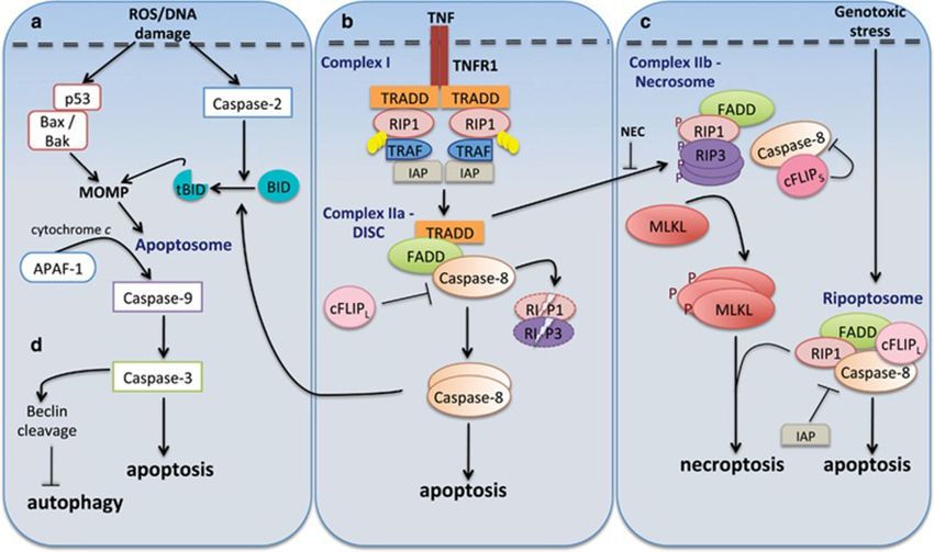 Figure 1 Caspases in cell death pathways. (Sahoo, 2023)
Representative Targets of Caspases
CASP3
CASP3, or Caspase-3, is a critical executioner caspase in the process of apoptosis, the programmed cell death mechanism that is essential for development, maintenance of tissue homeostasis, and elimination of damaged or diseased cells. Caspase-3 is synthesized as an inactive proenzyme that is activated through proteolytic cleavage by initiator caspases such as Caspase-8 and Caspase-9, typically in response to pro-apoptotic signals including those from the intrinsic (mitochondrial) and extrinsic (death receptor) pathways. Once activated, Caspase-3 cleaves various cellular substrates, leading to the characteristic morphological and biochemical features of apoptosis. These substrates include structural proteins in the cytoskeleton, such as actin and lamin, as well as key proteins involved in DNA repair and cell cycle regulation. The enzymatic activity of Caspase-3 results in cellular changes such as DNA fragmentation, degradation of cytoskeletal and nuclear proteins, formation of apoptotic bodies, and eventually, the phagocytosis of cell debris by neighboring cells or immune cells. Dysregulation of Caspase-3 activity is implicated in numerous diseases. Insufficient Caspase-3 activation can contribute to cancer progression by allowing abnormal cells to escape programmed cell death, whereas excessive activation of Caspase-3 is associated with conditions characterized by excessive cell death, such as Alzheimer's disease, stroke, and myocardial infarction. In these diseases, unintended activation of apoptosis can lead to tissue damage and functional decline.
Recommended Mouse Anti-CASP3 mAb (CAT#: ZG-0016J)
 Figure 2 Mouse Anti-CASP3 Recombinant Antibody (clone 5E1) in IHC. Immunohistochemical analysis of paraffin-embedded Rat-lung tissue. 1.Active Caspase-3 Monoclonal Antibody was diluted at 1:200 (4°C, overnight). 2, Sodium citrate pH 6.0 was used for Antibody retrieval(>98°C, 20min). 3, Secondary Antibody was diluted at 1:200 (room temperature, 30min). Negative control was used by secondary Antibody only.
Recommended Rabbit Anti-CASP3 mAb (CAT#: ZG-0012U)
 Figure 3 Rabbit Anti-CASP3 Antibody (ZG-0012U) in IHC. IHC image of ZG-0012U diluted at 1:100 and staining in paraffin-embedded human breast cancer performed on a Leica BondTM system. After dewaxing and hydration, antigen retrieval was mediated by high pressure in a citrate buffer (pH 6.0). Section was blocked with 10% normal goat serum 30min at RT. Then primary antibody (1% BSA) was incubated at 4°C overnight. The primary is detected by a Goat anti-rabbit IgG polymer labeled by HRP and visualized using 0.05% DAB.
Recommended Rabbit Anti-CASP3 mAb (CAT#: ZG-0580J)
 Figure 4 Rabbit Anti-CASP3 Antibody (ZG-0580J) in IF. Immunofluorescence staining of Hela Cells with ZG-0580J at 1:50, counter-stained with DAPI. The cells were fixed in 4% formaldehyde, permeated by 0.2% TritonX-100, and blocked in 10% normal Goat Serum. The cells were then incubated with the antibody overnight at 4°C. Nuclear DNA was labeled in blue with DAPI. The secondary antibody was FITC-conjugated Goat Anti-Rabbit IgG (H+L).
CASP8
CASP8, or Caspase-8, is a crucial initiator caspase in the apoptosis signaling pathways, primarily involved in the extrinsic pathway triggered by death receptors. This enzyme plays a pivotal role in cellular homeostasis and immune function by initiating the cascade of caspase activation that leads to programmed cell death. Caspase-8 is activated upon the engagement of death receptors on the cell surface, such as Fas (CD95) and the TNF (tumor necrosis factor) receptors. Following receptor activation by their respective ligands, such as Fas ligand (FasL) and TNF-related apoptosis-inducing ligand (TRAIL), the adaptor molecule FADD (Fas-associated death domain) recruits pro-caspase-8 to the death-inducing signaling complex (DISC). At the DISC, pro-caspase-8 undergoes dimerization and auto-cleavage to become active. Once activated, Caspase-8 can cleave and activate downstream effector caspases (such as Caspase-3 and Caspase-7), which execute the cell by degrading various cellular components. Additionally, Caspase-8 plays a role in the activation of the intrinsic (mitochondrial) apoptosis pathway through the cleavage of the BID protein, a Bcl-2 family member, which in turn promotes mitochondrial outer membrane permeabilization and cytochrome c release, further amplifying the apoptotic signals. The function of Caspase-8 is not limited to apoptosis; it is also involved in various non-apoptotic processes, including cell differentiation, proliferation, and the immune response, particularly in T cells and macrophages. Dysregulation of Caspase-8 activity is associated with various diseases, including cancer, where its deficiency can contribute to resistance to apoptosis, aiding in the survival and proliferation of cancer cells. Conversely, excessive activation of Caspase-8 is implicated in autoimmune and inflammatory conditions.
Recommended Mouse Anti-CASP8 mAb (CAT#: ZG-0566J)
 Figure 5 Mouse Anti-CASP8 Recombinant Antibody (clone 2G12) in IHC. Immunohistochemical analysis of paraffin-embedded Human-Tonsil tissue. 1.Caspase-8 Monoclonal Antibody was diluted at 1:200 (4°C, overnight). 2, Sodium citrate pH 6.0 was used for Antibody retrieval(>98°C, 20min). 3, Secondary Antibody was diluted at 1:200 (room temperature, 30min). Negative control was used by secondary Antibody only.
CASP9
CASP9, or Caspase-9, is another initiator caspase in the intrinsic apoptosis pathway. Caspase-9 plays a pivotal role in the cascade of signaling events that lead to apoptosis, which is essential for removing damaged or diseased cells and maintaining tissue homeostasis. Caspase-9 is activated in response to pro-apoptotic signals that result in mitochondrial outer membrane permeabilization and the release of cytochrome c into the cytosol. Once released, cytochrome c forms a complex with Apaf-1 (apoptotic protease activating factor 1) and dATP, which then recruits pro-caspase-9. This complex, known as the apoptosome, facilitates the auto-cleavage and activation of Caspase-9. Activated Caspase-9 then cleaves and activates downstream effector caspases, such as Caspase-3 and Caspase-7, which carry out the execution phase of apoptosis, leading to cellular disassembly. Dysregulation of Caspase-9 activity is implicated in a range of diseases. Overactivation can contribute to excessive cell death, which is detrimental in diseases such as Alzheimer's, stroke, and myocardial infarction, where it can exacerbate tissue damage. Conversely, reduced activation of Caspase-9 can lead to impaired apoptosis, allowing the survival and proliferation of potentially cancerous cells, thus contributing to the development and progression of cancer.
Recommended Rabbit Anti-CASP9 mAb (CAT#: ZG-0581J)
 Figure 6 Rabbit Anti-CASP9 Antibody (ZG-0581J) in IF. Immunofluorescence staining of HepG2 cells with ZG-0581J at 1:60, counter-stained with DAPI. The cells were fixed in 4% formaldehyde, permeabilized using 0.2% Triton X-100 and blocked in 10% normal Goat Serum. The cells were then incubated with the antibody overnight at 4°C. The secondary antibody was Alexa Fluor 488-congugated Goat Anti-Rabbit IgG (H+L).
Recommended Rabbit Anti-CASP9 mAb (CAT#: VS3-FY197)
 Figure 7 Recombinant Rabbit Anti-Casp9 Antibody (clone R04-8I4) in WB. Western blot analysis of Caspase9 in C6 lysates using Caspase9 Antibody.
Recommended Mouse Anti-CASP9 mAb (CAT#: ZG-0365F)
 Figure 8 Mouse Anti-CASP9 Recombinant Antibody (ZG-0365F) in WB. Western blot detection of CHO-K1 cell lysate (B) and CHO K1 transfected with Caspase-9 (A) cell lysate was performed using Caspase-9 mouse mAb (1:1000 dilution). Predicted band size: 49/37KDa. Observed band size: 49/37KDa.
Full List of Targets of Caspases
| Biomarker | Alternative Names | Gene ID | UniProt ID | Roles |
| Casp1 | Caspase 1; Caspase 1, Apoptosis-Related Cysteine Peptidase; Interleukin 1, Beta, Convertase; IL-1 Beta-Converting Enzyme; EC 3.4.22.36; Caspase-1; IL1BC; P45; ICE; Caspase 1, Apoptosis-Related Cysteine Peptidase (Interleukin 1, Beta, Convertase); Caspase 1, Apoptosis-Related Cysteine Protease (Interleukin 1, Beta, Convertase) | 12362 | P29452 | This gene encodes a protein which is a member of the cysteine-aspartic acid protease (caspase) family. Sequential activation of caspases plays a central role in the execution-phase of cell apoptosis. Caspases exist as inactive proenzymes which undergo proteolytic processing at conserved aspartic residues to produce 2 subunits, large and small, that dimerize to form the active enzyme. This gene was identified by its ability to proteolytically cleave and activate the inactive precursor of interleukin-1, a cytokine involved in the processes such as inflammation, septic shock, and wound healing. This gene has been shown to induce cell apoptosis and may function in various developmental stages. Studies of a similar gene in mouse suggest a role in the pathogenesis of Huntington disease. Alternative splicing results in transcript variants encoding distinct isoforms. |
| CASP10 | Caspase 10; FAS-Associated Death Domain Protein Interleukin-1B-Converting Enzyme 2; Caspase 10, Apoptosis-Related Cysteine Protease; ICE-Like Apoptotic Protease 4; CASP-10; FLICE2; MCH4; Caspase 10, Apoptosis-Related Cysteine Peptidase; Caspase 10 Apoptosis-Related Cysteine Peptidase | 843 | Q92851 | This gene encodes a protein which is a member of the cysteine-aspartic acid protease (caspase) family. Sequential activation of caspases plays a central role in the execution-phase of cell apoptosis. Caspases exist as inactive proenzymes which undergo proteolytic processing at conserved aspartic residues to produce two subunits, large and small, that dimerize to form the active enzyme. This protein cleaves and activates caspases 3 and 7, and the protein itself is processed by caspase 8. Mutations in this gene are associated with type IIA autoimmune lymphoproliferative syndrome, non-Hodgkin lymphoma and gastric cancer. Alternatively spliced transcript variants encoding different isoforms have been described for this gene. |
| CASP14 | ARCI12 | 23581 | P31944 | This gene encodes a member of the cysteine-aspartic acid protease (caspase) family. Sequential activation of caspases plays a central role in the execution-phase of cell apoptosis. Caspases exist as inactive proenzymes which undergo proteolytic processing at conserved aspartic residues to produce two subunits, large and small, that dimerize to form the active enzyme. This caspase has been shown to be processed and activated by caspase 8 and caspase 10 in vitro, and by anti-Fas agonist antibody or TNF-related apoptosis inducing ligand in vivo. The expression and processing of this caspase may be involved in keratinocyte terminal differentiation, which is important for the formation of the skin barrier. |
| CASP2 | ICH1; NEDD2; CASP-2; NEDD-2; PPP1R57 | 835 | P42575 | This gene encodes a member of the cysteine-aspartic acid protease (caspase) family. Caspases mediate cellular apoptosis through the proteolytic cleavage of specific protein substrates. The encoded protein may function in stress-induced cell death pathways, cell cycle maintenance, and the suppression of tumorigenesis. Increased expression of this gene may play a role in neurodegenerative disorders including Alzheimer's disease, Huntington's disease and temporal lobe epilepsy. Alternatively spliced transcript variants encoding multiple isoforms have been observed for this gene. |
| CASP3 | Caspase 3; Caspase 3, Apoptosis-Related Cysteine Peptidase; Caspase 3, Apoptosis-Related Cysteine Protease; SREBP Cleavage Activity 1; Cysteine Protease CPP32; Protein Yama; EC 3.4.22.56; Apopain; CASP-3 | 836 | P42574 | The protein encoded by this gene is a cysteine-aspartic acid protease that plays a central role in the execution-phase of cell apoptosis. The encoded protein cleaves and inactivates poly(ADP-ribose) polymerase while it cleaves and activates sterol regulatory element binding proteins as well as caspases 6, 7, and 9. This protein itself is processed by caspases 8, 9, and 10. It is the predominant caspase involved in the cleavage of amyloid-beta 4A precursor protein, which is associated with neuronal death in Alzheimer's disease. |
| Casp4 | CASP4; caspase 4, apoptosis-related cysteine peptidase; TX; ICH-2; Mih1/TX; ICEREL-II; ICE(rel)II; caspase-4; CASP-4; ICE(rel)-II; protease TX; protease ICH-2; apoptotic cysteine protease Mih1/TX; caspase 4, apoptosis-related cysteine protease; | 12363 | P70343 | This gene encodes a protein that is a member of the cysteine-aspartic acid protease (caspase) family. Sequential activation of caspases plays a central role in the execution-phase of cell apoptosis. Caspases exist as inactive proenzymes composed of a prodomain and a large and small protease subunit. Activation of caspases requires proteolytic processing at conserved internal aspartic residues to generate a heterodimeric enzyme consisting of the large and small subunits. This caspase is able to cleave and activate its own precursor protein, as well as caspase 1 precursor. When overexpressed, this gene induces cell apoptosis. Alternative splicing results in transcript variants encoding distinct isoforms. [provided by RefSeq, Jul 2008] |
| CASP5 | ICH-3; ICEREL-III; ICE(rel)III | 838 | P51878 | This gene encodes a member of the cysteine-aspartic acid protease (caspase) family. Sequential activation of caspases plays a central role in the execution-phase of cell apoptosis. Caspases exist as inactive proenzymes which undergo proteolytic processing at conserved aspartic residues to produce two subunits, large and small, that dimerize to form the active enzyme. Overexpression of the active form of this enzyme induces apoptosis in fibroblasts. Max, a central component of the Myc/Max/Mad transcription regulation network important for cell growth, differentiation, and apoptosis, is cleaved by this protein; this process requires Fas-mediated dephosphorylation of Max. The expression of this gene is regulated by interferon-gamma and lipopolysaccharide. Alternatively spliced transcript variants have been identified for this gene. |
| CASP6 | MCH2; CSP-6; caspase-6 | 839 | P55212 | This gene encodes a member of the cysteine-aspartic acid protease (caspase) family of enzymes. Sequential activation of caspases plays a central role in the execution-phase of cell apoptosis. Caspases exist as inactive proenzymes which undergo proteolytic processing at conserved aspartic acid residues to produce two subunits, large and small, that dimerize to form the active enzyme. This protein is processed by caspases 7, 8 and 10, and is thought to function as a downstream enzyme in the caspase activation cascade. Alternative splicing of this gene results in multiple transcript variants that encode different isoforms. |
| CASP7 | Caspase 7; Caspase 7, Apoptosis-Related Cysteine Peptidase; Caspase 7, Apoptosis-Related Cysteine Protease; ICE-Like Apoptotic Protease 3; ICE-LAP3; CASP-7; CMH-1; MCH3 | 840 | P55210 | This gene encodes a member of the cysteine-aspartic acid protease (caspase) family. Sequential activation of caspases plays a central role in the execution-phase of cell apoptosis. Caspases exist as inactive proenzymes which undergo proteolytic processing at conserved aspartic residues to produce two subunits, large and small, that dimerize to form the active enzyme. The precursor of the encoded protein is cleaved by caspase 3 and 10, is activated upon cell death stimuli and induces apoptosis. Alternatively spliced transcript variants encoding multiple isoforms have been observed for this gene. |
| CASP8 | Caspase 8; Caspase 8, Apoptosis-Related Cysteine Peptidase; Caspase 8, Apoptosis-Related Cysteine Protease; MORT1-Associated Ced-3 Homolog; ICE-Like Apoptotic Protease 5; Apoptotic Cysteine Protease; Apoptotic Protease Mch-5; FADD-Like ICE; Casp-8; FLICE; CAP4 | 841 | Q14790 | This gene encodes a member of the cysteine-aspartic acid protease (caspase) family. Sequential activation of caspases plays a central role in the execution-phase of cell apoptosis. Caspases exist as inactive proenzymes composed of a prodomain, a large protease subunit, and a small protease subunit. Activation of caspases requires proteolytic processing at conserved internal aspartic residues to generate a heterodimeric enzyme consisting of the large and small subunits. This protein is involved in the programmed cell death induced by Fas and various apoptotic stimuli. The N-terminal FADD-like death effector domain of this protein suggests that it may interact with Fas-interacting protein FADD. This protein was detected in the insoluble fraction of the affected brain region from Huntington disease patients but not in those from normal controls, which implicated the role in neurodegenerative diseases. Many alternatively spliced transcript variants encoding different isoforms have been described, although not all variants have had their full-length sequences determined. |
| CASP9 | Caspase 9; Caspase 9, Apoptosis-Related Cysteine Peptidase; Protein Phosphatase 1, Regulatory Subunit 56; ICE-Like Apoptotic Protease 6; ICE-LAP6; APAF-3; MCH6; Caspase 9, Apoptosis-Related Cysteine Protease; Apoptotic Protease Activating Factor 3; | 842 | P55211 | This gene encodes a member of the cysteine-aspartic acid protease (caspase) family. Sequential activation of caspases plays a central role in the execution-phase of cell apoptosis. Caspases exist as inactive proenzymes which undergo proteolytic processing at conserved aspartic residues to produce two subunits, large and small, that dimerize to form the active enzyme. This protein can undergo autoproteolytic processing and activation by the apoptosome, a protein complex of cytochrome c and the apoptotic peptidase activating factor 1; this step is thought to be one of the earliest in the caspase activation cascade. This protein is thought to play a central role in apoptosis and to be a tumor suppressor. Alternative splicing results in multiple transcript variants. |
Tested Data-Supported Products Targeting Caspases


Reference
- Sahoo, Gayatri, et al. "A review on caspases: key regulators of biological activities and apoptosis." Molecular neurobiology 60.10 (2023): 5805-5837.
For Research Use Only. Not For Clinical Use.