

Anti-CD19 (huB4)-SPDB-DM4 ADC
CAT#: ADC-020LCT
This ADC product is composed of an anti-CD19 antibody (clone huB4) conjuagated via a SPDB linker to DM4 (huB4-SPDB-DM4). It has demonstrated a response in NHL treatment by a MOA (Mechanism of Action) of Depolymerize Microtubules.
Gene Expression

Normal Tissue
Figure 1 Colon
Mucosal lymphoid cells
Staining:Medium
Intensity: Strong
Quantity: <25%
* Image credit: Image credit: Human Protein Atlas https://v21.proteinatlas.org/images/16110/36120_A_7_3.jpg

Normal Tissue
Figure 2 Testis
Leydig cells
Staining:Medium
Intensity: Moderate
Quantity:>75%
Location: Cytoplasmic/membranous
* Image credit: Image credit: Human Protein Atlas https://v21.proteinatlas.org/images/16110/36120_A_5_6.jpg

Normal Tissue
Figure 3 Appendix
Germinal center cells
Staining:Medium
Intensity: Moderate
Quantity: 75%-25%
Non-germinal center cells
Staining:High
Intensity: Strong
Quantity:>75%
* Image credit: Image credit: Human Protein Atlas https://v21.proteinatlas.org/images/16110/36120_A_2_2.jpg

Normal Tissue
Figure 4 Lymph node
Germinal center cells
Staining:High
Intensity: Strong
Quantity:>75%
Location: Cytoplasmic/membranous
Non-germinal center cells
Staining:High
Intensity: Strong
Quantity: 75%-25%
Location: Cytoplasmic/membranous
* Image credit: Image credit: Human Protein Atlas https://v21.proteinatlas.org/images/16110/36120_A_9_8.jpg

RNA Expression
Figure 5 RNA cell line category: Cell line enhanced (Daudi, REH, U-698)
Cell lines ordered by descending RNA expression order.
* Image credit: Image credit: Human Protein Atlas https://v21.proteinatlas.org/ENSG00000177455-CD19
❮


 ❯
❯
Specifications
- Antibody Overview
- Humanized IgG1 monoclonal Antibody, huB4 (Anti-CD19)
- Clone
- huB4
- Antibody Isotype
- IgG1κ
- Linker
- SPDB (N-succinimidyl 4-(2-pyridyldithio)butyrate)
- Linker Class/Description
- Class: Chemically cleavable Linker-Disulfide Linker
Description: Disulfide Linkers are extensively exploited as a chemically labile linkage. Since the release of disulfide-linked drugs requires a cytoplasmic thiol cofactor, such as glutathione (GSH). Disulfides maintain stable at physiological pH and only when ADCs are internalized inside cells, the cytosol provides reducing environment including intracellular enzyme protein disulfide isomerase, or similar enzymes, drugs can be released.
- Drug
- DM4 (N2'-Deacetyl-N2'-(4-mercapto-4-methyl-1-oxopentyl)maytansine)
- Drug Class/Description
- Class: Maytansinoid
Description: Maytansinoids are a group of cytotoxins structurally similar to rifamycin, geldanamycin, and ansatrienin. The eponymous natural cytotoxic agent maytansine is a 19-member lactam (ansa
macrolide) structure originally isolated from the Ethiopian shrub Maytenus ovatus. Maytansinoids can bind to tubulin at or near the vinblastine-binding site, which interfere the formation of microtubules and depolymerize already formed microtubules, inducing mitotic arrest in the intoxicated cells
Target
- Introduction
- Lymphocytes proliferate and differentiate in response to various concentrations of different antigens. The ability of the B cell to respond in a specific, yet sensitive manner to the various antigens is achieved with the use of low-affinity antigen receptors. This gene encodes a cell surface molecule which assembles with the antigen receptor of B lymphocytes in order to decrease the threshold for antigen receptor-dependent stimulation. [provided by RefSeq, Jul 2008]
- Alternative Names
- CD19; CD19 molecule; B4; CVID3; B-lymphocyte antigen CD19; differentiation antigen CD19; T-cell surface antigen Leu-12; B-lymphocyte surface antigen B4;
- Gene ID
- 930
- UniProt ID
- P15391
REVIEWS AND Q&AS
CITATIONS
RESOURCES
DOWNLOADS
RELATED PRODUCTS
Inquiry
Navs
Customer Review

There are currently no Customer reviews or questions for ADC-020LCT. Click the button above to contact us or submit your feedback about this product.
Submit Your Publication
Published with our product? Submit your paper and receive a 10% discount on your next order! Share your research to earn exclusive rewards.
Related Diseases
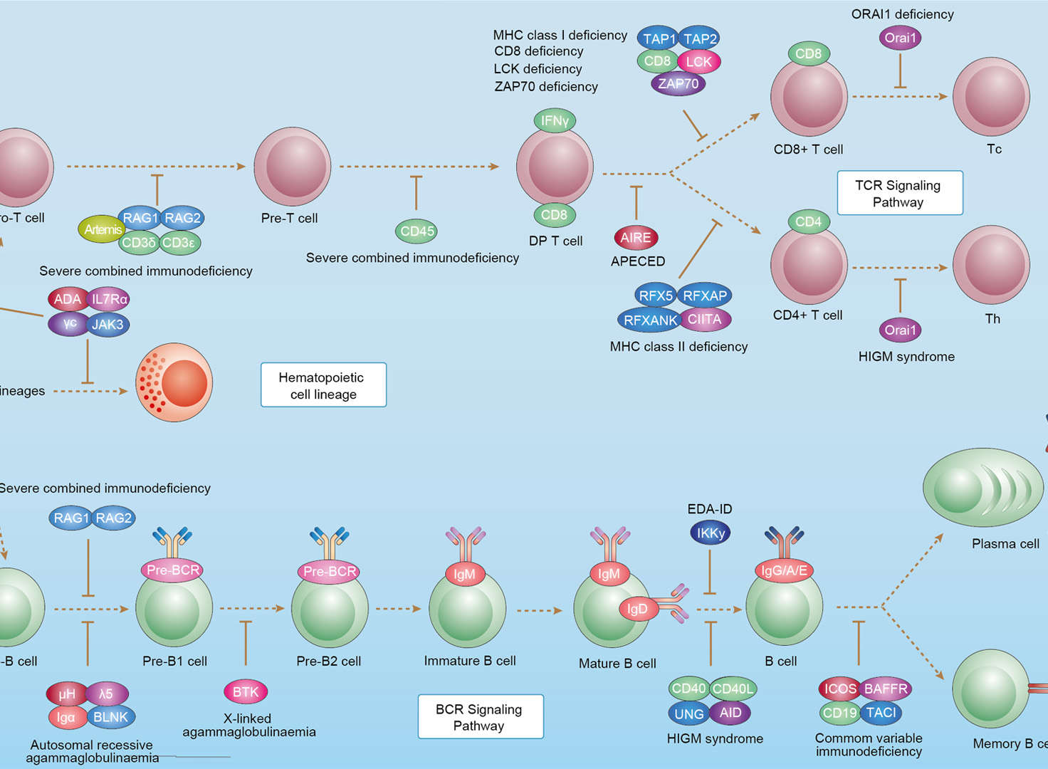 Primary Immunodeficiency
Downloadable Resources
Download resources about recombinant antibody development and antibody engineering to boost your research.
Datasheet
MSDS
COA
Certificate of Analysis LookupTo download a Certificate of Analysis, please enter a lot number in the search box below. Note: Certificate of Analysis not available for kit components.
Lot Number:
See other products for "Clone huB4"
- CAT
- Product Name
See other products for "CD19"
Select a product category from the dropdown menu below to view related products.
| CAT | Product Name | Application | Type |
|---|---|---|---|
| MOB-1087z | Mouse Anti-CD19 Recombinant Antibody (clone 32B9) | WB, FC, ICC, IF, FuncS | Mouse IgG1 |
| CAT | Product Name | Application | Type |
|---|---|---|---|
| TAB-174 | Mouse Anti-CD19 Recombinant Antibody (clone Taplitumomab) | ELISA, IP, FC, FuncS, Neut, IF, ICC | Mouse IgG1, κ |
| CAT | Product Name | Application | Type |
|---|---|---|---|
| TAB-H17 | Anti-Human CD19 Recombinant Antibody (TAB-H17) | IF, IP, Neut, FuncS, ELISA, FC, WB | IgG1 - kappa |
| CAT | Product Name | Application | Type |
|---|---|---|---|
| TAB-891 | Humanized Anti-CD19 Recombinant Antibody (TAB-891) | ELISA, FC, IP, FuncS, IF, Neut, ICC | Humanized IgG1, κ |
| CAT | Product Name | Application | Type |
|---|---|---|---|
| AGTO-G076E | Anti-CD19 immunotoxin HD37 (IgG)-PE | Cytotoxicity assay, Function study |
| CAT | Product Name | Application | Type |
|---|---|---|---|
| AGTO-G076D | Anti-CD19 immunotoxin HD37 (IgG)-DT | Cytotoxicity assay, Function study |
| CAT | Product Name | Application | Type |
|---|---|---|---|
| AGTO-G076R | Anti-CD19 immunotoxin HD37 (IgG)-RTA | Cytotoxicity assay, Function study |
| CAT | Product Name | Application | Type |
|---|---|---|---|
| AGTO-G076S | Anti-CD19 immunotoxin HD37 (IgG)-Sap | Cytotoxicity assay, Function study |
| CAT | Product Name | Application | Type |
|---|---|---|---|
| AGTO-G076P | Anti-CD19 immunotoxin HD37 (IgG)-PAP | Cytotoxicity assay, Function study |
| CAT | Product Name | Application | Type |
|---|---|---|---|
| TAB-108CL | Anti-Human CD19 Recombinant Antibody (TAB-108CL) | FC, ADCC, FuncS | Antibody |
| CAT | Product Name | Application | Type |
|---|---|---|---|
| TAB-1611CL | Mouse Anti-CD19 Recombinant Antibody (TAB-1611CL) | Depletion, FuncS | Mouse IgG2a, κ |
| CAT | Product Name | Application | Type |
|---|---|---|---|
| TAB-1612CL | Human Anti-CD19 Recombinant Antibody (TAB-1612CL) | Depletion, FuncS | Humanized IgG1 |
| CAT | Product Name | Application | Type |
|---|---|---|---|
| TAB-1613CL | Human Anti-CD19 Recombinant Antibody (TAB-1613CL) | Cyt, FC, FuncS, Inhib | Chimeric (mouse/human) IgG1 |
| CAT | Product Name | Application | Type |
|---|---|---|---|
| TAB-1614CL | Human Anti-CD19 Recombinant Antibody (TAB-1614CL) | FuncS | Chimeric (mouse/human) IgG |
| CAT | Product Name | Application | Type |
|---|---|---|---|
| TAB-1616CL | Human Anti-CD19 Recombinant Antibody (TAB-1616CL) | ELISA, WB | Human IgG |
| CAT | Product Name | Application | Type |
|---|---|---|---|
| TAB-1617CL | Human Anti-CD19 Recombinant Antibody (TAB-1617CL) | ELISA, WB | Human IgG |
| CAT | Product Name | Application | Type |
|---|---|---|---|
| TAB-1618CL | Human Anti-CD19 Recombinant Antibody (TAB-1618CL) | ELISA, WB | Human IgG |
| CAT | Product Name | Application | Type |
|---|---|---|---|
| TAB-1619CL | Human Anti-CD19 Recombinant Antibody (TAB-1619CL) | ELISA, WB | Human IgG |
| CAT | Product Name | Application | Type |
|---|---|---|---|
| TAB-1620CL | Anti-Human CD19 Recombinant Antibody (HD37) | FuncS |
| CAT | Product Name | Application | Type |
|---|---|---|---|
| TAB-1621CL | Anti-Human CD19 Recombinant Antibody (cHD37) | ADCC, FuncS | Chimeric antibody (mouse/human) |
| CAT | Product Name | Application | Type |
|---|---|---|---|
| TAB-1622CL | Mouse Anti-CD19 Recombinant Antibody (TAB-1622CL) | ELISA, FC, Apop, ADCC, FuncS | Mouse IgG |
| CAT | Product Name | Application | Type |
|---|---|---|---|
| TAB-1623CL | Mouse Anti-CD19 Recombinant Antibody (TAB-1623CL) | ELISA, FC | Mouse IgG |
| CAT | Product Name | Application | Type |
|---|---|---|---|
| TAB-1625CL | Anti-Human CD19 Recombinant Antibody (396) | Depletion, ELISA | Humanized antibody |
| CAT | Product Name | Application | Type |
|---|---|---|---|
| TAB-1612CL-S(P) | Human Anti-CD19 Recombinant Antibody; scFv Fragment (TAB-1612CL-S(P)) | Depletion, FuncS | Humanized scFv |
| CAT | Product Name | Application | Type |
|---|---|---|---|
| TAB-1613CL-S(P) | Mouse Anti-CD19 Recombinant Antibody; scFv Fragment (TAB-1613CL-S(P)) | Depletion, FuncS | Mouse scFv |
| CAT | Product Name | Application | Type |
|---|---|---|---|
| TAB-1614CL-S(P) | Mouse Anti-CD19 Recombinant Antibody; scFv Fragment (TAB-1614CL-S(P)) | FuncS | Mouse scFv |
| CAT | Product Name | Application | Type |
|---|---|---|---|
| Gly-061LC | Recombinant Anti-Human CD19 Antibody (Fc glycosylation/Low fucosylated) | ELISA, FC | Chimeric antibody (mouse/human) |
| Gly-061LC-1 | Recombinant Anti-Human CD19 Antibody (Fc glycosylation/Low fucosylated) | ELISA, FC | Chimeric antibody (mouse/human) |
| CAT | Product Name | Application | Type |
|---|---|---|---|
| Gly-105LC | Recombinant Anti-Human CD19 Antibody (Fc glycosylation) | ELISA | Mouse antibody |
| CAT | Product Name | Application | Type |
|---|---|---|---|
| Gly-141LC | Recombinant Anti-Human CD19 Antibody (Fc glycosylation/Non fucosylated) | ELISA | Humanized antibody |
| Gly-141LC-1 | Recombinant Anti-Human CD19 Antibody (Fc glycosylation/Non fucosylated) | ELISA | Humanized antibody |
| CAT | Product Name | Application | Type |
|---|---|---|---|
| MOB-0278MZ | Mouse Anti-CD19 Recombinant Antibody (clone ME-CD20) | IHC | Mouse IgG1 |
| CAT | Product Name | Application | Type |
|---|---|---|---|
| BRD-0100MZ | Chicken Anti-CD19 Polyclonal IgY | WB | Chicken antibody |
| CAT | Product Name | Application | Type |
|---|---|---|---|
| NEUT-286CQ | Mouse Anti-CD19 Recombinant Antibody (clone SJ25-C1) | Neut, FC, IHC, IHC-Fr, IP | Mouse IgG1 |
| CAT | Product Name | Application | Type |
|---|---|---|---|
| NEUT-287CQ | Mouse Anti-CD19 Recombinant Antibody (clone HIB19) | FC, CyTOF, IHC, Block | Mouse IgG1, κ |
| CAT | Product Name | Application | Type |
|---|---|---|---|
| NEUT-288CQ | Rat Anti-CD19 Recombinant Antibody (clone 1D3) | Depletion, Block, FC | Rat IgG2a |
| CAT | Product Name | Application | Type |
|---|---|---|---|
| MOR-0538 | Hi-Affi™ Rabbit Anti-CD19 Recombinant Antibody (clone DS538AB) | IHC-P | Rabbit IgG |
| CAT | Product Name | Application | Type |
|---|---|---|---|
| HPAB-0264-CN | Human Anti-CD19 Recombinant Antibody (HPAB-0264-CN) | ELISA, FC | Humanized IgG1, κ |
| CAT | Product Name | Application | Type |
|---|---|---|---|
| HPAB-0265-CN | Human Anti-CD19 Recombinant Antibody (HPAB-0265-CN) | ELISA, FC | Humanized IgG1, κ |
| CAT | Product Name | Application | Type |
|---|---|---|---|
| HPAB-0266-CN | Human Anti-CD19 Recombinant Antibody (HPAB-0266-CN) | ELISA, FC | Humanized IgG |
| CAT | Product Name | Application | Type |
|---|---|---|---|
| HPAB-0264-CN-S(P) | Human Anti-CD19 Recombinant Antibody; scFv Fragment (HPAB-0264-CN-S(P)) | ELISA, FC | Humanized scFv |
| CAT | Product Name | Application | Type |
|---|---|---|---|
| HPAB-0265-CN-S(P) | Human Anti-CD19 Recombinant Antibody; scFv Fragment (HPAB-0265-CN-S(P)) | ELISA, FC | Humanized scFv |
| CAT | Product Name | Application | Type |
|---|---|---|---|
| HPAB-0266-CN-S(P) | Human Anti-CD19 Recombinant Antibody; scFv Fragment (HPAB-0266-CN-S(P)) | ELISA, FC | Humanized scFv |
| CAT | Product Name | Application | Type |
|---|---|---|---|
| HPAB-0267-CN-S(P) | Human Anti-CD19 Recombinant Antibody; scFv Fragment (HPAB-0267-CN-S(P)) | ELISA, FC | Humanized scFv |
| CAT | Product Name | Application | Type |
|---|---|---|---|
| HPAB-0268-CN-S(P) | Human Anti-CD19 Recombinant Antibody; scFv Fragment (HPAB-0268-CN-S(P)) | ELISA, FC | Human scFv |
| CAT | Product Name | Application | Type |
|---|---|---|---|
| HPAB-0264-CN-F(E) | Human Anti-CD19 Recombinant Antibody; Fab Fragment (HPAB-0264-CN-F(E)) | ELISA, FC | Humanized Fab |
| CAT | Product Name | Application | Type |
|---|---|---|---|
| HPAB-0265-CN-F(E) | Human Anti-CD19 Recombinant Antibody; Fab Fragment (HPAB-0265-CN-F(E)) | ELISA, FC | Humanized Fab |
| CAT | Product Name | Application | Type |
|---|---|---|---|
| HPAB-0266-CN-F(E) | Human Anti-CD19 Recombinant Antibody; Fab Fragment (HPAB-0266-CN-F(E)) | ELISA, FC | Humanized Fab |
| CAT | Product Name | Application | Type |
|---|---|---|---|
| HPAB-0267-CN-F(E) | Human Anti-CD19 Recombinant Antibody; Fab Fragment (HPAB-0267-CN-F(E)) | ELISA, FC | Humanized Fab |
| CAT | Product Name | Application | Type |
|---|---|---|---|
| HPAB-0268-CN-F(E) | Human Anti-CD19 Recombinant Antibody; Fab Fragment (HPAB-0268-CN-F(E)) | ELISA, FC | Human Fab |
| CAT | Product Name | Application | Type |
|---|---|---|---|
| AFC-TAB-174 | Afuco™ Anti-CD19 ADCC Recombinant Antibody, ADCC Enhanced (AFC-TAB-174) | ELISA, IP, FC, FuncS, Neut, IF | ADCC enhanced antibody |
| CAT | Product Name | Application | Type |
|---|---|---|---|
| VS-0724-YC640 | AbPlus™ Anti-CD19 Magnetic Beads (VS-0724-YC640) | IP, Protein Purification |
| CAT | Product Name | Application | Type |
|---|---|---|---|
| VS-1024-XY83 | Mouse Anti-NHP CD19 Recombinant Antibody (clone PDR134) | WB, IF, FC | Mouse IgM, kappa |
| CAT | Product Name | Application | Type |
|---|---|---|---|
| VS-0225-XY20 | CytoStream™ Mouse Anti-CD19 Recombinant Antibody (VS-0225-XY20) | FC | Mouse IgG1, kappa |
| CAT | Product Name | Application | Type |
|---|---|---|---|
| VS-0325-FY59 | Human Anti-CD19 (clone 2A3) scFv-Fc Chimera | ELISA | Human IgG1, scFv-Fc |
| CAT | Product Name | Application | Type |
|---|---|---|---|
| VS13-YC154 | CytoStream™ Rabbit Anti-CD19 Recombinant Antibody (VS13-YC154) | WB, ICC, IF, IHC-P, FC | Rabbit IgG |
| CAT | Product Name | Application | Type |
|---|---|---|---|
| VS-0425-YC75 | Recombinant Anti-CD19 Vesicular Antibody, EV Displayed (VS-0425-YC75) | ELISA, FC, Neut, Cell-uptake |
| CAT | Product Name | Application | Type |
|---|---|---|---|
| VS-0525-XY1127 | Anti-Rat CD19 Immunohistochemistry Kit | IHC |
| CAT | Product Name | Application | Type |
|---|---|---|---|
| VS-0525-XY1128 | Anti-Mouse CD19 Immunohistochemistry Kit | IHC |
| CAT | Product Name | Application | Type |
|---|---|---|---|
| VS-0825-YC38 | SmartAb™ Recombinant Anti-CD19 pH-dependent Antibody (Clone Taplitumomab) | ELISA, IP, FC Neut, IF, ICC | Mouse IgG1 kappa |
| CAT | Product Name | Application | Type |
|---|---|---|---|
| VS-1025-YC139 | Anti-CD19 Antibody Prodrug, Protease Activated (VS-1025-YC139) | ISZ, Cyt, FuncS |
| CAT | Product Name | Application | Type |
|---|---|---|---|
| VS-1125-XY1 | Mouse Anti-CD19 Recombinant Antibody (VS-1125-XY1) | WB, IHC | Mouse IgG2b |
| CAT | Product Name | Application | Type |
|---|---|---|---|
| VS-1125-XY2 | Mouse Anti-CD19 Recombinant Antibody (VS-1125-XY2) | IHC, ICC, IF, FC | Mouse IgG1, kappa |
| CAT | Product Name | Application | Type |
|---|---|---|---|
| VS-1125-XY3 | Mouse Anti-CD19 Recombinant Antibody (VS-1125-XY3) | WB, IHC | Mouse IgG1, kappa |
| CAT | Product Name | Application | Type |
|---|---|---|---|
| VS-1125-XY4 | Mouse Anti-CD19 Recombinant Antibody (VS-1125-XY4) | WB, IHC, FC | Mouse IgG1 |
| CAT | Product Name | Application | Type |
|---|---|---|---|
| VS-1125-XY5 | Rabbit Anti-CD19 Recombinant Antibody (VS-1125-XY5) | WB, FC | Rabbit IgG |
Specific Inquiry
See Our Custom Production in Action


Popular Products


Application: ELISA, FC, IP, FuncS, IF, Neut, ICC

Application: ELISA, IP, FC, FuncS, Neut, IF, IHC

Application: FuncS, IF, Neut, ELISA, FC, IP, IHC

Application: WB, IP, IF, FuncS, FC, Neut, ELISA

Application: ELISA, FC, IP, FuncS, IF, Neut, ICC

Application: IF, IP, Neut, FuncS, ELISA, FC, WB

Application: IF, IP, Neut, FuncS, ELISA, FC, WB

Application: FC, IP, ELISA, Neut, FuncS, IF, ICC

Application: Neut, ELISA, IF, IP, FuncS, FC, ICC

Application: IF, IP, Neut, FuncS, ELISA, FC, ICC

Application: IF, IP, Neut, FuncS, ELISA, FC, ICC

Application: Neut, ELISA, IF, IP, FuncS, FC, ICC

Application: WB, FuncS, IF, Neut, ELISA, FC, IP

Application: FC, IP, ELISA, Neut, FuncS, IF, IHC


For research use only. Not intended for any clinical use. No products from Creative Biolabs may be resold, modified for resale or used to manufacture commercial products without prior written approval from Creative Biolabs.
Send Inquiry
This site is protected by reCAPTCHA and the Google Privacy Policy and Terms of Service apply.