

Anti-Mouse RFXANK Immunohistochemistry Kit (CAT#: VS-0525-XY6089)
The RFXANK IHC Kit provides a complete set of reagents for detecting RFXANK in Human, Mouse, and Rat tissue samples. This kit offers an easy and efficient staining process suitable for both paraffin-embedded and frozen sections. With demonstrated cross-species reactivity, it supports research on immune function, transcriptional regulation, and related biological processes across various models.
Specific Inquiry
Subcellular Location
 Normal Tissue


 RNA Expression

Figure 1 IF staining of human cell line MCF7
(Immunofluorescent staining of human cell line MCF7 shows localization to nucleoplasm.)
* Image credit: Human Protein Atlas v21.proteinatlas.org/images/49157/if_selected.jpg
(Immunofluorescent staining of human cell line MCF7 shows localization to nucleoplasm.)
* Image credit: Human Protein Atlas v21.proteinatlas.org/images/49157/if_selected.jpg
Figure 2 Cerebral cortex
(Endothelial cells Staining: Low Intensity: Weak Quantity: 75%-25% Location: Nuclear Glial cells Staining: Medium Intensity: Moderate Quantity: 75%-25% Location: nuclear Neuronal cells Staining: Medium Intensity: Moderate Quantity:>75% Location: Cytoplasmic/ membranous nuclear Neuropil Staining: Low Intensity: Weak Quantity:>75% Location: Cytoplasmic/ membranous)
* Image credit: Human Protein Atlas v21.proteinatlas.org/images/49157/111833_B_8_5.jpg
Figure 3 Colon
(Endothelial cells Staining: Medium Intensity: Moderate Quantity:>75% Location: Cytoplasmic/ membranous Nuclear Glandular cells Staining: Medium Intensity: Moderate Quantity:>75% Location: Cytoplasmic/ membranous nuclear Peripheral nerve/ganglion Staining: Medium Intensity: Moderate Quantity:>75% Location: Cytoplasmic/ membranous nuclear)
* Image credit: Human Protein Atlas v21.proteinatlas.org/images/49157/111833_A_7_3.jpg
Figure 4 Liver
(Cholangiocytes Staining: Low Intensity: Weak Quantity: 75%-25% Location: Nuclear Hepatocytes Staining: Medium Intensity: Moderate Quantity: 75%-25% Location: Cytoplasmic/ membranous nuclear)
* Image credit: Human Protein Atlas v21.proteinatlas.org/images/49157/111833_A_9_4.jpg
(Cholangiocytes Staining: Low Intensity: Weak Quantity: 75%-25% Location: Nuclear Hepatocytes Staining: Medium Intensity: Moderate Quantity: 75%-25% Location: Cytoplasmic/ membranous nuclear)
* Image credit: Human Protein Atlas v21.proteinatlas.org/images/49157/111833_A_9_4.jpg
Figure 5 Kidney
(Cells in glomeruli Staining: High Intensity: Strong Quantity: 75%-25% Location: nuclear Cells in tubules Staining: High Intensity: Strong Quantity: 75%-25% Location: Cytoplasmic/ membranous nuclear)
* Image credit: Human Protein Atlas v21.proteinatlas.org/images/49157/111833_A_7_5.jpg
(Cells in glomeruli Staining: High Intensity: Strong Quantity: 75%-25% Location: nuclear Cells in tubules Staining: High Intensity: Strong Quantity: 75%-25% Location: Cytoplasmic/ membranous nuclear)
* Image credit: Human Protein Atlas v21.proteinatlas.org/images/49157/111833_A_7_5.jpg
Figure 6 Testis
(Cells in seminiferous ducts Staining: Medium Intensity: Moderate Quantity:>75% Location: Cytoplasmic/ membranous nuclear Leydig cells Staining: Medium Intensity: Moderate Quantity:>75% Location: Cytoplasmic/ membranous nuclear)
* Image credit: Human Protein Atlas v21.proteinatlas.org/images/49157/111833_A_4_6.jpg
Figure 7 Lymph node
(Germinal center cells Staining: Medium Intensity: Moderate Quantity:>75% Location: Nuclear Non-germinal center cells Staining: Medium Intensity: Moderate Quantity: 75%-25% Location: Nuclear)
* Image credit: Human Protein Atlas v21.proteinatlas.org/images/49157/111833_A_9_8.jpg
(Germinal center cells Staining: Medium Intensity: Moderate Quantity:>75% Location: Nuclear Non-germinal center cells Staining: Medium Intensity: Moderate Quantity: 75%-25% Location: Nuclear)
* Image credit: Human Protein Atlas v21.proteinatlas.org/images/49157/111833_A_9_8.jpg
Figure 8 RNA cell line category: Low cell line specificity
(Cell lines ordered by descending RNA expression order)
* Image credit: Human Protein Atlas v21.proteinatlas.org/ENSG00000064490-RFXANK
(Cell lines ordered by descending RNA expression order)
* Image credit: Human Protein Atlas v21.proteinatlas.org/ENSG00000064490-RFXANK
Specifications
- Application
- IHC
- Size
- 50 Tests
- Species Reactivity
- Human, Mouse, Rat
- Target
- RFXANK
- Primary Antibody
- Mouse Anti-RFXANK Antibody
- Secondary Antibody
- Goat anti-Mouse Antibody, HRP
- Sample Type
- FFPE tissue; Frozen section tissue
- Kit Storage
- All reagents should be kept at 2-8°C. The kit remains stable for up to 6 months after arrival.
Related Resources
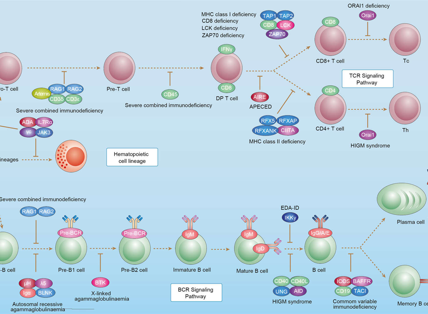 Primary Immunodeficiency
Downloads
Download resources about recombinant antibody development and antibody engineering to boost your research.
See other products for "RFXANK"
Select a product category from the dropdown menu below to view related products.
Please select product type
IgG Antibody Products
Antibody Magnetic Beads
IHC Kit and Antibody Products: Precision Tools for Immunohistochemistry
| CAT | Product Name | Application | Type |
|---|---|---|---|
| MOB-1553z | Mouse Anti-RFXANK Recombinant Antibody (clone 8H10) | WB, ICC, IF, IHC | Mouse IgG2b |
Customer Reviews and Q&As

There are currently no Customer reviews or questions for VS-0525-XY6089. Click the button above to contact us or submit your feedback about this product.
Popular products with customers

Application: FC, IP, ELISA, Neut, FuncS, IF, IHC

Application: ELISA, IP, FC, FuncS, Neut, IF, ICC

Application: IF, IP, Neut, FuncS, ELISA, FC, WB

Application: IF, IP, Neut, FuncS, ELISA, FC, ICC

Application: IF, IP, Neut, FuncS, ELISA, FC, ICC

Application: Neut, ELISA, IF, IP, FuncS, FC, ICC

Application: FuncS, IF, Neut, ELISA, FC, IP, WB

-2.png)
Application: IB, ELISA, FC, FuncS

Application: ELISA, FC, WB, FuncS


Application: Neut, ELISA, FuncS

Application: Neut, FC, IHC-Fr, IP, BA

Application: ELISA, IHC, FC, IP, IF, Inhib

Application: ELISA, WB, FC, IHC, IP


Application: IF, IP, Neut, FuncS, ELISA, FC


For Research Use Only. Not For Clinical Use.
Send Inquiry
This site is protected by reCAPTCHA and the Google Privacy Policy and Terms of Service apply.