

AbPlus™ Anti-RFXANK Magnetic Beads (VS-0724-YC554) (CAT#: VS-0724-YC554)
The AbPlus Anti-RFXANK Magnetic Beads (VS-0724-YC554) is an innovative affinity resin which is bound with anti-RFXANK specific antibody. The beads were designed for small-scale affinity purification and immunoprecipitation (IP) of RFXANK protein under native and denaturing conditions.
Specific Inquiry
Subcellular Location
 Normal Tissue


 RNA Expression

Figure 1 IF staining of human cell line MCF7
(Immunofluorescent staining of human cell line MCF7 shows localization to nucleoplasm.)
* Image credit: Human Protein Atlas v21.proteinatlas.org/images/49157/if_selected.jpg
(Immunofluorescent staining of human cell line MCF7 shows localization to nucleoplasm.)
* Image credit: Human Protein Atlas v21.proteinatlas.org/images/49157/if_selected.jpg
Figure 2 Cerebral cortex
(Endothelial cells Staining: Low Intensity: Weak Quantity: 75%-25% Location: Nuclear Glial cells Staining: Medium Intensity: Moderate Quantity: 75%-25% Location: nuclear Neuronal cells Staining: Medium Intensity: Moderate Quantity:>75% Location: Cytoplasmic/ membranous nuclear Neuropil Staining: Low Intensity: Weak Quantity:>75% Location: Cytoplasmic/ membranous)
* Image credit: Human Protein Atlas v21.proteinatlas.org/images/49157/111833_B_8_5.jpg
Figure 3 Colon
(Endothelial cells Staining: Medium Intensity: Moderate Quantity:>75% Location: Cytoplasmic/ membranous Nuclear Glandular cells Staining: Medium Intensity: Moderate Quantity:>75% Location: Cytoplasmic/ membranous nuclear Peripheral nerve/ganglion Staining: Medium Intensity: Moderate Quantity:>75% Location: Cytoplasmic/ membranous nuclear)
* Image credit: Human Protein Atlas v21.proteinatlas.org/images/49157/111833_A_7_3.jpg
Figure 4 Liver
(Cholangiocytes Staining: Low Intensity: Weak Quantity: 75%-25% Location: Nuclear Hepatocytes Staining: Medium Intensity: Moderate Quantity: 75%-25% Location: Cytoplasmic/ membranous nuclear)
* Image credit: Human Protein Atlas v21.proteinatlas.org/images/49157/111833_A_9_4.jpg
(Cholangiocytes Staining: Low Intensity: Weak Quantity: 75%-25% Location: Nuclear Hepatocytes Staining: Medium Intensity: Moderate Quantity: 75%-25% Location: Cytoplasmic/ membranous nuclear)
* Image credit: Human Protein Atlas v21.proteinatlas.org/images/49157/111833_A_9_4.jpg
Figure 5 Kidney
(Cells in glomeruli Staining: High Intensity: Strong Quantity: 75%-25% Location: nuclear Cells in tubules Staining: High Intensity: Strong Quantity: 75%-25% Location: Cytoplasmic/ membranous nuclear)
* Image credit: Human Protein Atlas v21.proteinatlas.org/images/49157/111833_A_7_5.jpg
(Cells in glomeruli Staining: High Intensity: Strong Quantity: 75%-25% Location: nuclear Cells in tubules Staining: High Intensity: Strong Quantity: 75%-25% Location: Cytoplasmic/ membranous nuclear)
* Image credit: Human Protein Atlas v21.proteinatlas.org/images/49157/111833_A_7_5.jpg
Figure 6 Testis
(Cells in seminiferous ducts Staining: Medium Intensity: Moderate Quantity:>75% Location: Cytoplasmic/ membranous nuclear Leydig cells Staining: Medium Intensity: Moderate Quantity:>75% Location: Cytoplasmic/ membranous nuclear)
* Image credit: Human Protein Atlas v21.proteinatlas.org/images/49157/111833_A_4_6.jpg
Figure 7 Lymph node
(Germinal center cells Staining: Medium Intensity: Moderate Quantity:>75% Location: Nuclear Non-germinal center cells Staining: Medium Intensity: Moderate Quantity: 75%-25% Location: Nuclear)
* Image credit: Human Protein Atlas v21.proteinatlas.org/images/49157/111833_A_9_8.jpg
(Germinal center cells Staining: Medium Intensity: Moderate Quantity:>75% Location: Nuclear Non-germinal center cells Staining: Medium Intensity: Moderate Quantity: 75%-25% Location: Nuclear)
* Image credit: Human Protein Atlas v21.proteinatlas.org/images/49157/111833_A_9_8.jpg
Figure 8 RNA cell line category: Low cell line specificity
(Cell lines ordered by descending RNA expression order)
* Image credit: Human Protein Atlas v21.proteinatlas.org/ENSG00000064490-RFXANK
(Cell lines ordered by descending RNA expression order)
* Image credit: Human Protein Atlas v21.proteinatlas.org/ENSG00000064490-RFXANK
Specifications
- Applications
- Immunoprecipitation, Protein Purification
- Matrix
- Magnetic bead
- Bead Ligand
- Anti-RFXANK specific antibody
- Target
- RFXANK
- Immunogen
- Recombinant human RFXANK fragment derived from E. coli.
- Target Species
- Human
- Bead Capacity
- 40 mg/mL
- Bead size
- 25 μm
- Format
- Suspension
- Concentration
- 2 mg/mL
- Buffer
- PBS, pH 7.4
- Preservative
- 0.1% Sodium azide
- Storage
- Stored at 4°C, and is stable for up to 2 years. Do not centrifuge, dry or freeze the magnetic beads.
Applications
- Application Notes
- The beads are in suspension and will settle upon storage. Prior to use, mix the vial gently (do not vortex) to ensure delivery of proper bead volume.
Target
- Introduction
- Major histocompatibility (MHC) class II molecules are transmembrane proteins that have a central role in development and control of the immune system. The protein encoded by this gene, along with regulatory factor X-associated protein and regulatory factor-5, forms a complex that binds to the X box motif of certain MHC class II gene promoters and activates their transcription. Once bound to the promoter, this complex associates with the non-DNA-binding factor MHC class II transactivator, which controls the cell type specificity and inducibility of MHC class II gene expression. This protein contains ankyrin repeats involved in protein-protein interactions. Mutations in this gene have been linked to bare lymphocyte syndrome type II, complementation group B. Multiple alternatively spliced transcript variants encoding different isoforms have been described for this gene. [provided by RefSeq, Jul 2013]
- Alternative Names
- Regulatory Factor X Associated Ankyrin Containing Protein; Regulatory Factor X Subunit B; Ankyrin Repeat-Containing Regulatory Factor X-Associated Protein; Regulatory Factor X-Associated Ankyrin-Containing Protein; Ankyrin Repeat Family A Protein 1; DNA-Binding Protein RFXANK; RFX-Bdelta4;
- Gene ID
- 8625
- UniProt ID
- O14593
Related Resources
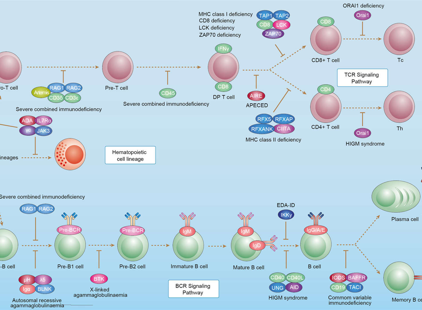 Primary Immunodeficiency
Downloads
Download resources about recombinant antibody development and antibody engineering to boost your research.
See other products for "RFXANK"
Select a product category from the dropdown menu below to view related products.
Please select product type
IgG Antibodies
IHC Kits & Antibodies
| CAT | Product Name | Application | Type |
|---|---|---|---|
| MOB-1553z | Mouse Anti-RFXANK Recombinant Antibody (clone 8H10) | WB, ICC, IF, IHC | Mouse IgG2b |
Customer Reviews and Q&As

There are currently no Customer reviews or questions for VS-0724-YC554. Click the button above to contact us or submit your feedback about this product.
Popular products with customers

Application: IP, IF, FuncS, FC, Neut, ELISA, IHC

Application: ELISA, FC, IP, FuncS, IF, Neut, ICC

Application: Neut, ELISA, IF, IP, FuncS, FC, ICC

Application: ELISA, Neut, IF, IP, FC, FuncS

Application: Inhib, Cyt

Application: Neut, ELISA, IF, IP, FuncS, FC, ICC

Application: FC, IP, ELISA, Neut, FuncS, IF, ICC


Application: ELISA, IHC, FC, IP, IF, Inhib


Application: Neut, IHC, Activ, FuncS, IF, ELISA
-4.jpg)


For Research Use Only. Not For Clinical Use.
Send Inquiry
This site is protected by reCAPTCHA and the Google Privacy Policy and Terms of Service apply.