

Anti-HER2 (clone hu4D5Fabv8)-MC-vc-PAB-MMAF ADC
CAT#: ADC-183CL
This ADC product is composed of an anti-HER2 antibody (clone hu4D5Fabv8) conjugated via MC-vc-PAB linker to MMAF(ABP-hu4D5Fabv8-(HC A121C)-MC-val-cit-PAB-MMAF). It has demonstrated a response in treatment by a MOA (Mechanism of Action) of microtubules depolymerizing.
Gene Expression

Subcellular Location
Figure 1 IF staining of human cell line U-251 MG
Immunofluorescent staining of human cell line U-251 MG shows localization to plasma membrane.
* Image credit: Image credit: Human Protein Atlas v21.proteinatlas.org/images/1060/169_D12_1_selected.jpg

Subcellular Location
Figure 2 IF staining of human cell line A-431
Immunofluorescent staining of human cell line A-431 shows localization to plasma membrane.
* Image credit: Image credit: Human Protein Atlas v21.proteinatlas.org/images/1338/if_selected.jpg

Normal Tissue
Figure 3 High expression in esophagus
* Image credit: Image credit: Human Protein Atlas v21.proteinatlas.org/images/1383/4682_B_5_1_val_selected.jpg

Normal Tissue
Figure 4 IHC staining of human rectum
Immunohistochemical staining of human rectum shows weak cytoplasmic positivity in glandular cells.
* Image credit: Image credit: Human Protein Atlas v21.proteinatlas.org/images/43/ihc_selected.jpg

Normal Tissue
Figure 5 IHC staining of human urinary bladder
Immunohistochemical staining of human urinary bladder shows moderate cytoplasmic and membranous positivity in urothelial cells.
* Image credit: Image credit: Human Protein Atlas v21.proteinatlas.org/images/20416/ihc_selected.jpg

Normal Tissue
Figure 6 Kidney
Collecting ducts Staining: Low Intensity: Moderate Quantity: <25% Distal tubules Staining: Low Intensity: Moderate Quantity: <25%
* Image credit: Image credit: Human Protein Atlas v21.proteinatlas.org/images/43/1054_A_7_5.jpg

Normal Tissue
Figure 7 Breast
Glandular cells Staining: Medium Intensity: Moderate Quantity: 75%-25% Location: Cytoplasmic/ membranous Myoepithelial cells Staining: Medium Intensity: Moderate Quantity: 75%-25% Location: Cytoplasmic/ membranous
* Image credit: Image credit: Human Protein Atlas v21.proteinatlas.org/images/20416/45831_B_3_4.jpg

RNA Expression
Figure 8 RNA cell line category: Group enriched (OE19, SK-BR-3)
Cell lines ordered by descending RNA expression order
* Image credit: Image credit: Human Protein Atlas v21.proteinatlas.org/ENSG00000141736-ERBB2
❮


 ❯
❯
Specifications
- Antibody Overview
- Anti-HER2 hu4D5Fabv8 Fab fragment Antibody, Albumin Binding (ABP)-hu4D5Fabv8-(HC A121C)
- Clone
- hu4D5Fabv8
- Linker
- MC-VC-PABC (maleimidocaproyl-valine-citrulline-p-aminobenzoyloxycarbonyl)
- Linker Class/Description
- Class: Enzymatically cleavable linkers-Peptide linkers
Description: peptide linkers, belonging to Enzymatically cleavable linkers, combine greater systemic stability with rapid enzymatic release of the drug in the target cell. The scission of peptidic bonds relies on lysosomal proteolytic enzymes, which have very low activities in blood due to endogenous inhibitors and the unfavorably high pH value of blood.
- Drug
- MMAF (Monomethyl auristatin F)
- Drug Class/Description
- Class: Auristatin
Description: Auristatins are water-soluble dolastatin analogs of dolastatin 10. Dolastatin 10 belongs to dolastatin family and it can powerfully bind to tubulin, thus inhibiting polymerization mediated through the binding to the vinca alkaloid binding domain, and causes cell to accumulate in metaphase arrest.
Target
- Introduction
- This gene encodes a member of the epidermal growth factor (EGF) receptor family of receptor tyrosine kinases. This protein has no ligand binding domain of its own and therefore cannot bind growth factors. However, it does bind tightly to other ligand-bound EGF receptor family members to form a heterodimer, stabilizing ligand binding and enhancing kinase-mediated activation of downstream signalling pathways, such as those involving mitogen-activated protein kinase and phosphatidylinositol-3 kinase. Allelic variations at amino acid positions 654 and 655 of isoform a (positions 624 and 625 of isoform b) have been reported, with the most common allele, Ile654/Ile655, shown here. Amplification and/or overexpression of this gene has been reported in numerous cancers, including breast and ovarian tumors. Alternative splicing results in several additional transcript variants, some encoding different isoforms and others that have not been fully characterized. [provided by RefSeq, Jul 2008]
- Alternative Names
- ERBB2; erb-b2 receptor tyrosine kinase 2; NEU; NGL; HER2; TKR1; CD340; HER-2; MLN 19; HER-2/neu; receptor tyrosine-protein kinase erbB-2; herstatin; p185erbB2; proto-oncogene Neu; c-erb B2/neu protein; proto-oncogene c-ErbB-2; metastatic lymph node gene 19 protein; human epidermal growth factor receptor 2; neuro/glioblastoma derived oncogene homolog; tyrosine kinase-type cell surface receptor HER2; neuroblastoma/glioblastoma derived oncogene homolog; v-erb-b2 avian erythroblastic leukemia viral oncoprotein 2; v-erb-b2 avian erythroblastic leukemia viral oncogene homolog 2; v-erb-b2 erythroblastic leukemia viral oncogene homolog 2, neuro/glioblastoma derived oncogene homolog;
- Gene ID
- 2064
- UniProt ID
- P04626
REVIEWS AND Q&AS
CITATIONS
RESOURCES
DOWNLOADS
RELATED PRODUCTS
Inquiry
Navs
Customer Review

There are currently no Customer reviews or questions for ADC-183CL. Click the button above to contact us or submit your feedback about this product.
Submit Your Publication
Published with our product? Submit your paper and receive a 10% discount on your next order! Share your research to earn exclusive rewards.
Related Diseases
 Bladder Cancer
 Non-small Cell Lung Cancer
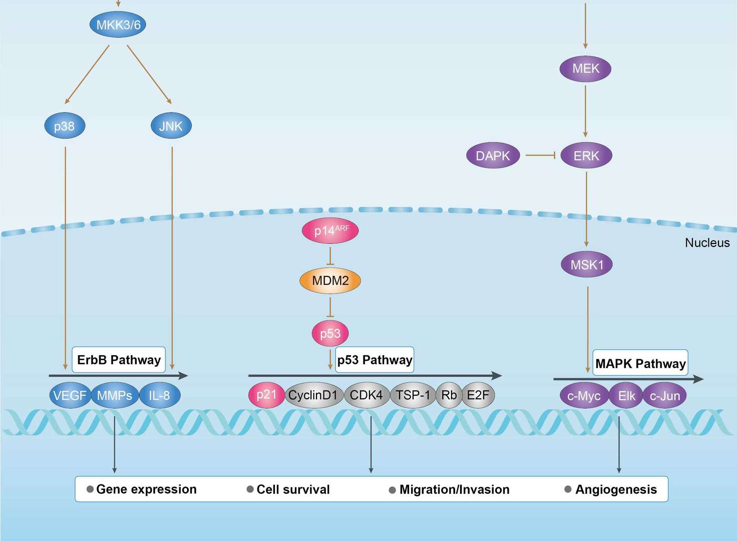
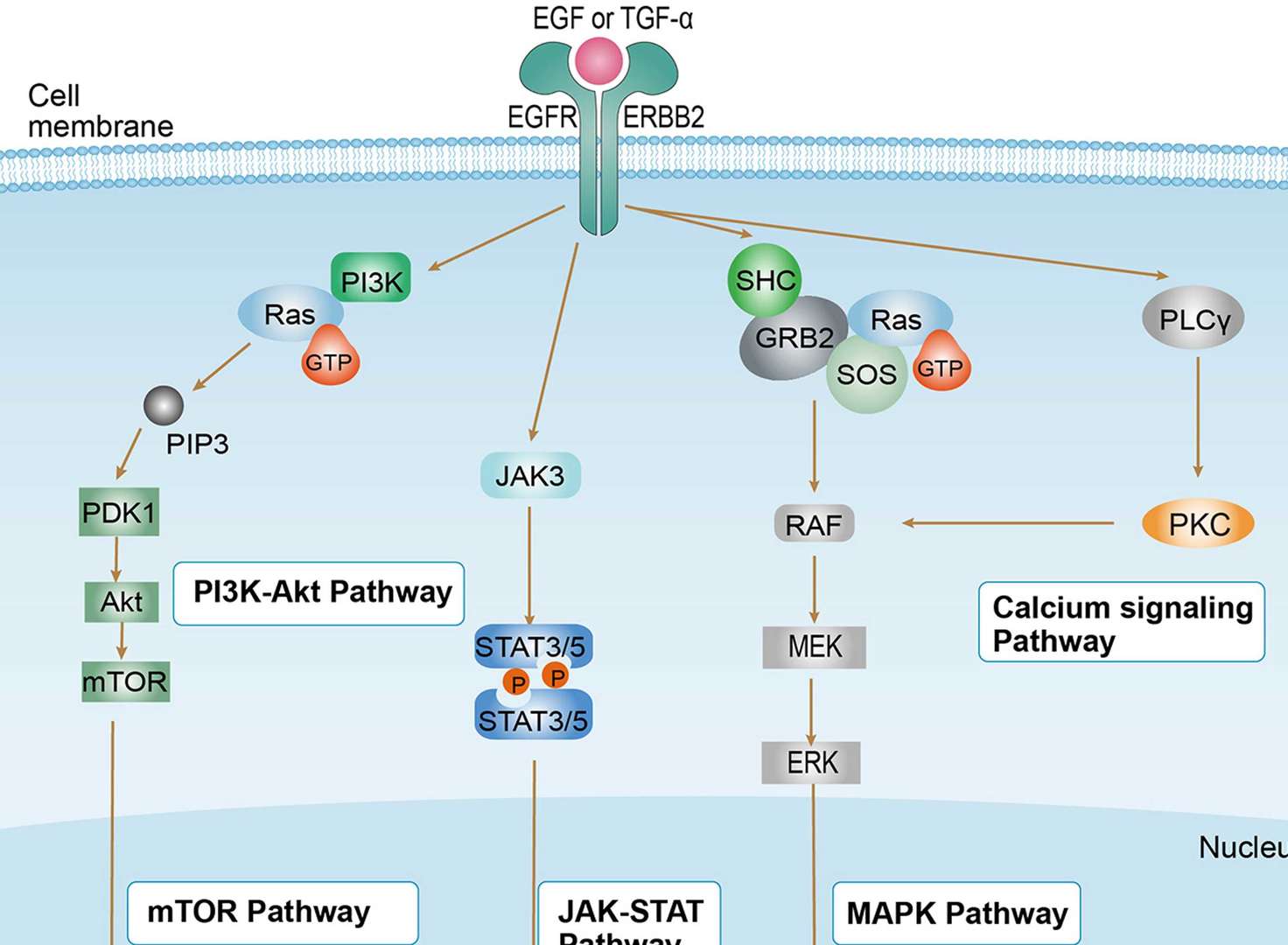
Related Signaling Pathways
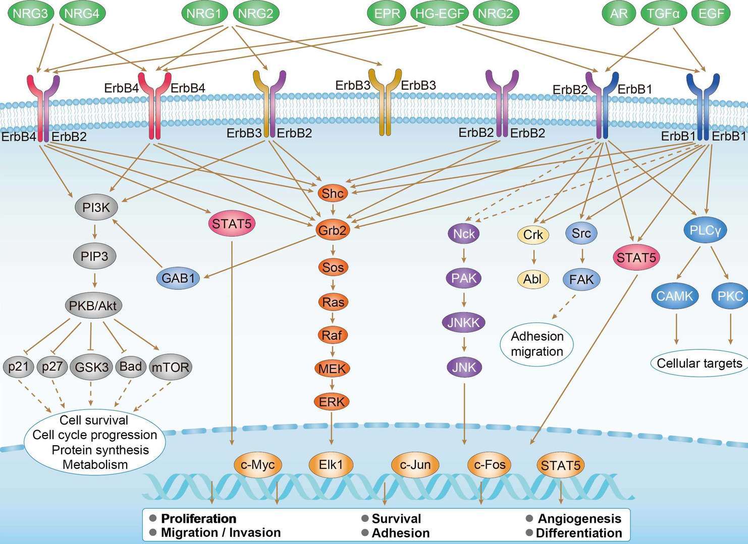 ErbB Signaling Pathway
Downloadable Resources
Download resources about recombinant antibody development and antibody engineering to boost your research.
Datasheet
MSDS
COA
Certificate of Analysis LookupTo download a Certificate of Analysis, please enter a lot number in the search box below. Note: Certificate of Analysis not available for kit components.
Lot Number:
See other products for "Clone hu4D5Fabv8"
- CAT
- Product Name
See other products for "ERBB2"
Select a product category from the dropdown menu below to view related products.
| CAT | Product Name | Application | Type |
|---|---|---|---|
| NABG-059 | Recombinant Anti-Human ERBB2 VHH Single Domain Antibody | IHC, FC, CA, FuncS | Llama VHH |
| CAT | Product Name | Application | Type |
|---|---|---|---|
| IAB-B008(A) | Recombinant Anti-human ERBB2 Intrabody [(D-Arg)9] | IF, FC, FuncS | scFv-(D-Arg)9 |
| CAT | Product Name | Application | Type |
|---|---|---|---|
| IAB-B008(G) | Recombinant Anti-human ERBB2 Intrabody [+36 GFP] | WB, ICC, FuncS | scFv-(+36GFP) |
| CAT | Product Name | Application | Type |
|---|---|---|---|
| IAB-B008(T) | Recombinant Anti-human ERBB2 Intrabody [Tat] | ICC, Neut, FuncS | scFv-Tat |
| CAT | Product Name | Application | Type |
|---|---|---|---|
| TAB-005 | Anti-Human ErbB2 Recombinant Antibody (TAB-005) | FC, IP, ELISA, Neut, FuncS, IF, IHC | IgG1 - kappa |
| CAT | Product Name | Application | Type |
|---|---|---|---|
| TAB-761 | Anti-ERBB2 Recombinant Antibody (Margetuximab) | IF, IP, Neut, FuncS, ELISA, FC, WB | IgG1 - kappa |
| CAT | Product Name | Application | Type |
|---|---|---|---|
| TAB-H70 | Anti-Human ERBB2 Recombinant Antibody (TAB-H70) | ELISA, IP, FC, FuncS, Neut, IF, ICC | IgG1 - kappa |
| CAT | Product Name | Application | Type |
|---|---|---|---|
| TAB-053 | Anti-Human ERBB2 Recombinant Antibody (TAB-053) | FuncS, IF, Neut, ELISA, FC, IP, WB | IgG1 - kappa |
| CAT | Product Name | Application | Type |
|---|---|---|---|
| AGTO-G022E | Anti-ERBB2 immunotoxin 4D5 (scFv)-PE | Cytotoxicity assay, Function study |
| CAT | Product Name | Application | Type |
|---|---|---|---|
| AGTO-G022D | Anti-ERBB2 immunotoxin 4D5 (scFv)-DT | Cytotoxicity assay, Function study |
| CAT | Product Name | Application | Type |
|---|---|---|---|
| AGTO-G022G | Anti-ERBB2 immunotoxin 4D5 (scFv)-Gel | Cytotoxicity assay, Function study |
| CAT | Product Name | Application | Type |
|---|---|---|---|
| AGTO-G022R | Anti-ERBB2 immunotoxin 4D5 (scFv)-RTA | Cytotoxicity assay, Function study |
| CAT | Product Name | Application | Type |
|---|---|---|---|
| AGTO-G022S | Anti-ERBB2 immunotoxin 4D5 (scFv)-Sap | Cytotoxicity assay, Function study |
| CAT | Product Name | Application | Type |
|---|---|---|---|
| PABL-085 | Human Anti-ERBB2 Recombinant Antibody (PABL-085) | ELISA, WB, FuncS | Human IgG |
| CAT | Product Name | Application | Type |
|---|---|---|---|
| PABL-126 | Human Anti-ERBB2 Recombinant Antibody (PABL-126) | ELISA, WB, FuncS | Human IgG |
| CAT | Product Name | Application | Type |
|---|---|---|---|
| PSBL-085 | Human Anti-ERBB2 Recombinant Antibody; scFv Fragment (PSBL-085) | ELISA, WB, FuncS | Human scFv |
| CAT | Product Name | Application | Type |
|---|---|---|---|
| PSBL-126 | Human Anti-ERBB2 Recombinant Antibody; scFv Fragment (PSBL-126) | ELISA, WB, FuncS | Human scFv |
| CAT | Product Name | Application | Type |
|---|---|---|---|
| PFBL-085 | Human Anti-ERBB2 Recombinant Antibody (PFBL-085) | ELISA, WB, FuncS | Human Fab |
| CAT | Product Name | Application | Type |
|---|---|---|---|
| PFBL-126 | Human Anti-ERBB2 Recombinant Antibody; Fab Fragment (PFBL-126) | ELISA, WB, FuncS | Human Fab |
| CAT | Product Name | Application | Type |
|---|---|---|---|
| PNBL-066 | Recombinant Anti-HER2 VHH Single Domain Antibody (PNBL-066) | SPR | Llama VHH |
| CAT | Product Name | Application | Type |
|---|---|---|---|
| PNBL-067 | Recombinant Anti-HER2 VHH Single Domain Antibody (PNBL-067) | SPR | Llama VHH |
| CAT | Product Name | Application | Type |
|---|---|---|---|
| PNBL-069 | Recombinant Anti-HER2 VHH Single Domain Antibody (PNBL-069) | ELISA | Llama VHH |
| CAT | Product Name | Application | Type |
|---|---|---|---|
| PABZ-041 | Human Anti-ERBB2 Recombinant Antibody (clone bH1) | WB, IF, FuncS | Human IgG |
| CAT | Product Name | Application | Type |
|---|---|---|---|
| PABC-089 | Mouse Anti-ERBB2 Recombinant Antibody (clone A21) | ELISA, WB, FC | Mouse IgG |
| CAT | Product Name | Application | Type |
|---|---|---|---|
| PABL-466 | Human Anti-ERBB2 Recombinant Antibody (clone mAb37) | WB | Human IgG |
| CAT | Product Name | Application | Type |
|---|---|---|---|
| PFBZ-041 | Human Anti-ERBB2 Recombinant Antibody (clone bH1); Fab Fragment | WB, IF, FuncS | Human Fab |
| CAT | Product Name | Application | Type |
|---|---|---|---|
| PFBC-089 | Mouse Anti-ERBB2 Recombinant Antibody (clone A21); Fab Fragment | ELISA, WB, FC | Mouse Fab |
| CAT | Product Name | Application | Type |
|---|---|---|---|
| PFBL-463 | Human Anti-ERBB2 Recombinant Antibody Fab Fragment (PFBL-463) | WB | Human Fab |
| CAT | Product Name | Application | Type |
|---|---|---|---|
| PFBW-161 | Mouse Anti-ERBB2 Recombinant Antibody (clone chA21); Fab Fragment | FC, WB | Mouse Fab |
| CAT | Product Name | Application | Type |
|---|---|---|---|
| PSBZ-041 | Human Anti-ERBB2 Recombinant Antibody (clone bH1); scFv Fragment | WB, IF, FuncS | Human scFv |
| CAT | Product Name | Application | Type |
|---|---|---|---|
| PSBC-089 | Mouse Anti-ERBB2 Recombinant Antibody (clone A21); scFv Fragment | ELISA, WB, FC | Mouse scFv |
| CAT | Product Name | Application | Type |
|---|---|---|---|
| TAB-0191CL | Human Anti-ERBB2 Recombinant Antibody (TAB-0191CL) | ELISA, Cyt, Internalization | Human IgG |
| CAT | Product Name | Application | Type |
|---|---|---|---|
| TAB-0192CL | Human Anti-ERBB2 Recombinant Antibody (TAB-0192CL) | ELISA, ADCC, Cyt, Internalization | Human IgG |
| CAT | Product Name | Application | Type |
|---|---|---|---|
| TAB-0193CL | Human Anti-ERBB2 Recombinant Antibody (TAB-0193CL) | ELISA, ADCC, Cyt, Internalization | Human IgG |
| CAT | Product Name | Application | Type |
|---|---|---|---|
| TAB-0194CL | Human Anti-ERBB2 Recombinant Antibody (TAB-0194CL) | ELISA, ADCC, Cyt, Internalization | Human IgG |
| CAT | Product Name | Application | Type |
|---|---|---|---|
| TAB-0191CL-S(P) | Human Anti-ERBB2 Recombinant Antibody; scFv Fragment (TAB-0191CL-S(P)) | ELISA, Cyt, Internalization | Human scFv |
| CAT | Product Name | Application | Type |
|---|---|---|---|
| TAB-033CT | Human Anti-ERBB2 Recombinant Antibody (TAB-033CT) | ELISA, WB | Human IgG1, κ |
| CAT | Product Name | Application | Type |
|---|---|---|---|
| TAB-034CT | Anti-Human HER2/neu Recombinant Antibody (TAB-034CT) | WB |
| CAT | Product Name | Application | Type |
|---|---|---|---|
| TAB-036CT | Anti-Human HER2/neu Recombinant Antibody (6B2) | ELISA, WB |
| CAT | Product Name | Application | Type |
|---|---|---|---|
| TAB-037CT | Anti-Human HER2/neu Recombinant Antibody (7C2) | ELISA, WB |
| CAT | Product Name | Application | Type |
|---|---|---|---|
| TAB-038CT | Human Anti-ERBB2 Recombinant Antibody (TAB-038CT) | FuncS, FC | Humanized IgG1 |
| CAT | Product Name | Application | Type |
|---|---|---|---|
| TAB-049CT | Anti-Human HER2/neu Recombinant Antibody (ChA21) | IHC, Inhib, WB, Activ, FC | Chimeric antibody (mouse/human) |
| CAT | Product Name | Application | Type |
|---|---|---|---|
| TAB-034CT-S(P) | Anti-Human HER2/neu Recombinant Antibody scFv Fragment (TAB-034CT-S(P)) | WB |
| CAT | Product Name | Application | Type |
|---|---|---|---|
| TAB-036CT-S(P) | Anti-Human HER2/neu Recombinant Antibody scFv Fragment (6B2) | ELISA, WB |
| CAT | Product Name | Application | Type |
|---|---|---|---|
| TAB-049CT-S(P) | Anti-Human HER2/neu Recombinant Antibody scFv Fragment (ChA21) | WB | Chimeric antibody (mouse/human) |
| CAT | Product Name | Application | Type |
|---|---|---|---|
| TAB-049CT-F(E) | Anti-Human HER2/neu Recombinant Antibody Fab Fragment (ChA21) | WB | Chimeric antibody (mouse/human) |
| CAT | Product Name | Application | Type |
|---|---|---|---|
| TAB-032CT | Anti-Human HER2/neu Single Domain Antibody, Research Grade Biosimilar | WB | Single domain antibody |
| CAT | Product Name | Application | Type |
|---|---|---|---|
| Gly-024LC | Recombinant Anti-Human ERBB2 Antibody (Fab glycosylation) | ELISA | Mouse antibody |
| CAT | Product Name | Application | Type |
|---|---|---|---|
| Gly-117LC | Recombinant Anti-Human ERBB2 Antibody (Fc glycosylation) | ELISA | Humanized antibody |
| Gly-145LC | Recombinant Anti-Human ERBB2 Antibody (Fc glycosylation) | ELISA | Human antibody |
| Gly-146LC | Recombinant Anti-Human ERBB2 Antibody (Fc glycosylation) | ELISA | Human antibody |
| Gly-147LC | Recombinant Anti-Human ERBB2 Antibody (Fc glycosylation) | ELISA | Human antibody |
| Gly-148LC | Recombinant Anti-Human ERBB2 Antibody (Fc glycosylation) | ELISA | Human antibody |
| CAT | Product Name | Application | Type |
|---|---|---|---|
| Gly-177LC | Recombinant Anti-Human ERBB2 Antibody (Non-glycosylated) | ELISA | Humanized antibody |
| CAT | Product Name | Application | Type |
|---|---|---|---|
| BRD-0195MZ | Chicken Anti-ERBB2 Polyclonal IgY | WB | Chicken antibody |
| CAT | Product Name | Application | Type |
|---|---|---|---|
| MHC-LC004 | PE-A*02:01/Human Her-2/neu E75 (KIFGSLAFL) MHC Tetramer | FCM |
| CAT | Product Name | Application | Type |
|---|---|---|---|
| MHC-LC005 | APC-A*02:01/Human Her-2/neu E75 (KIFGSLAFL) MHC Tetramer | FCM |
| CAT | Product Name | Application | Type |
|---|---|---|---|
| MHC-LC006 | BV421-A*02:01/Human Her-2/neu E75 (KIFGSLAFL) MHC Tetramer | FCM |
| CAT | Product Name | Application | Type |
|---|---|---|---|
| MHC-LC007 | PE-A*02:01/Human Her-2/neu (RLLQETELV) MHC Tetramer | FCM |
| CAT | Product Name | Application | Type |
|---|---|---|---|
| MHC-LC008 | APC-A*02:01/Human Her-2/neu (RLLQETELV) MHC Tetramer | FCM |
| CAT | Product Name | Application | Type |
|---|---|---|---|
| NEUT-737CQ | Mouse Anti-ERBB2 Recombinant Antibody (clone 7.16.4) | Block, IP, IF, FC | Mouse IgG2a |
| CAT | Product Name | Application | Type |
|---|---|---|---|
| MOR-1178 | Hi-Affi™ Rabbit Anti-ERBB2 Recombinant Antibody (clone DS1178AB) | IHC-P, IHC-Fr | Rabbit IgG |
| CAT | Product Name | Application | Type |
|---|---|---|---|
| MOR-4586 | Hi-Affi™ Rabbit Anti-ERBB2 Recombinant Antibody (clone TH99DS) | WB | Rabbit IgG |
| CAT | Product Name | Application | Type |
|---|---|---|---|
| MOR-4587 | Hi-Affi™ Rabbit Anti-ERBB2 Recombinant Antibody (clone TH100DS) | WB | Rabbit IgG |
| CAT | Product Name | Application | Type |
|---|---|---|---|
| AFC-TAB-468CQ | Afuco™ Anti-ERBB2 ADCC Recombinant Antibody, ADCC Enhanced (AFC-TAB-468CQ) | ELISA, IHC, FC, IP, IF, FuncS | ADCC enhanced antibody |
| CAT | Product Name | Application | Type |
|---|---|---|---|
| AFC-TAB-053 | Afuco™ Anti-ERBB2 ADCC Recombinant Antibody, ADCC Enhanced (AFC-TAB-053) | FuncS, IF, Neut, ELISA, FC, IP | ADCC enhanced antibody |
| CAT | Product Name | Application | Type |
|---|---|---|---|
| AFC-TAB-761 | Afuco™ Anti-ERBB2 ADCC Recombinant Antibody, ADCC Enhanced (AFC-TAB-761) | IF, IP, Neut, FuncS, ELISA, FC | ADCC enhanced antibody |
| CAT | Product Name | Application | Type |
|---|---|---|---|
| AFC-TAB-005 | Afuco™ Anti-ERBB2 ADCC Recombinant Antibody, ADCC Enhanced (AFC-TAB-005) | FC, IP, ELISA, Neut, FuncS, IF | ADCC enhanced antibody |
| CAT | Product Name | Application | Type |
|---|---|---|---|
| VS-0424-XY91 | AbPlus™ Anti-ERBB2 Magnetic Beads (3.F2) | IP, Protein Purification |
| CAT | Product Name | Application | Type |
|---|---|---|---|
| VS-0724-YC1516 | AbPlus™ Anti-Erbb2 Magnetic Beads (VS-0724-YC1516) | IP, Protein Purification |
| CAT | Product Name | Application | Type |
|---|---|---|---|
| VS-0125-FY24 | Human Anti-ERBB2 (clone 1.18.1) scFv-Fc Chimera | FC, BI | Human IgG1, scFv-Fc |
| CAT | Product Name | Application | Type |
|---|---|---|---|
| VS-0225-XY108 | CytoStream™ Mouse Anti-ERBB2 Recombinant Antibody (clone 24D2) | FC | Mouse IgG1, kappa |
| CAT | Product Name | Application | Type |
|---|---|---|---|
| VS-0425-YC430 | Recombinant Anti-ERBB2 Vesicular Antibody, EV Displayed (VS-0425-YC430) | ELISA, FC, Neut, Cell-uptake |
| CAT | Product Name | Application | Type |
|---|---|---|---|
| VS-0525-XY2335 | Anti-ERBB2 Immunohistochemistry Kit | IHC |
| CAT | Product Name | Application | Type |
|---|---|---|---|
| VS-0525-XY2336 | Anti-Mouse ERBB2 Immunohistochemistry Kit | IHC |
| CAT | Product Name | Application | Type |
|---|---|---|---|
| VS-0525-YC3 | Recombinant Anti-ERBB2 Biparatopic Antibody, Symmetric scFv-Fab | ELISA | Symmetric scFv-Fab |
| CAT | Product Name | Application | Type |
|---|---|---|---|
| VS-0525-YC6 | Recombinant Anti-ERBB2 Biparatopic Antibody, Asymmetric hetero Fab | ELISA | Asymmetric hetero Fab |
| CAT | Product Name | Application | Type |
|---|---|---|---|
| VS-0525-YC7 | Recombinant Anti-ERBB2 Biparatopic Antibody, Asymmetric hetero HC-LC | ELISA, Inhib | Asymmetric hetero HC, common LC |
| CAT | Product Name | Application | Type |
|---|---|---|---|
| VS-0825-YC22 | SmartAb™ Recombinant Anti-ERBB2 pH-dependent Antibody (VS-0825-YC22) | IF, Neut, ELISA, FC, IP, WB | Human IgG1 kappa |
| CAT | Product Name | Application | Type |
|---|---|---|---|
| VS-0825-YC24 | SmartAb™ Recombinant Anti-ERBB2 pH-dependent Antibody (Clone bH1) | WB, IF | Human IgG |
| CAT | Product Name | Application | Type |
|---|---|---|---|
| VS-0825-YC117 | SmartAb™ Recombinant Anti-ERBB2 pH-dependent Antibody (VS-0825-YC117) | FC, IP, ELISA, Neut, IF, IHC | Human IgG1 kappa |
| CAT | Product Name | Application | Type |
|---|---|---|---|
| VS-1025-YC2 | Anti-ERBB2 Antibody Prodrug, Protease Activated (VS-1025-YC2) | ISZ, Cyt, FuncS |
| CAT | Product Name | Application | Type |
|---|---|---|---|
| VS-1025-YC19 | Anti-ERBB2 Antibody Prodrug, Protease Activated (VS-1025-YC19) | ISZ, Cyt, FuncS |
Specific Inquiry
See Our Custom Production in Action


Popular Products

Application: WB, FuncS, IF, Neut, ELISA, FC, IP

Application: FC, IP, ELISA, Neut, FuncS, IF, IHC

Application: WB, FuncS, IF, Neut, ELISA, FC, IP

Application: ELISA, Neut, IF, IP, FC, FuncS

Application: FC, IP, ELISA, Neut, FuncS, IF, ICC

Application: FC, IP, ELISA, Neut, FuncS, IF, ICC

Application: IP, IF, FuncS, FC, Neut, ELISA, IHC

Application: IF, IP, Neut, FuncS, ELISA, FC, ICC

Application: ELISA, FC, IP, FuncS, IF, Neut, ICC

Application: ELISA, FC, IP, FuncS, IF, Neut, WB

Application: FC, IP, ELISA, Neut, FuncS, IF, IHC


Application: ELISA, WB, BLI, SPR


For research use only. Not intended for any clinical use. No products from Creative Biolabs may be resold, modified for resale or used to manufacture commercial products without prior written approval from Creative Biolabs.
Send Inquiry
This site is protected by reCAPTCHA and the Google Privacy Policy and Terms of Service apply.