

AbPlus™ Anti-JAK2 Magnetic Beads (CBACN-632)
CAT#: VS-0424-XY162
The AbPlus Anti-JAK2 Magnetic Beads (CBACN-632) is an innovative affinity resin which is bound with anti-JAK2 specific antibody (CBACN-632). The beads were designed for small-scale affinity purification and immunoprecipitation (IP) of JAK2 protein under native and denaturing conditions.
Gene Expression

Subcellular Location
Figure 1 IF staining of human cell line U-2 OS
Immunofluorescent staining of human cell line U-2 OS shows localization to nucleoplasm, plasma membrane & focal adhesion sites.
* Image credit: Image credit: Human Protein Atlas v21.proteinatlas.org/images/43870/1338_A2_1_selected.jpg

Normal Tissue
Figure 2 IHC staining of human bone marrow
Immunohistochemical staining of human bone marrow shows moderate cytoplasmic and nuclear positivity in hematopoietic cells.
* Image credit: Image credit: Human Protein Atlas v21.proteinatlas.org/images/40820/ihc_selected.jpg

Normal Tissue
Figure 3 IHC staining of human lymph node
Immunohistochemical staining of human lymph node shows strong cytoplasmic positivity in lymphoid tissue.
* Image credit: Image credit: Human Protein Atlas v21.proteinatlas.org/images/13089/ihc_selected.jpg

Normal Tissue
Figure 4 Cerebral cortex
Endothelial cells
Staining: Medium
Intensity: Moderate
Quantity:>75%
Location: Cytoplasmic/membranous
* Image credit: Image credit: Human Protein Atlas v21.proteinatlas.org/images/13089/29841_B_7_5.jpg

Normal Tissue
Figure 5 Colon
Endothelial cells
Staining: High
Intensity: Strong
Quantity:>75%
Location: Cytoplasmic/membranous
* Image credit: Image credit: Human Protein Atlas v21.proteinatlas.org/images/13089/29841_A_9_3.jpg

Normal Tissue
Figure 6 Liver
Hepatocytes
Staining: High
Intensity: Strong
Quantity:>75%
Location: Cytoplasmic/membranous
* Image credit: Image credit: Human Protein Atlas v21.proteinatlas.org/images/13089/29841_A_8_4.jpg

Normal Tissue
Figure 7 Kidney
Cells in glomeruli
Staining: High
Intensity: Strong
Quantity: 75%-25%
Location: Cytoplasmic/membranous
Cells in tubules
Staining: High
Intensity: Strong
Quantity: 75%-25%
Location: Cytoplasmic/membranous
* Image credit: Image credit: Human Protein Atlas v21.proteinatlas.org/images/13089/29841_A_9_5.jpg

Normal Tissue
Figure 8 Testis
Cells in seminiferous ducts
Staining: Medium
Intensity: Strong
Quantity: <25%
Location: Cytoplasmic/membranous
Leydig cells
Staining: Medium
Intensity: Moderate
Quantity:>75%
Location: Cytoplasmic/membranous
* Image credit: Image credit: Human Protein Atlas v21.proteinatlas.org/images/13089/29841_A_4_6.jpg

RNA Expression
Figure 9 RNA cell line category: Group enriched (ASC diff, HDLM-2, HEL)
Cell lines ordered by descending RNA expression order.
* Image credit: Image credit: Human Protein Atlas v21.proteinatlas.org/ENSG00000096968-JAK2
❮


 ❯
❯
Specifications
- Applications
- Immunoprecipitation, Protein Purification
- Matrix
- Magrose bead (> 50 μmol/mL gel)
- Bead Ligand
- Anti-JAK2 specific antibody (CBACN-632)
- Target
- JAK2
- Immunogen
- Synthetic phospho-peptide corresponding to residues surrounding Tyr1007 and 1008 of human JAK2
- Target Species
- Human, Mouse, Rat
- Antibody Clone
- CBACN-632
- Bead Capacity
- 20-30 mg/mL binding antibody
- Bead size
- 10-37 μm
- Stability
- pH 2-14
- Format
- 20% Suspension
- Buffer
- PBS, pH 7.4, with 1% BSA
- Preservative
- 0.03% Proclin 300
- Storage
- Stored at 4°C, and is stable for up to 2 years. Do not centrifuge, dry or freeze the magnetic beads.
Applications
- Application Notes
- The beads are in suspension and will settle upon storage. Prior to use, mix the vial gently (do not vortex) to ensure delivery of proper bead volume.
Target
- Introduction
- JAK2 has several functions. Non-receptor tyrosine kinase involved in various processes such as cell growth, development, differentiation or histone modifications. mediates essential signaling events in both innate and adaptive immunity. In the cytoplasm, plays a pivotal role in signal transduction via its association with type I receptors such as growth hormone (GHR), prolactin (PRLR), leptin (LEPR), erythropoietin (EPOR), thrombopoietin (THPO); or type II receptors including IFN-alpha, IFN-beta, IFN-gamma and multiple interleukins (PubMed:7615558). Following ligand-binding to cell surface receptors, phosphorylates specific tyrosine residues on the cytoplasmic tails of the receptor, creating docking sites for STATs proteins (PubMed:9618263). Subsequently, phosphorylates the STATs proteins once they are recruited to the receptor. Phosphorylated STATs then form homodimer or heterodimers and translocate to the nucleus to activate gene transcription. For example, cell stimulation with erythropoietin (EPO) during erythropoiesis leads to JAK2 autophosphorylation, activation, and its association with erythropoietin receptor (EPOR) that becomes phosphorylated in its cytoplasmic domain. Then, STAT5 (STAT5A or STAT5B) is recruited, phosphorylated and activated by JAK2. Once activated, dimerized STAT5 translocates into the nucleus and promotes the transcription of several essential genes involved in the modulation of erythropoiesis. Part of a signaling cascade that is activated by increased cellular retinol and that leads to the activation of STAT5 (STAT5A or STAT5B) (PubMed:21368206). In addition, JAK2 mediates angiotensin-2-induced ARHGEF1 phosphorylation (PubMed:20098430). It plays a role in cell cycle by phosphorylating CDKN1B (PubMed:21423214). Cooperates with TEC through reciprocal phosphorylation to mediate cytokine-driven activation of FOS transcription. In the nucleus, plays a key role in chromatin by specifically mediating phosphorylation of 'Tyr-41' of histone H3 (H3Y41ph), a specific tag that promotes exclusion of CBX5 (HP1 alpha) from chromatin.
- Alternative Names
- JTK10; THCYT3
- Gene ID
- 3717
- UniProt ID
- O60674
REVIEWS AND Q&AS
CITATIONS
RESOURCES
DOWNLOADS
RELATED PRODUCTS
Inquiry
Navs
Customer Review

There are currently no Customer reviews or questions for VS-0424-XY162. Click the button above to contact us or submit your feedback about this product.
Submit Your Publication
Published with our product? Submit your paper and receive a 10% discount on your next order! Share your research to earn exclusive rewards.
Related Signaling Pathways
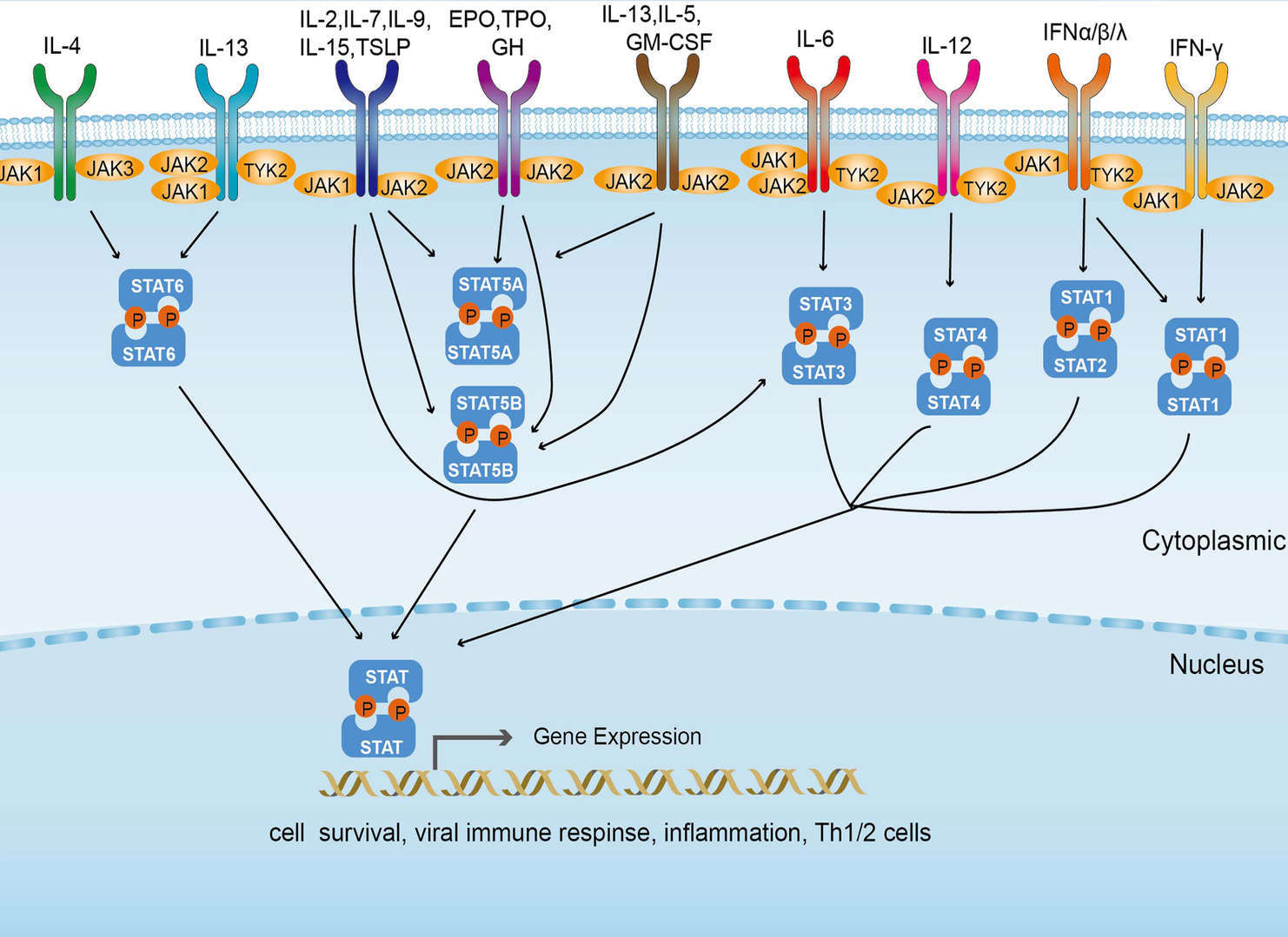 JAK-STAT Signaling Pathway
Related Diseases
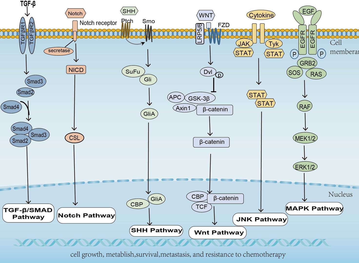 Pancreatic Cancer
 AGE-RAGE Signaling Pathway in Diabetic Complications
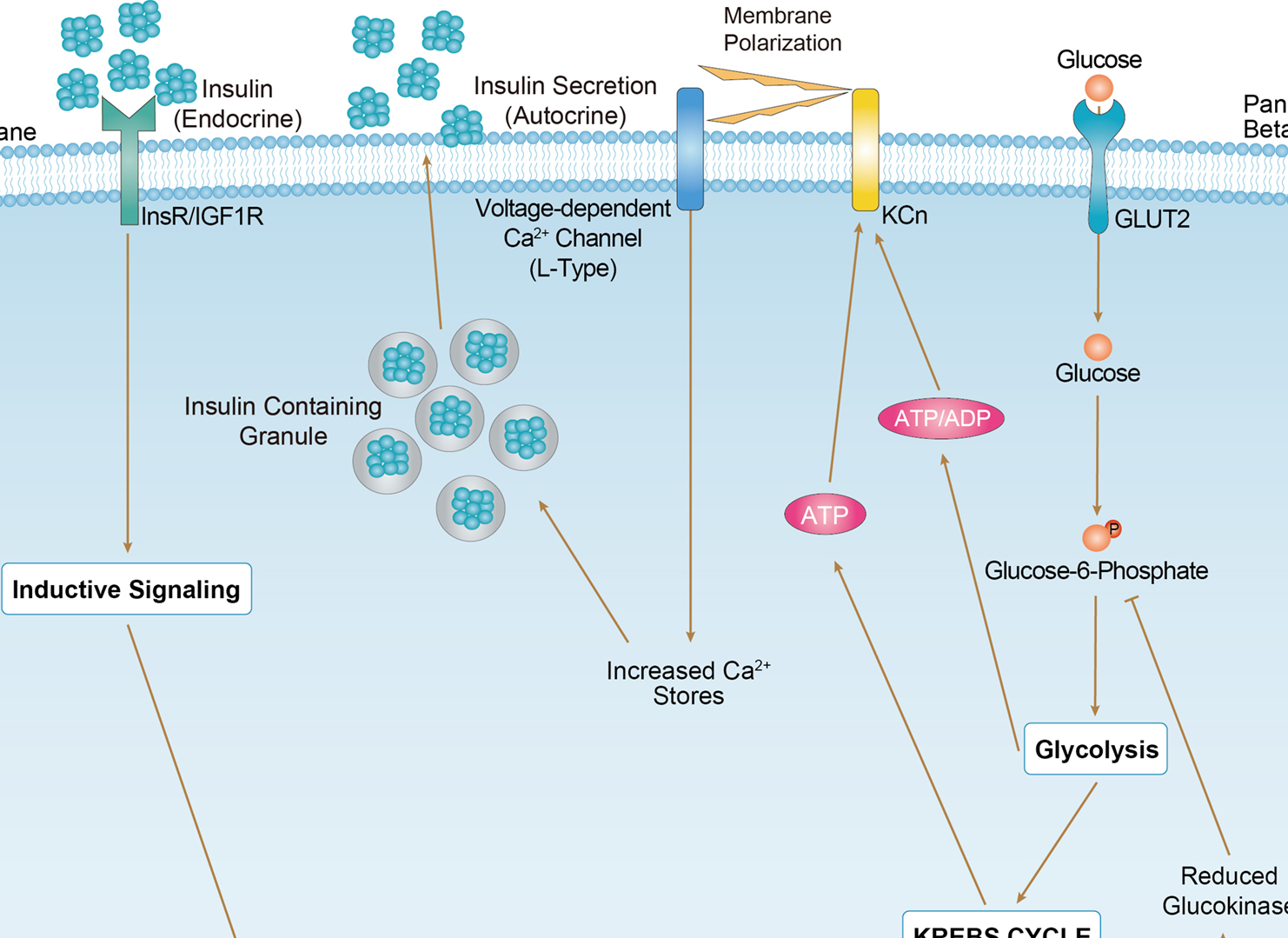 Maturity Onset Diabetes of the Young
Downloadable Resources
Download resources about recombinant antibody development and antibody engineering to boost your research.
Datasheet
MSDS
COA
Certificate of Analysis LookupTo download a Certificate of Analysis, please enter a lot number in the search box below. Note: Certificate of Analysis not available for kit components.
Lot Number:
See other products for "JAK2"
Select a product category from the dropdown menu below to view related products.
| CAT | Product Name | Application | Type |
|---|---|---|---|
| MOB-1854z | Mouse Anti-JAK2 Recombinant Antibody (clone 16B1) | WB, ELISA, FC, ICC, IF, IHC | Mouse IgG1 |
| CAT | Product Name | Application | Type |
|---|---|---|---|
| MOB-2300CT | Recombinant Mouse anti-Human JAK2 Monoclonal antibody (Z377) | WB |
| CAT | Product Name | Application | Type |
|---|---|---|---|
| MOR-1898 | Hi-Affi™ Recombinant Rabbit Anti-JAK2 Monoclonal Antibody (DS1898AB) | WB, IHC-Fr, IHC-P, ICC, IP, Dot | IgG |
| CAT | Product Name | Application | Type |
|---|---|---|---|
| MRO-0880-CN | Recombinant Mouse Anti-JAK2 Monoclonal Antibody (6-D3) | WB, IF, IHC, FC | Mouse IgG2b |
| CAT | Product Name | Application | Type |
|---|---|---|---|
| MRO-0881-CN | Recombinant Rabbit Anti-JAK2 Monoclonal Antibody (CBACN-328) | WB, IF | Rabbit IgG |
| CAT | Product Name | Application | Type |
|---|---|---|---|
| MRO-1986-CN | Rabbit Anti-JAK2 Polyclonal Antibody (MRO-1986-CN) | IF, IHC | Rabbit IgG |
| CAT | Product Name | Application | Type |
|---|---|---|---|
| VS13-YC609 | CytoStream™ Mouse Anti-JAK2 Recombinant Antibody (VS13-YC609) | WB, ICC, IF, IHC-P, FC | Mouse IgG2b |
| CAT | Product Name | Application | Type |
|---|---|---|---|
| VS-0525-XY3687 | Anti-JAK2 Immunohistochemistry Kit | IHC |
| CAT | Product Name | Application | Type |
|---|---|---|---|
| VS-0525-XY3688 | Anti-Mouse JAK2 Immunohistochemistry Kit | IHC |
| CAT | Product Name | Application | Type |
|---|---|---|---|
| VS-0525-XY3689 | Anti-Rat JAK2 Immunohistochemistry Kit | IHC |
Specific Inquiry
See Our Custom Production in Action


Popular Products


Application: FC, IP, ELISA, Neut, FuncS, IF, IHC

Application: Neut, ELISA, IF, IP, FuncS, FC, ICC

Application: ELISA, IP, FC, FuncS, Neut, IF, WB

Application: WB, FuncS, IF, Neut, ELISA, FC, IP

Application: WB, ELISA, IP, FC, FuncS, Neut, IF

Application: IP, IF, FuncS, FC, Neut, ELISA, ICC

Application: IP, IF, FuncS, FC, Neut, ELISA, ICC

Application: IF, IP, Neut, FuncS, ELISA, FC, ICC

Application: FuncS, IF, Neut, ELISA, FC, IP, ICC
-2.png)
Application: ELISA, FC, IP, FuncS, IF, Neut, ICC

Application: FuncS, IF, Neut, ELISA, FC, IP, ICC


Application: ELISA

Application: ELISA, IP, WB, IHC, IF, FuncS


For research use only. Not intended for any clinical use. No products from Creative Biolabs may be resold, modified for resale or used to manufacture commercial products without prior written approval from Creative Biolabs.
Send Inquiry
This site is protected by reCAPTCHA and the Google Privacy Policy and Terms of Service apply.