

Anti-Mouse DVL2 Immunohistochemistry Kit (CAT#: VS-0525-XY2119)
Our DVL2 IHC kit provides complete reagents for detecting this key Wnt signaling mediator involved in cellular communication and development. It works on paraffin and frozen sections across various species, delivering consistent results for studies on tissue patterning and cancer progression.
Specific Inquiry
Subcellular Location and Protein Expression


 Normal Tissue


 RNA Expression

Figure 1 IF staining of human cell line SH-SY5Y
(Immunofluorescent staining of human cell line SH-SY5Y shows localization to nucleoplasm.)
* Image credit: Human Protein Atlas https://v21.proteinatlas.org/images/21611/1220_G5_2_selected.jpg
(Immunofluorescent staining of human cell line SH-SY5Y shows localization to nucleoplasm.)
* Image credit: Human Protein Atlas https://v21.proteinatlas.org/images/21611/1220_G5_2_selected.jpg
Figure 2 IHC staining of human fallopian tube
(Immunohistochemical staining of human fallopian tube shows strong cytoplasmic positivity in glandular cells.)
* Image credit: Human Protein Atlas https://v21.proteinatlas.org/images/22914/53430_A_7_7_selected.jpg
(Immunohistochemical staining of human fallopian tube shows strong cytoplasmic positivity in glandular cells.)
* Image credit: Human Protein Atlas https://v21.proteinatlas.org/images/22914/53430_A_7_7_selected.jpg
Figure 3 IHC staining of human duodenum
(Immunohistochemical staining of human duodenum shows strong cytoplasmic positivity in glandular cells.)
* Image credit: Human Protein Atlas https://v21.proteinatlas.org/images/9312/ihc_selected.jpg
(Immunohistochemical staining of human duodenum shows strong cytoplasmic positivity in glandular cells.)
* Image credit: Human Protein Atlas https://v21.proteinatlas.org/images/9312/ihc_selected.jpg
Figure 4 IF staining of human cell line A549
(Immunofluorescent staining of human cell line A549 shows localization to nucleoplasm.)
* Image credit: Human Protein Atlas https://v21.proteinatlas.org/images/21611/1139_H3_1_red_green.jpg
(Immunofluorescent staining of human cell line A549 shows localization to nucleoplasm.)
* Image credit: Human Protein Atlas https://v21.proteinatlas.org/images/21611/1139_H3_1_red_green.jpg
Figure 5 IF staining of human cell line U-2 OS
(Immunofluorescent staining of human cell line U-2 OS shows localization to nucleoplasm.)
* Image credit: Human Protein Atlas https://v21.proteinatlas.org/images/21611/1132_H3_1_red_green.jpg
(Immunofluorescent staining of human cell line U-2 OS shows localization to nucleoplasm.)
* Image credit: Human Protein Atlas https://v21.proteinatlas.org/images/21611/1132_H3_1_red_green.jpg
Figure 6 Cerebral cortex
(Neuronal cells
Staining:Medium
Intensity: Moderate
Quantity:>75%
Location: Cytoplasmic/membranous nuclear
Neuropil
Staining:Medium
Intensity: Moderate
Quantity:>75%
Location: Cytoplasmic/membranous)
* Image credit: Human Protein Atlas https://v21.proteinatlas.org/images/22914/53430_B_9_5.jpg
(Neuronal cells
Staining:Medium
Intensity: Moderate
Quantity:>75%
Location: Cytoplasmic/membranous nuclear
Neuropil
Staining:Medium
Intensity: Moderate
Quantity:>75%
Location: Cytoplasmic/membranous)
* Image credit: Human Protein Atlas https://v21.proteinatlas.org/images/22914/53430_B_9_5.jpg
Figure 7 Colon
(Glandular cells
Staining:Medium
Intensity: Moderate
Quantity:>75%
Location: Cytoplasmic/membranous
Peripheral nerve/ganglion
Staining:Medium
Intensity: Moderate
Quantity:>75%
Location: Cytoplasmic/membranous nuclear)
* Image credit: Human Protein Atlas https://v21.proteinatlas.org/images/22914/53430_A_7_3.jpg
Figure 8 Liver
(Hepatocytes
Staining:High
Intensity: Strong
Quantity:>75%
Location: Cytoplasmic/membranous nuclear)
* Image credit: Human Protein Atlas https://v21.proteinatlas.org/images/22914/53430_A_8_4.jpg
(Hepatocytes
Staining:High
Intensity: Strong
Quantity:>75%
Location: Cytoplasmic/membranous nuclear)
* Image credit: Human Protein Atlas https://v21.proteinatlas.org/images/22914/53430_A_8_4.jpg
Figure 9 Kidney
(Cells in glomeruli
Staining:Medium
Intensity: Moderate
Quantity: 75%-25%
Location: Nuclear
Cells in tubules
Staining:Medium
Intensity: Moderate
Quantity:>75%
Location: Cytoplasmic/membranous)
* Image credit: Human Protein Atlas https://v21.proteinatlas.org/images/22914/53430_A_8_5.jpg
(Cells in glomeruli
Staining:Medium
Intensity: Moderate
Quantity: 75%-25%
Location: Nuclear
Cells in tubules
Staining:Medium
Intensity: Moderate
Quantity:>75%
Location: Cytoplasmic/membranous)
* Image credit: Human Protein Atlas https://v21.proteinatlas.org/images/22914/53430_A_8_5.jpg
Figure 10 Testis
(Cells in seminiferous ducts
Staining:Medium
Intensity: Moderate
Quantity:>75%
Location: Cytoplasmic/membranous nuclear
Leydig cells
Staining:Medium
Intensity: Moderate
Quantity:>75%
Location: Cytoplasmic/membranous nuclear)
* Image credit: Human Protein Atlas https://v21.proteinatlas.org/images/22914/53430_A_5_6.jpg
Figure 11 Lymph node
(Germinal center cells
Staining:Medium
Intensity: Moderate
Quantity:>75%
Location: Cytoplasmic/membranous
Non-germinal center cells
Staining:Medium
Intensity: Moderate
Quantity: 75%-25%
Location: Cytoplasmic/membranous)
* Image credit: Human Protein Atlas https://v21.proteinatlas.org/images/22914/53430_A_7_8.jpg
Figure 12 RNA cell line category: Low cell line specificity
(Cell lines ordered by descending RNA expression order.)
* Image credit: Human Protein Atlas https://v21.proteinatlas.org/ENSG00000004975-DVL2
(Cell lines ordered by descending RNA expression order.)
* Image credit: Human Protein Atlas https://v21.proteinatlas.org/ENSG00000004975-DVL2
Specifications
- Application
- IHC
- Size
- 50 Tests
- Species Reactivity
- Human, Mouse, Rat
- Target
- DVL2
- Primary Antibody
- Mouse Anti-DVL2 Antibody
- Secondary Antibody
- Goat anti-Mouse Antibody, HRP
- Sample Type
- FFPE tissue; Frozen section tissue
- Kit Storage
- All reagents should be kept at 2-8°C. The kit remains stable for up to 6 months after arrival.
Related Resources
 Breast Cancer
 Colorectal Cancer
 Endometrial Cancer
 Gastric Cancer
 Hepatocellular Carcinoma
 Prostate Cancer
 Throid Cancer
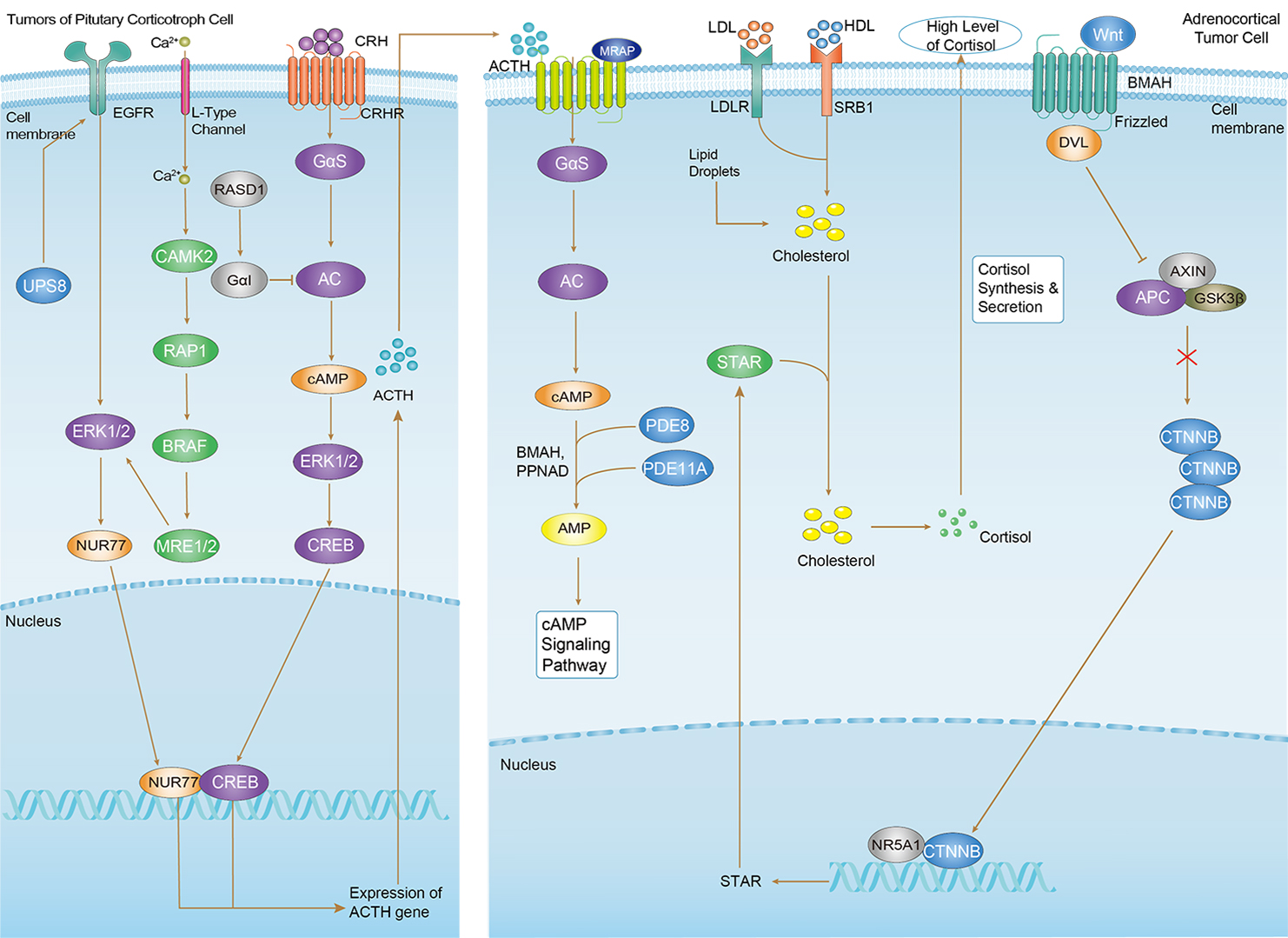 Cushing Syndrome
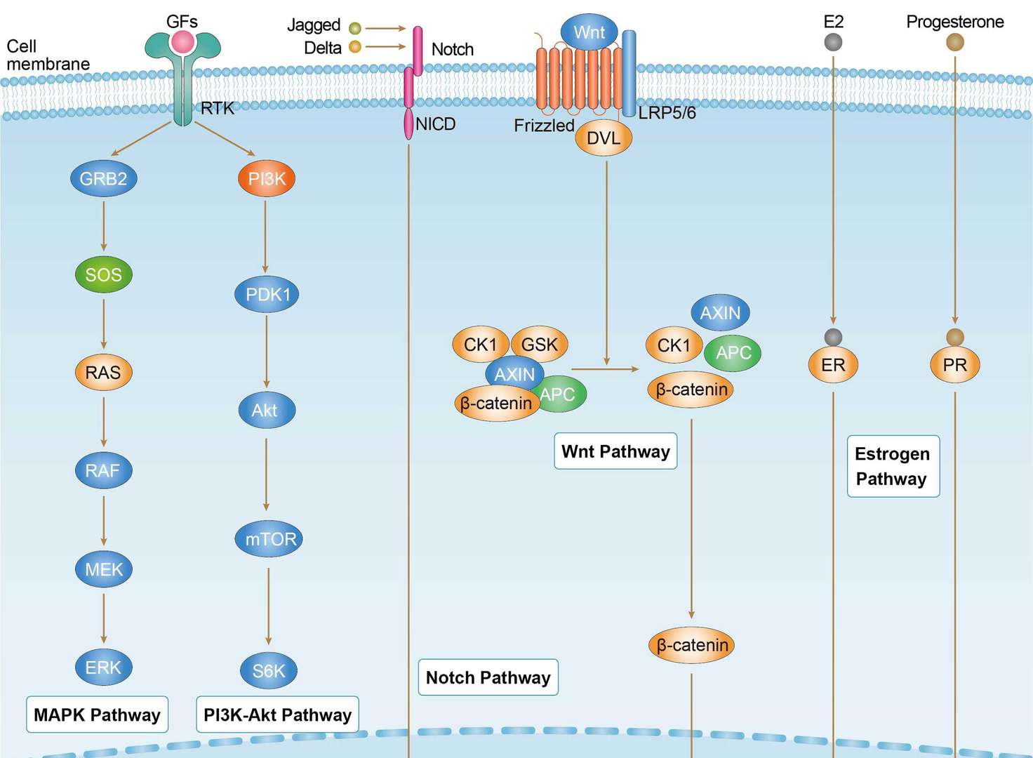
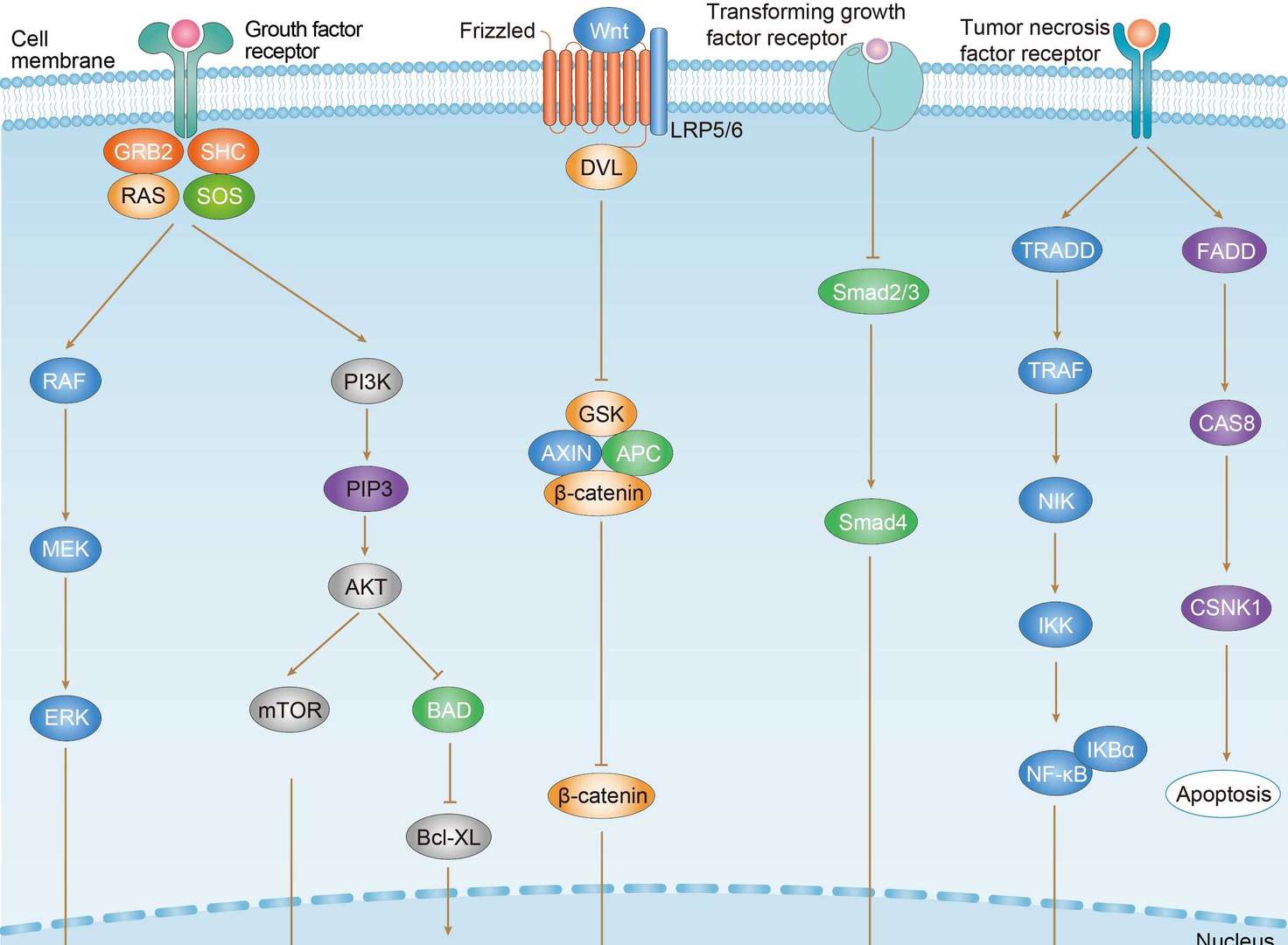
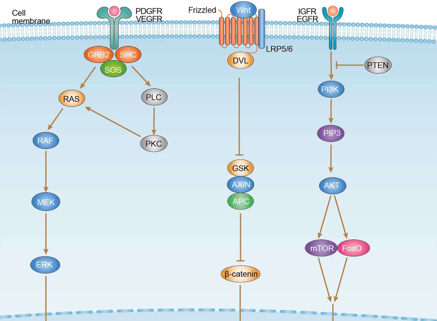
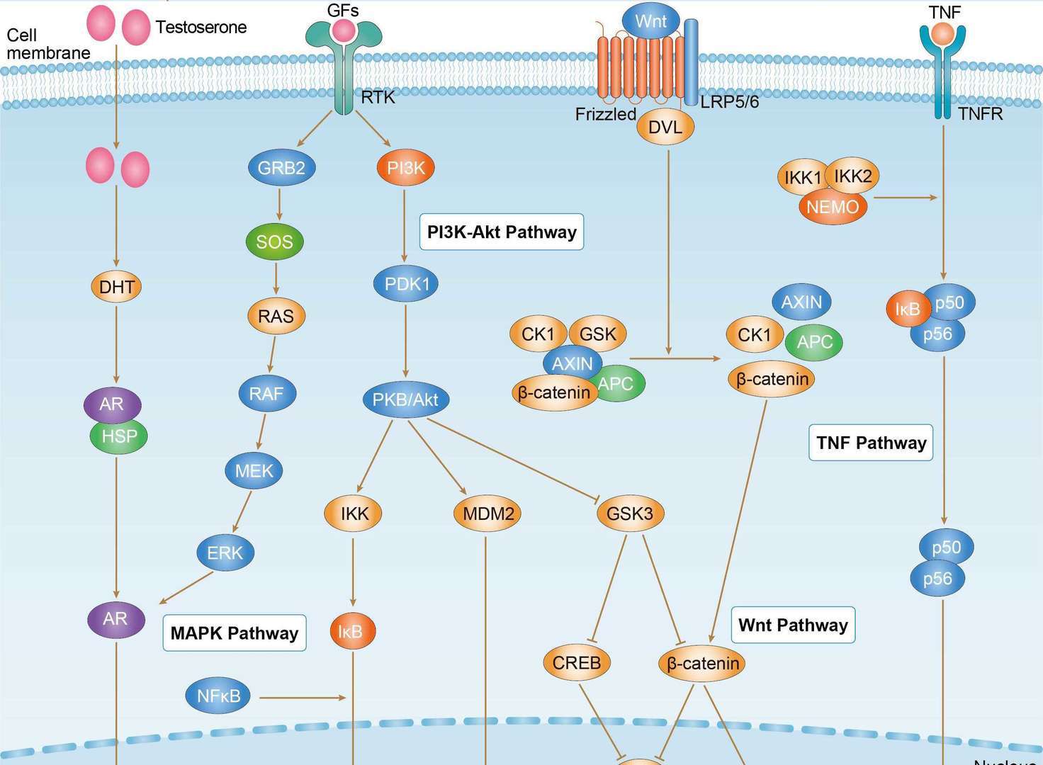
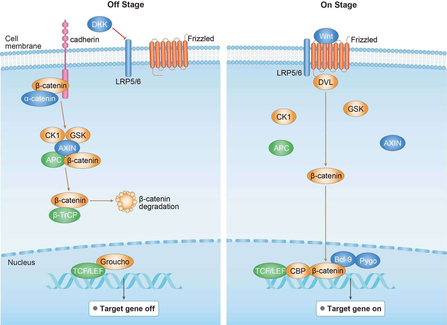 Canonical Wnt Signaling Pathway
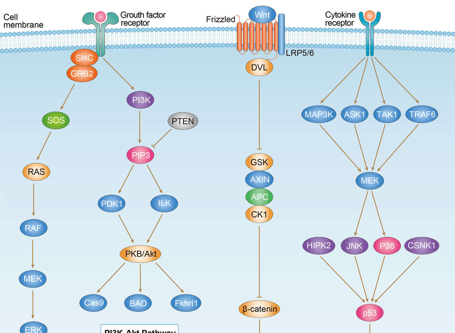
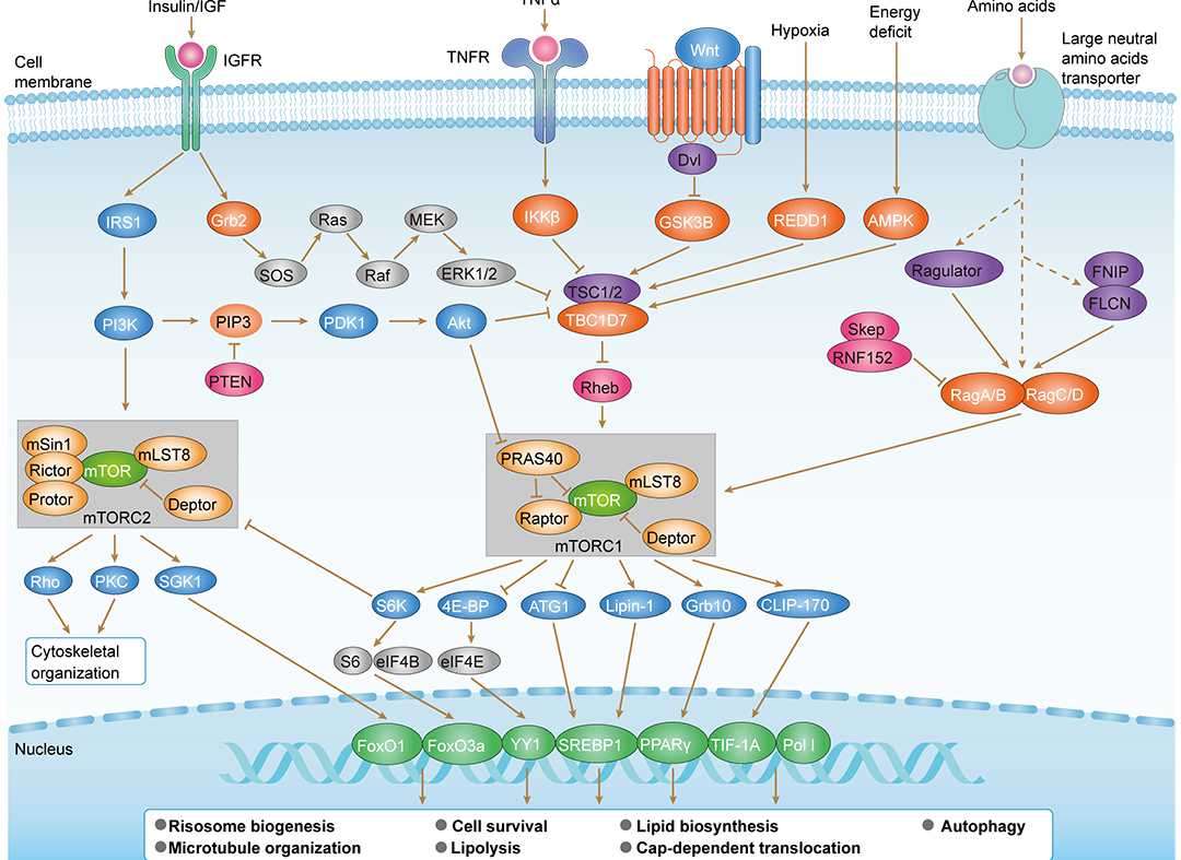 mTOR Signaling Pathway
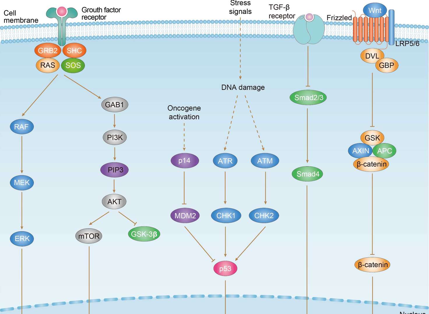
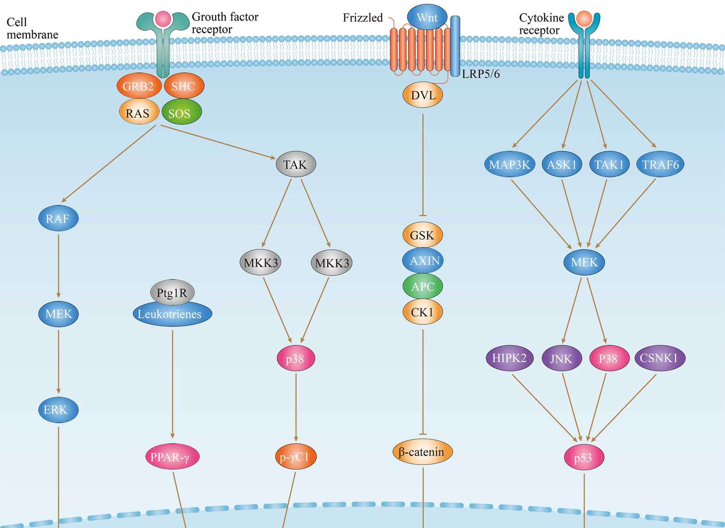
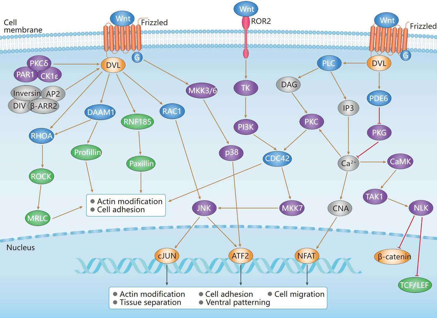 Non-Canonical Wnt Signaling Pathway
Downloads
Download resources about recombinant antibody development and antibody engineering to boost your research.
See other products for "DVL2"
Select a product category from the dropdown menu below to view related products.
Please select product type
IgG Antibody Products
Rabbit Monoclonal Antibody Products
IHC Kit and Antibody Products: Precision Tools for Immunohistochemistry
| CAT | Product Name | Application | Type |
|---|---|---|---|
| DrMAB-312 | Human Anti-DVL2 Recombinant Antibody (clone U8) | IF, IP-MS | Human IgG |
| VS3-WK2001 | Rabbit Anti-DVL2 (pS143) Recombinant Antibody (clone R02-6I1) | WB | Rabbit IgG |
Customer Reviews and Q&As

There are currently no Customer reviews or questions for VS-0525-XY2119. Click the button above to contact us or submit your feedback about this product.
Popular products with customers


Application: Neut, ELISA, IF, IP, FuncS, FC, IHC

Application: FC, IP, ELISA, Neut, FuncS, IF, WB


Application: WB, ELISA, FC, IHC, IP


Application: WB, ELISA, Neut, FuncS

Application: ELISA, IHC, FC, IP, IF, Inhib


Application: Neut, IHC, Activ, FuncS, IF, ELISA


For Research Use Only. Not For Clinical Use.
For research use only. Not intended for any clinical use. No products from Creative Biolabs may be resold, modified for resale or used to manufacture commercial products without prior written approval from Creative Biolabs.
Send Inquiry
This site is protected by reCAPTCHA and the Google Privacy Policy and Terms of Service apply.