

Rabbit Anti-CTNNB1 Recombinant Antibody (VS3-CJ894)
CAT#: VS3-CJ894
This product is a rabbit antibody that recognizes human, mouse, and rat CTNNB1.
Gene Expression

Subcellular Location
Figure 1 IF staining of human cell line U-2 OS
Immunofluorescent staining of human cell line U-2 OS shows localization to plasma membrane.
* Image credit: Image credit: Human Protein Atlas v21.proteinatlas.org/images/29159/if_selected.jpg

Normal Tissue
Figure 2 IHC staining of human stomach
Immunohistochemical staining of human stomach shows strong membranous and cytoplasmic positivity in glandular cells.
* Image credit: Image credit: Human Protein Atlas v21.proteinatlas.org/images/29159/ihc_selected.jpg

Normal Tissue
Figure 3 IHC staining of human colon
Immunohistochemical staining of human colon shows strong cytoplasmic and membranous positivity in glandular cells.
* Image credit: Image credit: Human Protein Atlas v21.proteinatlas.org/images/108/ihc_selected.jpg

Normal Tissue
Figure 4 IHC staining of human liver
Immunohistochemical staining of human liver shows strong positivity of cytoplasm and nuclei in hepatocytes.
* Image credit: Image credit: Human Protein Atlas v21.proteinatlas.org/images/1950/ihc_selected.jpg

Normal Tissue
Figure 5 Cerebral cortex
Endothelial cells
Staining: Medium
Intensity: Moderate
Quantity:>75%
Location: Cytoplasmic/membranous
Neuropil
Staining: Medium
Intensity: Moderate
Quantity:>75%
Location: Cytoplasmic/membranous
* Image credit: Image credit: Human Protein Atlas v21.proteinatlas.org/images/29160/62939_B_8_5.jpg

Normal Tissue
Figure 6 Colon
Endothelial cells
Staining: Medium
Intensity: Moderate
Quantity:>75%
Location: Cytoplasmic/membranous
Glandular cells
Staining: High
Intensity: Strong
Quantity:>75%
Location: Cytoplasmic/membranous
Peripheral nerve/ganglion
Staining: Medium
Intensity: Moderate
Quantity:>75%
Location: Cytoplasmic/membranous
* Image credit: Image credit: Human Protein Atlas v21.proteinatlas.org/images/29160/62939_A_9_3.jpg

Normal Tissue
Figure 7 Liver
Cholangiocytes
Staining: High
Intensity: Strong
Quantity:>75%
Location: Cytoplasmic/membranous
Hepatocytes
Staining: High
Intensity: Strong
Quantity:>75%
Location: Cytoplasmic/membranous
* Image credit: Image credit: Human Protein Atlas v21.proteinatlas.org/images/29160/62939_A_9_4.jpg

Normal Tissue
Figure 8 Pancreas
Exocrine glandular cells
Staining: High
Intensity: Strong
Quantity:>75%
Location: Cytoplasmic/membranous
Pancreatic endocrine cells
Staining: Medium
Intensity: Moderate
Quantity:>75%
Location: Cytoplasmic/membranous
* Image credit: Image credit: Human Protein Atlas v21.proteinatlas.org/images/29160/62939_A_1_3.jpg

Normal Tissue
Figure 9 Kidney
Cells in tubules
Staining: High
Intensity: Strong
Quantity:>75%
Location: Cytoplasmic/membranous
* Image credit: Image credit: Human Protein Atlas v21.proteinatlas.org/images/29160/62939_A_9_5.jpg

Normal Tissue
Figure 10 Testis
Sertoli cells
Staining: Medium
Intensity: Strong
Quantity: <25%
Spermatogonia cells
Staining: High
Intensity: Strong
Quantity: 75%-25%
* Image credit: Image credit: Human Protein Atlas v21.proteinatlas.org/images/29160/62939_A_4_6.jpg

RNA Expression
Figure 11 RNA cell line category: Low cell line specificity
Cell lines ordered by descending RNA expression order.
* Image credit: Image credit: Human Protein Atlas v21.proteinatlas.org/ENSG00000168036-CTNNB1
❮


 ❯
❯
Specifications
- Immunogen
- Recombinant protein
- Host Species
- Rabbit
- Type
- Rabbit IgG
- Specificity
- Human, Mouse, Rat CTNNB1
- Species Reactivity
- Human, Mouse, Rat
- Applications
- WB, ICC, IHC, IP
- Conjugate
- Unconjugated
Product Property
- Purification
- Protein A affinity purified
- Purity
- >95% as determined by SDS-PAGE
- Format
- Liquid
- Buffer
- 40% Glycerol, 1% BSA, TBS, pH7.4.
- Preservative
- 0.05% Sodium Azide
- Storage
- Store at 4°C for short term. Aliquot and store at -20°C for long term. Avoid repeated freeze/thaw cycles.
Applications
- Application Notes
- This antibody has been tested for use in Western Blot, Immunocytochemistry, Immunohistochemistry, Immunoprecipitation.
Target
- Alternative Names
- EVR7; CTNNB; MRD19; NEDSDV; armadillo
- Gene ID
- 1499
- UniProt ID
- P35222
- Sequence Similarities
- Belongs to the beta-catenin family.
- Cellular Localization
- Cell junction, Cell membrane, Cytoplasm, Cytoskeleton, Membrane, Nucleus, Synapse
- Post Translation Modifications
- Phosphorylation at Ser-552 by AMPK promotes stabilizion of the protein, enhancing TCF/LEF-mediated transcription (By similarity).
Phosphorylation by GSK3B requires prior phosphorylation of Ser-45 by another kinase (PubMed:10966653, PubMed:12051714, PubMed:12027456).
Phosphorylation proceeds then from Thr-41 to Ser-37 and Ser-33 (PubMed:12077367, PubMed:25169422).
Phosphorylated by NEK2 (PubMed:18086858).
EGF stimulates tyrosine phosphorylation (PubMed:10187801).
Phosphorylated on Ser-33 and Ser-37 by HIPK2 and GSK3B, this phosphorylation triggers proteasomal degradation (PubMed:20307497).
Phosphorylation on Ser-191 and Ser-246 by CDK5 (PubMed:17009320).
Phosphorylation by CDK2 regulates insulin internalization (PubMed:21262353).
Phosphorylation by PTK6 at Tyr-64, Tyr-142, Tyr-331 and/or Tyr-333 with the predominant site at Tyr-64 is not essential for inhibition of transcriptional activity (PubMed:20026641).
Phosphorylation by SRC at Tyr-333 promotes interaction with isoform M2 of PKM (PKM2); promoting transcription activation (PubMed:22056988).
Ubiquitinated by the SCF(BTRC) E3 ligase complex when phosphorylated by GSK3B, leading to its degradation (PubMed:12077367).
Ubiquitinated by a E3 ubiquitin ligase complex containing UBE2D1, SIAH1, CACYBP/SIP, SKP1, APC and TBL1X, leading to its subsequent proteasomal degradation (PubMed:11389839, PubMed:11389840, PubMed:20307497).
Ubiquitinated and degraded following interaction with SOX9 (By similarity).
S-nitrosylation at Cys-619 within adherens junctions promotes VEGF-induced, NO-dependent endothelial cell permeability by disrupting interaction with E-cadherin, thus mediating disassembly adherens junctions.
O-glycosylation at Ser-23 decreases nuclear localization and transcriptional activity, and increases localization to the plasma membrane and interaction with E-cadherin CDH1.
Deacetylated at Lys-49 by SIRT1.
Phosphorylated at Thr-556 by herpes virus 1/HHV-1 leading to CTNNB1 inhibition.
- Protein Refseq
- NP_001091679.1; NP_001091680.1; NP_001895.1
- Function
- Key downstream component of the canonical Wnt signaling pathway. In the absence of Wnt, forms a complex with AXIN1, AXIN2, APC, CSNK1A1 and GSK3B that promotes phosphorylation on N-terminal Ser and Thr residues and ubiquitination of CTNNB1 via BTRC and its subsequent degradation by the proteasome. In the presence of Wnt ligand, CTNNB1 is not ubiquitinated and accumulates in the nucleus, where it acts as a coactivator for transcription factors of the TCF/LEF family, leading to activate Wnt responsive genes. Involved in the regulation of cell adhesion. Acts as a negative regulator of centrosome cohesion. Involved in the CDK2/PTPN6/CTNNB1/CEACAM1 pathway of insulin internalization. Blocks anoikis of malignant kidney and intestinal epithelial cells and promotes their anchorage-independent growth by down-regulating DAPK2. Disrupts PML function and PML-NB formation by inhibiting RANBP2-mediated sumoylation of PML (PubMed:17524503, PubMed:18077326, PubMed:18086858, PubMed:18957423, PubMed:21262353, PubMed:22647378, PubMed:22699938, PubMed:22155184). Promotes neurogenesis by maintaining sympathetic neuroblasts within the cell cycle (By similarity). Involved in chondrocyte differentiation via interaction with SOX9: SOX9-binding competes with the binding sites of TCF/LEF within CTNNB1, thereby inhibiting the Wnt signaling (By similarity).
REVIEWS AND Q&AS
CITATIONS
RESOURCES
DOWNLOADS
RELATED PRODUCTS
Inquiry
Navs
Customer Review

There are currently no Customer reviews or questions for VS3-CJ894. Click the button above to contact us or submit your feedback about this product.
Submit Your Publication
Published with our product? Submit your paper and receive a 10% discount on your next order! Share your research to earn exclusive rewards.
Related Diseases
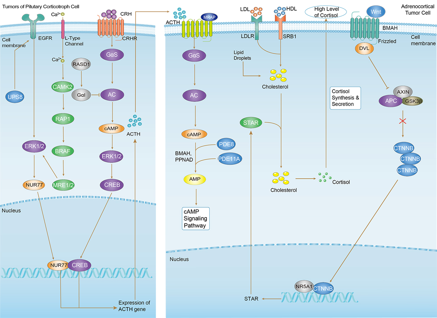 Cushing Syndrome
Downloadable Resources
Download resources about recombinant antibody development and antibody engineering to boost your research.
Product Notes
This is a product of Creative Biolabs' Hi-Affi™ recombinant antibody portfolio, which has several benefits including:
• Increased sensitivity
• Confirmed specificity
• High repeatability
• Excellent batch-to-batch consistency
• Sustainable supply
• Animal-free production
See more details about Hi-Affi™ recombinant antibody benefits.
Datasheet
MSDS
COA
Certificate of Analysis LookupTo download a Certificate of Analysis, please enter a lot number in the search box below. Note: Certificate of Analysis not available for kit components.
Lot Number:
Protocol & Troubleshooting
We have outlined the assay protocols, covering reagents, solutions, procedures, and troubleshooting tips for common issues in order to better assist clients in conducting experiments with our products. View the full list of Protocol & Troubleshooting.
Isotype Control
- CAT
- Product Name
Secondary Antibody
- CAT
- Product Name
See other products for "CTNNB1"
Select a product category from the dropdown menu below to view related products.
| CAT | Product Name | Application | Type |
|---|---|---|---|
| MOB-247 | Recombinant Anti-human CTNNB1 Antibody | WB, FC, FuncS | IgG |
| CAT | Product Name | Application | Type |
|---|---|---|---|
| MOB-247-F(E) | Recombinant Anti-human CTNNB1 Antibody Fab Fragment | ELISA, Nert, FuncS | Fab |
| CAT | Product Name | Application | Type |
|---|---|---|---|
| MOB-247-S(P) | Recombinant Anti-human CTNNB1 Antibody scFv Fragment | ELISA, WB, IP, FuncS | scFv |
| CAT | Product Name | Application | Type |
|---|---|---|---|
| NABG-049 | Recombinant Anti-Mouse Ctnnb1 VHH Single Domain Antibody | ELISA, IHC, FC, FuncS | Llama VHH |
| CAT | Product Name | Application | Type |
|---|---|---|---|
| MHH-247 | Recombinant Human Anti-human CTNNB1 Antibody | ELISA, FuncS | IgG |
| CAT | Product Name | Application | Type |
|---|---|---|---|
| MHH-247-F(E) | Recombinant Human Anti-human CTNNB1 Antibody Fab Fragment | ELISA, WB, Neut, FuncS | Fab |
| CAT | Product Name | Application | Type |
|---|---|---|---|
| MHH-247-S(P) | Recombinant Human Anti-human CTNNB1 Antibody scFv Fragment | ELISA, IP, FuncS | scFv |
| CAT | Product Name | Application | Type |
|---|---|---|---|
| PABZ-140 | Mouse Anti-CTNNB1 Recombinant Antibody (clone BC-22) | ELISA, Block | Mouse IgG |
| CAT | Product Name | Application | Type |
|---|---|---|---|
| PFBZ-140 | Mouse Anti-CTNNB1 Recombinant Antibody (clone BC-22); Fab Fragment | ELISA, Block | Mouse Fab |
| CAT | Product Name | Application | Type |
|---|---|---|---|
| PSBZ-140 | Mouse Anti-CTNNB1 Recombinant Antibody (clone BC-22); scFv Fragment | ELISA, Block | Mouse scFv |
| CAT | Product Name | Application | Type |
|---|---|---|---|
| MOB-0264MZ | Recombinant Mouse Anti-Human CTNNB1 Antibody (clone γ-Catenin-1) | IHC | Mouse antibody |
| CAT | Product Name | Application | Type |
|---|---|---|---|
| MHC-LC1439 | PE-HLA-A2/Human CTNNB1 (YLDSGIHSGA) MHC Tetramer | FCM |
| CAT | Product Name | Application | Type |
|---|---|---|---|
| MHC-LC1440 | APC-HLA-A2/Human CTNNB1 (YLDSGIHSGA) MHC Tetramer | FCM |
| CAT | Product Name | Application | Type |
|---|---|---|---|
| MHC-LC1441 | BV421-HLA-A2/Human CTNNB1 (YLDSGIHSGA) MHC Tetramer | FCM |
| CAT | Product Name | Application | Type |
|---|---|---|---|
| MOR-0859 | Hi-Affi™ Rabbit Anti-CTNNB1 Recombinant Antibody (clone DS859AB) | WB | Rabbit IgG |
| CAT | Product Name | Application | Type |
|---|---|---|---|
| MHC-LC1946 | PE-A2/Human CTNNB1 (GLLGTLVQ) MHC Tetramer | FCM |
| CAT | Product Name | Application | Type |
|---|---|---|---|
| MHC-LC1947 | PE-A2/Human CTNNB1 (GLLGTLVQL) MHC Tetramer | FCM |
| CAT | Product Name | Application | Type |
|---|---|---|---|
| NS-008CN | Mouse Anti-CTNNB1 Recombinant Antibody (clone 13C6) | ELISA, IHC | Mouse IgG1 |
| CAT | Product Name | Application | Type |
|---|---|---|---|
| NS-008CN-F(E) | Mouse Anti-CTNNB1 Recombinant Antibody (clone 13C6); Fab Fragment | ELISA, IHC | Mouse Fab |
| CAT | Product Name | Application | Type |
|---|---|---|---|
| NS-008CN-S(P) | Mouse Anti-CTNNB1 Recombinant Antibody (clone 13C6); scFv Fragment | ELISA, IHC | Mouse scFv |
| CAT | Product Name | Application | Type |
|---|---|---|---|
| MRO-0175-CN | Mouse Anti-CTNNB1 Recombinant Antibody (clone 10-C0-B7) | WB, IF, IHC, FC | Mouse IgG2b |
| CAT | Product Name | Application | Type |
|---|---|---|---|
| MOR-0039-FY | Rabbit Anti-CTNNB1 Recombinant Antibody (clone AFY0010) | ICC, IHC-P, WB | Rabbit IgG |
| CAT | Product Name | Application | Type |
|---|---|---|---|
| VS3-QX299 | Mouse Anti-CTNNB1 Recombinant Antibody (clone 516CT4.3.3) | WB | Mouse IgM |
| CAT | Product Name | Application | Type |
|---|---|---|---|
| VS-1024-XY50 | Mouse Anti-NHP CTNNB1 Recombinant Antibody (clone 15B8) | WB, IHC, IF | Mouse IgG |
| CAT | Product Name | Application | Type |
|---|---|---|---|
| VS-0325-XY577 | Anti-CTNNB1 Immunohistochemistry Kit | IHC |
| CAT | Product Name | Application | Type |
|---|---|---|---|
| VS-0525-XY1770 | Anti-Mouse CTNNB1 Immunohistochemistry Kit | IHC |
| CAT | Product Name | Application | Type |
|---|---|---|---|
| VS-0525-XY1769 | Anti-Human CTNNB1 Immunohistochemistry Kit | IHC |
| CAT | Product Name | Application | Type |
|---|---|---|---|
| VS-0525-XY1771 | Anti-Sheep CTNNB1 Immunohistochemistry Kit | IHC |
| CAT | Product Name | Application | Type |
|---|---|---|---|
| VS-1025-YC17 | Anti-CTNNB1 Antibody Prodrug, Protease Activated (13C6) | ISZ, Cyt, FuncS |
| CAT | Product Name | Application | Type |
|---|---|---|---|
| VS-0126-XL214 | Rabbit Anti-beta Catenin (phospho S33/S37) Monoclonal Antibody | Phospho Antibodies for Cell Signaling Research |
Specific Inquiry

See Our Custom Production in Action


Popular Products

Application: WB, FuncS, IF, Neut, ELISA, FC, IP

Application: Neut, ELISA, IF, IP, FuncS, FC, ICC

Application: Neut, ELISA, IF, IP, FuncS, FC, ICC

Application: ELISA, FC, IP, FuncS, IF, Neut, ICC

Application: IF, IP, Neut, FuncS, ELISA, FC, ICC

Application: FC, IHC, FuncS, Inhib, Cyt

Application: FuncS, IF, Neut, ELISA, FC, IP, WB

Application: Neut, ELISA, IF, IP, FuncS, FC, ICC

Application: ELISA, FC, IP, FuncS, IF, Neut, ICC

Application: ELISA, FC, IP, FuncS, IF, Neut, ICC

Application: FC, IP, ELISA, Neut, FuncS, IF, ICC

Application: ELISA, IP, FC, FuncS, Neut, IF, ICC


Application: WB, ELISA, FuncS


Application: ELISA, IP, WB, IHC, IF, FuncS

Application: FuncS, Inhib, IP, ELISA


For research use only. Not intended for any clinical use. No products from Creative Biolabs may be resold, modified for resale or used to manufacture commercial products without prior written approval from Creative Biolabs.
Send Inquiry
This site is protected by reCAPTCHA and the Google Privacy Policy and Terms of Service apply.