

AbPlus™ Anti-EGFR Magnetic Beads (pSEX81-6)
CAT#: VS-0424-XY84
The AbPlus Anti-EGFR Magnetic Beads (pSEX81-6) is an innovative affinity resin which is bound with anti-EGFR specific antibody (pSEX81-6). The beads were designed for small-scale affinity purification and immunoprecipitation (IP) of EGFR protein under native and denaturing conditions.
Gene Expression

Subcellular Location and Protein Expression
Figure 1 IF staining of human cell line A-431
Immunofluorescent staining of human cell line A-431 shows localization to plasma membrane & cell junctions.
* Image credit: Image credit: Human Protein Atlas https://v21.proteinatlas.org/images/18530/144_E9_2_selected.jpg

Subcellular Location and Protein Expression
Figure 2 IF staining of human cell line U-251 MG
Immunofluorescent staining of human cell line U-251 MG shows localization to plasma membrane.
* Image credit: Image credit: Human Protein Atlas https://v21.proteinatlas.org/images/18530/143_E9_1_blue_red_green.jpg

Normal Tissue
Figure 3 Colon
Glandular cells
Staining:Medium
Intensity: Moderate
Quantity:>75%
Location: Cytoplasmic/membranous
* Image credit: Image credit: Human Protein Atlas https://v21.proteinatlas.org/images/18530/41191_A_9_3.jpg

Normal Tissue
Figure 4 Liver
Cholangiocytes
Staining:Medium
Intensity: Moderate
Quantity: 75%-25%
Location: Cytoplasmic/membranous
* Image credit: Image credit: Human Protein Atlas https://v21.proteinatlas.org/images/18530/41191_A_7_4.jpg

Normal Tissue
Figure 5 Kidney
Bowman's capsule
Staining:Medium
Intensity: Moderate
Quantity: 75%-25%
Collecting ducts
Staining:Medium
Intensity: Strong
Quantity: <25%
Distal tubules
Staining:Medium
Intensity: Strong
Quantity: <25%
Proximal tubules (cell body)
Staining:Medium
Intensity: Strong
Quantity: <25%
* Image credit: Image credit: Human Protein Atlas https://v21.proteinatlas.org/images/18530/41191_A_9_5.jpg

Normal Tissue
Figure 6 Testis
Leydig cells
Staining:Medium
Intensity: Moderate
Quantity:>75%
Pachytene spermatocytes
Staining:Medium
Intensity: Moderate
Quantity: 75%-25%
Round or early spermatids
Staining:Medium
Intensity: Moderate
Quantity: 75%-25%
* Image credit: Image credit: Human Protein Atlas https://v21.proteinatlas.org/images/18530/41191_A_6_6.jpg

Normal Tissue
Figure 7 Placenta
Cytotrophoblasts
Staining:High
Intensity: Strong
Quantity:>75%
Decidual cells
Staining:Medium
Intensity: Moderate
Quantity:>75%
Hofbauer cells
Staining:High
Intensity: Strong
Quantity:>75%
Syncytiotrophoblasts - cell body
Staining:High
Intensity: Strong
Quantity:>75%
Syncytiotrophoblasts - microvilli
Staining:High
Intensity: Strong
* Image credit: Image credit: Human Protein Atlas https://v21.proteinatlas.org/images/18530/41191_A_1_7.jpg

Normal Tissue
Figure 8 Lymph node
Non-germinal center cells
Staining:Medium
Intensity: Strong
Quantity: <25%
Location: Cytoplasmic/membranous
* Image credit: Image credit: Human Protein Atlas https://v21.proteinatlas.org/images/18530/41191_A_7_8.jpg

RNA Expression
Figure 9 RNA cell line category: Cell line enriched (A-431)
Cell lines ordered by descending RNA expression order.
* Image credit: Image credit: Human Protein Atlas https://v21.proteinatlas.org/ENSG00000146648-EGFR
❮


 ❯
❯
Specifications
- Applications
- Immunoprecipitation, Protein Purification
- Matrix
- Magrose bead (> 50 μmol/mL gel)
- Bead Ligand
- Anti-EGFR specific antibody (pSEX81-6)
- Target
- EGFR
- Immunogen
- Purified EGFR
- Target Species
- Human
- Antibody Clone
- pSEX81-6
- Bead Capacity
- 20-30 mg/mL binding antibody
- Bead size
- 10-37 μm
- Stability
- pH 2-14
- Format
- 20% Suspension
- Buffer
- PBS, pH 7.4, with 1% BSA
- Preservative
- 0.03% Proclin 300
- Storage
- Stored at 4°C, and is stable for up to 2 years. Do not centrifuge, dry or freeze the magnetic beads.
Applications
- Application Notes
- The beads are in suspension and will settle upon storage. Prior to use, mix the vial gently (do not vortex) to ensure delivery of proper bead volume.
Target
- Introduction
- The protein encoded by this gene is a transmembrane glycoprotein that is a member of the protein kinase superfamily. This protein is a receptor for members of the epidermal growth factor family. EGFR is a cell surface protein that binds to epidermal growth factor. Binding of the protein to a ligand induces receptor dimerization and tyrosine autophosphorylation and leads to cell proliferation. Mutations in this gene are associated with lung cancer. Multiple alternatively spliced transcript variants that encode different protein isoforms have been found for this gene.
- Alternative Names
- EGFR; epidermal growth factor receptor; ERBB; HER1; mENA; ERBB1; PIG61; NISBD2; proto-oncogene c-ErbB-1; cell growth inhibiting protein 40; erb-b2 receptor tyrosine kinase 1; cell proliferation-inducing protein 61; receptor tyrosine-protein kinase erbB-1; avian erythroblastic leukemia viral (v-erb-b) oncogene homolog
- Gene ID
- 1956
- UniProt ID
- P00533
REVIEWS AND Q&AS
CITATIONS
RESOURCES
DOWNLOADS
RELATED PRODUCTS
Inquiry
Navs
Customer Review

There are currently no Customer reviews or questions for VS-0424-XY84. Click the button above to contact us or submit your feedback about this product.
Submit Your Publication
Published with our product? Submit your paper and receive a 10% discount on your next order! Share your research to earn exclusive rewards.
Related Diseases
 Bladder Cancer
 Non-small Cell Lung Cancer
 Pancreatic Cancer
 Hepatocellular Carcinoma
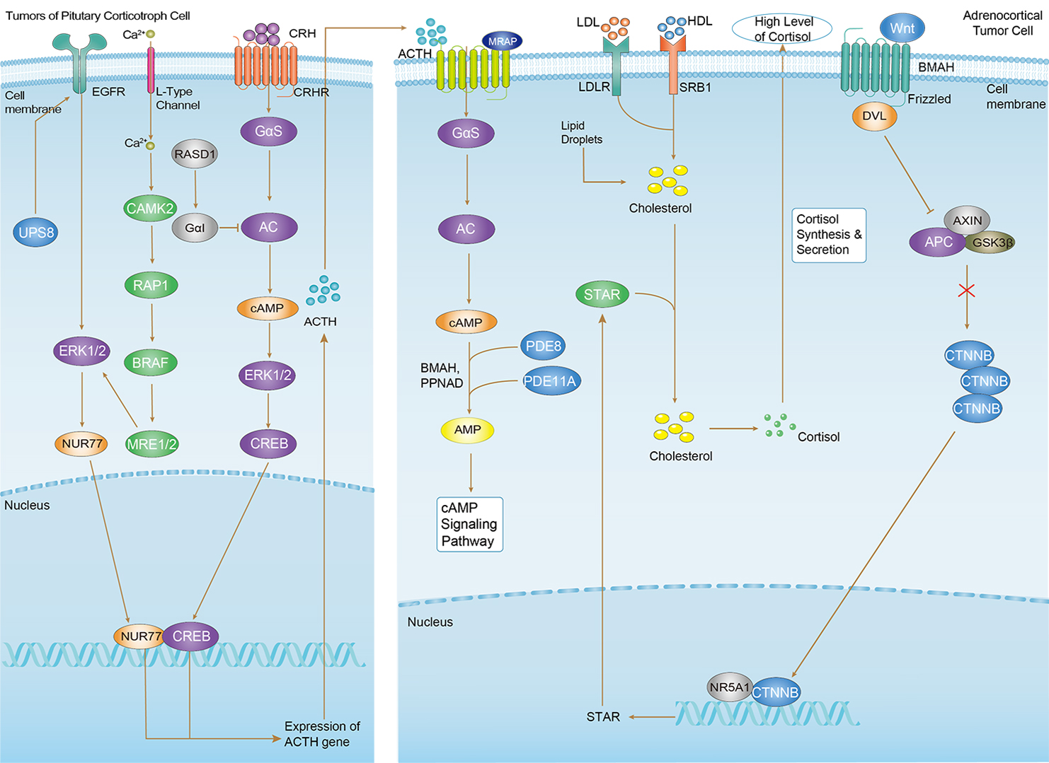 Cushing Syndrome
Related Signaling Pathways
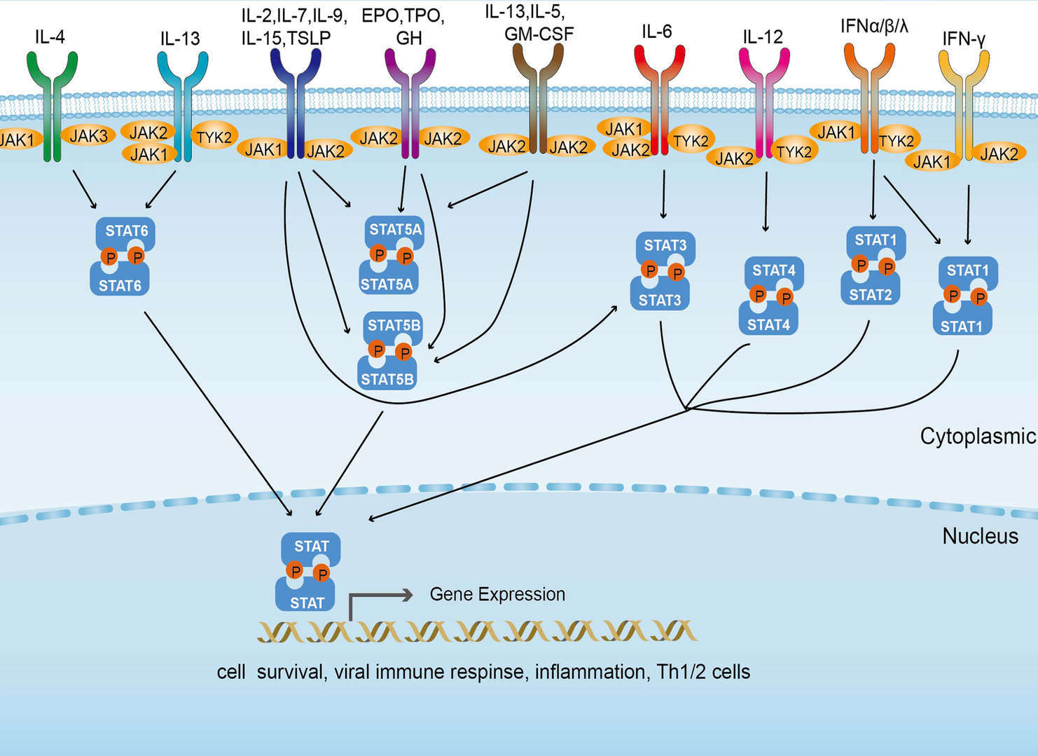 JAK-STAT Signaling Pathway
Downloadable Resources
Download resources about recombinant antibody development and antibody engineering to boost your research.
Datasheet
MSDS
COA
Certificate of Analysis LookupTo download a Certificate of Analysis, please enter a lot number in the search box below. Note: Certificate of Analysis not available for kit components.
Lot Number:
See other products for "EGFR"
Select a product category from the dropdown menu below to view related products.
| CAT | Product Name | Application | Type |
|---|---|---|---|
| TAB-750 | Anti-EGFR/HER1 Recombinant Antibody (TAB-750) | Neut, ELISA, IF, IP, FuncS, FC, WB | IgG1 - kappa |
| CAT | Product Name | Application | Type |
|---|---|---|---|
| MOB-1078z | Mouse Anti-EGFR Recombinant Antibody (clone 42C11) | WB, ELISA, FC, IF, IHC, FuncS | Mouse IgG1, κ |
| CAT | Product Name | Application | Type |
|---|---|---|---|
| NABG-056 | Recombinant Anti-Mouse Egfr VHH Single Domain Antibody | ELISA, IHC, FC, FuncS | Llama VHH |
| CAT | Product Name | Application | Type |
|---|---|---|---|
| TAB-H35 | Anti-Human EGFR Recombinant Antibody (Futuximab) | IF, WB, Inhib | IgG1 - kappa |
| CAT | Product Name | Application | Type |
|---|---|---|---|
| TAB-020 | Anti-Human EGFR Recombinant Antibody (Panitumumab) | ELISA, IP, FC, FuncS, Neut, IF, ICC | IgG2 - kappa |
| CAT | Product Name | Application | Type |
|---|---|---|---|
| TAB-165 | Anti-Human EGFR Recombinant Antibody (Matuzumab) | Neut, ELISA, IF, IP, FuncS, FC, ICC | IgG1 |
| CAT | Product Name | Application | Type |
|---|---|---|---|
| TAB-710 | Anti-EGFR Recombinant Antibody (Nimotuzumab) | ELISA, IP, FC, FuncS, Neut, IF, IHC | IgG1 - kappa |
| CAT | Product Name | Application | Type |
|---|---|---|---|
| TAB-040 | Anti-Human EGFR Recombinant Antibody (TAB-040) | ELISA, FC, IP, FuncS, IF, Neut, ICC | IgG1 - kappa |
| CAT | Product Name | Application | Type |
|---|---|---|---|
| TAB-119 | Anti-Human EGFR Recombinant Antibody (TAB-119) | FC, IP, ELISA, Neut, FuncS, IF, WB | IgG1 - kappa |
| CAT | Product Name | Application | Type |
|---|---|---|---|
| TAB-753 | Anti-EGFR Recombinant Antibody (Imgatuzumab) | Neut, ELISA, IF, IP, FuncS, FC, WB | IgG1 - kappa |
| CAT | Product Name | Application | Type |
|---|---|---|---|
| TAB-003 | Anti-Human EGFR Recombinant Antibody (Cetuximab) | IF, IP, Neut, FuncS, ELISA, FC, ICC | IgG1 - kappa |
| CAT | Product Name | Application | Type |
|---|---|---|---|
| TAB-H49 | Anti-Human EGFR Recombinant Antibody (Modotuximab) | FuncS, IF, Neut, ELISA, FC, IP, IHC | IgG1 - kappa |
| CAT | Product Name | Application | Type |
|---|---|---|---|
| TAB-228CL | Anti-Human EGFR Recombinant Antibody (ABT-806) | WB, IHC | Antibody |
| CAT | Product Name | Application | Type |
|---|---|---|---|
| MOB-0242MC | Rabbit Anti-Human EGFR (phospho Y1092) Antibody | IHC, WB |
| CAT | Product Name | Application | Type |
|---|---|---|---|
| MOB-0243MC | Rabbit Anti-Human EGFR (phospho Y1068) Antibody | IHC, WB |
| CAT | Product Name | Application | Type |
|---|---|---|---|
| PABL-080 | Human Anti-EGFR Recombinant Antibody (PABL-080) | ELISA, WB, FuncS | Human IgG |
| CAT | Product Name | Application | Type |
|---|---|---|---|
| PSBL-080 | Human Anti-EGFR Recombinant Antibody; scFv Fragment (PSBL-080) | ELISA, WB, FuncS | Human scFv |
| CAT | Product Name | Application | Type |
|---|---|---|---|
| PFBL-080 | Human Anti-EGFR Recombinant Antibody; Fab Fragment (PFBL-080) | ELISA, WB, FuncS | Human Fab |
| CAT | Product Name | Application | Type |
|---|---|---|---|
| PNBL-016 | Recombinant Anti-Human EGFR VHH Single Domain Antibody (PNBL-016) | WB, ELISA | Llama VHH |
| CAT | Product Name | Application | Type |
|---|---|---|---|
| PNBL-017 | Recombinant Anti-Human EGFR VHH Single Domain Antibody (PNBL-017) | FuncS, ELISA, IF | Llama VHH |
| CAT | Product Name | Application | Type |
|---|---|---|---|
| PNBL-018 | Recombinant Anti-Human EGFR VHH Single Domain Antibody (PNBL-018) | FuncS, SPR | Llama VHH |
| CAT | Product Name | Application | Type |
|---|---|---|---|
| PABZ-039 | Mouse Anti-EGFR Recombinant Antibody (clone mAb528) | FC | Mouse IgG |
| CAT | Product Name | Application | Type |
|---|---|---|---|
| PFBZ-039 | Mouse Anti-EGFR Recombinant Antibody (clone mAb528); Fab Fragment | FC | Mouse Fab |
| CAT | Product Name | Application | Type |
|---|---|---|---|
| PFBW-039 | Human Anti-EGFR Recombinant Antibody Fab Fragment (PFBW-039) | FuncS | Chimeric (mouse/human) Fab |
| CAT | Product Name | Application | Type |
|---|---|---|---|
| PFBC-040 | Human Anti-EGFR Recombinant Antibody (clone MR1); Fab Fragment | Block | Human Fab |
| CAT | Product Name | Application | Type |
|---|---|---|---|
| PFBL-459 | Human Anti-EGFR Recombinant Antibody (clone C225); Fab Fragment | FC | Human Fab |
| CAT | Product Name | Application | Type |
|---|---|---|---|
| PFBW-171 | Mouse Anti-EGFR Recombinant Antibody; Fab Fragment (PFBW-171) | WB | Mouse Fab |
| CAT | Product Name | Application | Type |
|---|---|---|---|
| PSBZ-039 | Mouse Anti-EGFR Recombinant Antibody (clone mAb528); scFv Fragment | FC | Mouse scFv |
| CAT | Product Name | Application | Type |
|---|---|---|---|
| PSBW-039 | Mouse Anti-EGFR Recombinant Antibody scFv Fragment (PSBW-039) | Block | Mouse scFv |
| CAT | Product Name | Application | Type |
|---|---|---|---|
| PSBC-040 | Human Anti-EGFR Recombinant Antibody (clone MR1); scFv Fragment | Block | Human scFv |
| CAT | Product Name | Application | Type |
|---|---|---|---|
| TAB-0225CL | Human Anti-EGFR Recombinant Antibody (TAB-0225CL) | Block, Inhib, FuncS, Apop, In vivo | Chimeric (Mouse/Human) IgG1 |
| CAT | Product Name | Application | Type |
|---|---|---|---|
| TAB-0564CL | Mouse Anti-EGFR Recombinant Antibody (TAB-0564CL) | ELISA | Mouse IgG |
| CAT | Product Name | Application | Type |
|---|---|---|---|
| TAB-0565CL | Mouse Anti-EGFR Recombinant Antibody (TAB-0565CL) | ELISA | Mouse IgG |
| CAT | Product Name | Application | Type |
|---|---|---|---|
| TAB-0564CL-S(P) | Mouse Anti-EGFR Recombinant Antibody; scFv Fragment (TAB-0564CL-S(P)) | ELISA | Mouse scFv |
| CAT | Product Name | Application | Type |
|---|---|---|---|
| TAB-0565CL-S(P) | Mouse Anti-EGFR Recombinant Antibody; scFv Fragment (TAB-0565CL-S(P)) | ELISA | Mouse scFv |
| CAT | Product Name | Application | Type |
|---|---|---|---|
| TAB-0564CL-F(E) | Mouse Anti-EGFR Recombinant Antibody; Fab Fragment (TAB-0564CL-F(E)) | ELISA | Mouse Fab |
| CAT | Product Name | Application | Type |
|---|---|---|---|
| TAB-270MZ | Human Anti-EGFR Recombinant Antibody (TAB-270MZ) | ELISA | Human antibody |
| CAT | Product Name | Application | Type |
|---|---|---|---|
| TAB-274MZ | Human Anti-EGFR Recombinant Antibody (TAB-274MZ) | FC | Humanized IgG |
| CAT | Product Name | Application | Type |
|---|---|---|---|
| TAB-278MZ | Human Anti-EGFR Recombinant Antibody (TAB-278MZ) | Cyt, ELISA, Inhib | Human IgG |
| CAT | Product Name | Application | Type |
|---|---|---|---|
| TAB-015MZ-VHH | Anti-Human EGFR Recombinant Antibody (TAB-015MZ-VHH) | sELISA | Single domain antibody |
| CAT | Product Name | Application | Type |
|---|---|---|---|
| Gly-055LC | Recombinant Anti-Human EGFR Antibody (Fc glycosylation/High-mannose glycosylated) | ELISA | Chimeric antibody (mouse/human) |
| Gly-055LC-1 | Recombinant Anti-Human EGFR Antibody (Fc glycosylation/High-mannose glycosylated) | ELISA | Chimeric antibody (mouse/human) |
| CAT | Product Name | Application | Type |
|---|---|---|---|
| Gly-144LC | Recombinant Anti-Human EGFR Antibody (Fc glycosylation) | ELISA | Humanized antibody |
| CAT | Product Name | Application | Type |
|---|---|---|---|
| Gly-167LC | Recombinant Anti-Human EGFR Antibody (Non-glycosylated) | ELISA | Human antibody |
| CAT | Product Name | Application | Type |
|---|---|---|---|
| BRD-0183MZ | Chicken Anti-EGFR Polyclonal IgY | WB | Chicken antibody |
| CAT | Product Name | Application | Type |
|---|---|---|---|
| MHC-LC773 | A*0201/Human EGFR (YLNTVQPTCV) MHC Tetramer | FCM |
| CAT | Product Name | Application | Type |
|---|---|---|---|
| NEUT-722CQ | Rabbit Anti-EGFR Recombinant Antibody (clone CBL1011) | Neut | Rabbit IgG |
| CAT | Product Name | Application | Type |
|---|---|---|---|
| NEUT-723CQ | Mouse Anti-EGFR Recombinant Antibody (clone CBL931) | WB, IP, IHC, ICC, Neut | Mouse IgG1 |
| CAT | Product Name | Application | Type |
|---|---|---|---|
| NEUT-724CQ | Rabbit Anti-EGFR Recombinant Antibody (NEUT-724CQ) | IF, FC, WB, IP, Neut | Rabbit IgG |
| CAT | Product Name | Application | Type |
|---|---|---|---|
| MOR-1101 | Hi-Affi™ Rabbit Anti-EGFR Recombinant Antibody (clone DS1101AB) | IHC-P | Rabbit IgG |
| CAT | Product Name | Application | Type |
|---|---|---|---|
| MOR-4520 | Hi-Affi™ Rabbit Anti-EGFR Recombinant Antibody (clone TH28DS) | IF, ICC, FC | Rabbit IgG |
| CAT | Product Name | Application | Type |
|---|---|---|---|
| MOR-4570 | Hi-Affi™ Rabbit Anti-EGFR Recombinant Antibody (clone TH82DS) | ELISA | Rabbit IgG |
| CAT | Product Name | Application | Type |
|---|---|---|---|
| MOR-4571 | Hi-Affi™ Rabbit Anti-EGFR Recombinant Antibody (clone TH83DS) | WB, IF, ICC, FC | Rabbit IgG |
| CAT | Product Name | Application | Type |
|---|---|---|---|
| MOR-4675 | Hi-Affi™ Rabbit Anti-EGFR Recombinant Antibody (clone TH189DS) | WB, IF, ICC, FC | Rabbit IgG |
| CAT | Product Name | Application | Type |
|---|---|---|---|
| MHC-LC4545 | PE-DQB1*03:02/Human EGFR (SRALEEKKGNYVVTHG) MHC Tetramer | FCM |
| CAT | Product Name | Application | Type |
|---|---|---|---|
| AFC-TAB-165 | Afuco™ Anti-EGFR ADCC Recombinant Antibody, ADCC Enhanced (AFC-TAB-165) | Neut, ELISA, IF, IP, FuncS, FC | ADCC enhanced antibody |
| CAT | Product Name | Application | Type |
|---|---|---|---|
| AFC-TAB-464CQ | Afuco™ Anti-EGFR ADCC Recombinant Antibody, ADCC Enhanced (AFC-TAB-464CQ) | ELISA, IHC, FC, IP, IF, FuncS | ADCC enhanced antibody |
| CAT | Product Name | Application | Type |
|---|---|---|---|
| AFC-TAB-003 | Afuco™ Anti-EGFR ADCC Recombinant Antibody, ADCC Enhanced (AFC-TAB-003) | IF, IP, Neut, FuncS, ELISA, FC | ADCC enhanced antibody |
| CAT | Product Name | Application | Type |
|---|---|---|---|
| AFC-TAB-040 | Afuco™ Anti-EGFR ADCC Recombinant Antibody, ADCC Enhanced (AFC-TAB-040) | ELISA, FC, IP, FuncS, IF, Neut | ADCC enhanced antibody |
| CAT | Product Name | Application | Type |
|---|---|---|---|
| AFC-TAB-119 | Afuco™ Anti-EGFR ADCC Recombinant Antibody, ADCC Enhanced (AFC-TAB-119) | FC, IP, ELISA, Neut, FuncS, IF | ADCC enhanced antibody |
| CAT | Product Name | Application | Type |
|---|---|---|---|
| VS-0924-YC32 | Mouse Anti-EGFR Recombinant Antibody (VS-0924-YC32) - Cancer Stem Cell Marker | IHC, WB | Mouse IgG1 |
| CAT | Product Name | Application | Type |
|---|---|---|---|
| VS-0924-YC35 | Rabbit Anti-EGFR Antibody (VS-0924-YC35) - Cancer Stem Cell Marker | IHC, WB, IF | Rabbit IgG |
| CAT | Product Name | Application | Type |
|---|---|---|---|
| VS-1024-XY177 | Mouse Anti-NHP EGFR Recombinant Antibody (clone 225) | IF, IP | Mouse IgG1 |
| CAT | Product Name | Application | Type |
|---|---|---|---|
| VS-0125-FY28 | Human Anti-EGFR (clone ABT-806) scFv-Fc Chimera | FC, Cyt | Human IgG1, scFv-Fc |
| CAT | Product Name | Application | Type |
|---|---|---|---|
| VS-0225-XY102 | CytoStream™ Mouse Anti-EGFR Recombinant Antibody (VS-0225-XY102) | FC | Mouse IgG1, kappa |
| CAT | Product Name | Application | Type |
|---|---|---|---|
| VS-0325-XY735 | Anti-EGFR Immunohistochemistry Kit | IHC |
| CAT | Product Name | Application | Type |
|---|---|---|---|
| VS-0425-YC340 | Recombinant Anti-EGFR Vesicular Antibody, EV Displayed (VS-0425-YC340) | ELISA, FC, Neut, Cell-uptake |
| CAT | Product Name | Application | Type |
|---|---|---|---|
| VS-0525-XY2183 | Anti-Mouse EGFR Immunohistochemistry Kit | IHC |
| CAT | Product Name | Application | Type |
|---|---|---|---|
| VS-0525-YC65 | Recombinant Anti-EGFR (AA 269-278 x AA 526-535) Biparatopic Antibody, Tandem scFv (Clone Pep 2 x Clone Pep 3) | FC | Tandem scFv |
| CAT | Product Name | Application | Type |
|---|---|---|---|
| VS-0525-YC66 | Recombinant Anti-EGFR (AA 582-591 x AA 606-614) Biparatopic Antibody, Tandem scFv (Clone Pep 4 x Clone Pep 1) | FC | Tandem scFv |
| CAT | Product Name | Application | Type |
|---|---|---|---|
| VS-0525-YC68 | Recombinant Anti-EGFR (AA 526-535 x AA 600-605) Biparatopic Antibody, Tandem scFv (Clone Pep 3 x Clone Pep 5) | FC | Tandem scFv |
| CAT | Product Name | Application | Type |
|---|---|---|---|
| VS-0525-YC213 | Recombinant Anti-EGFR (Domain II x Domain III) Biparatopic Antibody, Tandem scFv | ELISA, FC, IF, IHC, IP | Tandem scFv |
| CAT | Product Name | Application | Type |
|---|---|---|---|
| VS-0525-XY2182 | Anti-Human EGFR Immunohistochemistry Kit | IHC |
| CAT | Product Name | Application | Type |
|---|---|---|---|
| VS-0825-YC110 | SmartAb™ Recombinant Anti-EGFR pH-dependent Antibody (VS-0825-YC110) | Neut, ELISA, IF, IP, FC, WB | Human IgG1 kappa |
| CAT | Product Name | Application | Type |
|---|---|---|---|
| VS-1025-YC4 | Anti-EGFR Antibody Prodrug, Protease Activated (clone 528) | ISZ, Cyt, FuncS |
| CAT | Product Name | Application | Type |
|---|---|---|---|
| VS-1025-YC5 | Anti-EGFR Antibody Prodrug, Protease Activated (Cetuximab) | ISZ, Cyt, FuncS |
| CAT | Product Name | Application | Type |
|---|---|---|---|
| VS-1025-YC6 | Anti-EGFR Antibody Prodrug, Protease Activated (Panitumumab) | ISZ, Cyt, FuncS |
| CAT | Product Name | Application | Type |
|---|---|---|---|
| VS-1125-XY308 | Rabbit Anti-EGFR Recombinant Antibody (VS-1125-XY308) | WB, IHC, ICC, IF, FC, IP | Rabbit IgG |
| CAT | Product Name | Application | Type |
|---|---|---|---|
| VS-1125-XY309 | Rabbit Anti-EGFR Recombinant Antibody (VS-1125-XY309) | WB, ICC, IF, FC, IP | Rabbit IgG |
| CAT | Product Name | Application | Type |
|---|---|---|---|
| VS-1125-XY310 | Mouse Anti-EGFR Recombinant Antibody (VS-1125-XY310) | WB, IHC, ICC, IF, FC | Mouse IgG2a |
| CAT | Product Name | Application | Type |
|---|---|---|---|
| VS-1125-XY311 | Mouse Anti-EGFR Recombinant Antibody (VS-1125-XY311) | WB | Mouse IgG |
| CAT | Product Name | Application | Type |
|---|---|---|---|
| VS-1125-XY312 | Mouse Anti-EGFR Recombinant Antibody (VS-1125-XY312) | WB, IHC, ICC, IF, ELISA, IP | Mouse IgG1 |
Specific Inquiry
See Our Custom Production in Action


Popular Products

Application: WB, FuncS, IF, Neut, ELISA, FC, IP

Application: FC, IP, ELISA, Neut, FuncS, IF, IHC

Application: ELISA, FC, IP, FuncS, IF, Neut, ICC

Application: ELISA, IP, FC, FuncS, Neut, IF, ICC


Application: FuncS, IF, Neut, ELISA, FC, IP, IHC

Application: FC, IP, ELISA, Neut, FuncS, IF, WB

Application: IP, IF, FuncS, FC, Neut, ELISA, ICC

Application: IF, IP, Neut, FuncS, ELISA, FC, WB

Application: Neut, ELISA, IF, IP, FuncS, FC, ICC

Application: IP, IF, FuncS, FC, Neut, ELISA, IHC


Application: WB, Neut, FuncS

Application: WB, ELISA, FuncS


Application: WB, IF, FuncS


For research use only. Not intended for any clinical use. No products from Creative Biolabs may be resold, modified for resale or used to manufacture commercial products without prior written approval from Creative Biolabs.
Send Inquiry
This site is protected by reCAPTCHA and the Google Privacy Policy and Terms of Service apply.